. 2014 Aug;49(8):1206-16.
doi: 10.1007/s00535-013-0865-3. Epub 2013 Aug 8.

Oral caffeine administration ameliorates acute colitis by suppressing chitinase 3-like 1 expression in intestinal epithelial cells

Affiliations

Oral caffeine administration ameliorates acute colitis by suppressing chitinase 3-like 1 expression in intestinal epithelial cells

In-Ah Lee et al. J Gastroenterol. 2014 Aug.

Abstract

Background: The initial trigger of inflammatory bowel disease (IBD) can be partly attributed towards the interaction and invasion of intestinal epithelial cells (IECs) and submucosal compartments. Identifying safe and economical methods to block these interactions may help prevent the onset of early colitis. Chitinase 3-like 1 (CHI3L1) is an inducible host protein that facilitates bacterial attachment and invasion on/into IECs. Therefore, we test the hypothesis of inhibiting CHI3L1 using the pan-chitinase inhibitor caffeine to reduce the likelihood of early colitis onset.

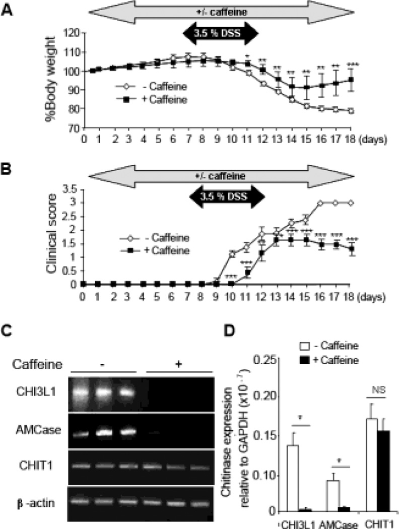
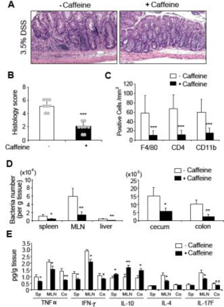
Methods: IEC lines were treated with caffeine (2.5 or 5 mM) and analyzed for CHI3L1 expression and the impact on bacterial invasion. In vivo, mice were treated with 2.5 mM caffeine and induced with 3.5 % dextran sulfate sodium (DSS)-mediated colitis and subsequently analyzed colitis development.

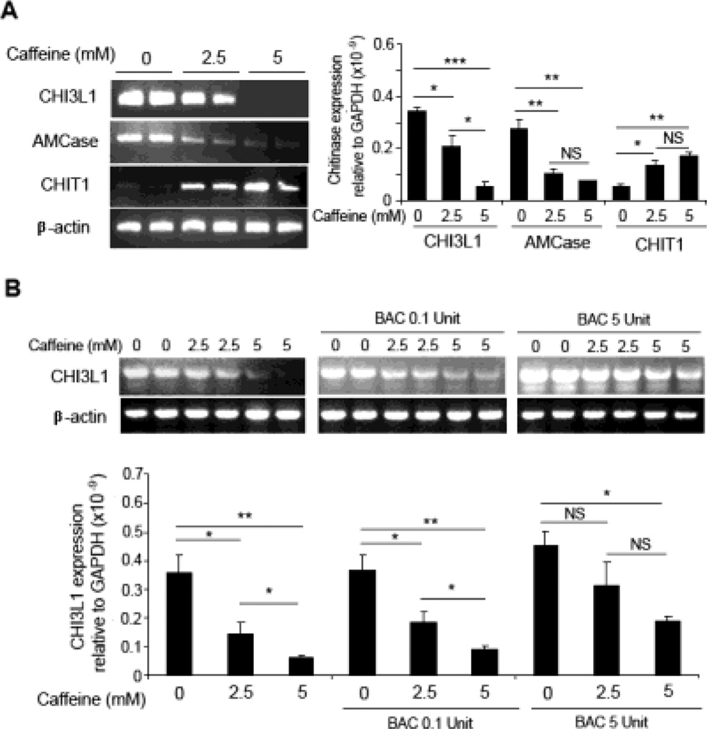
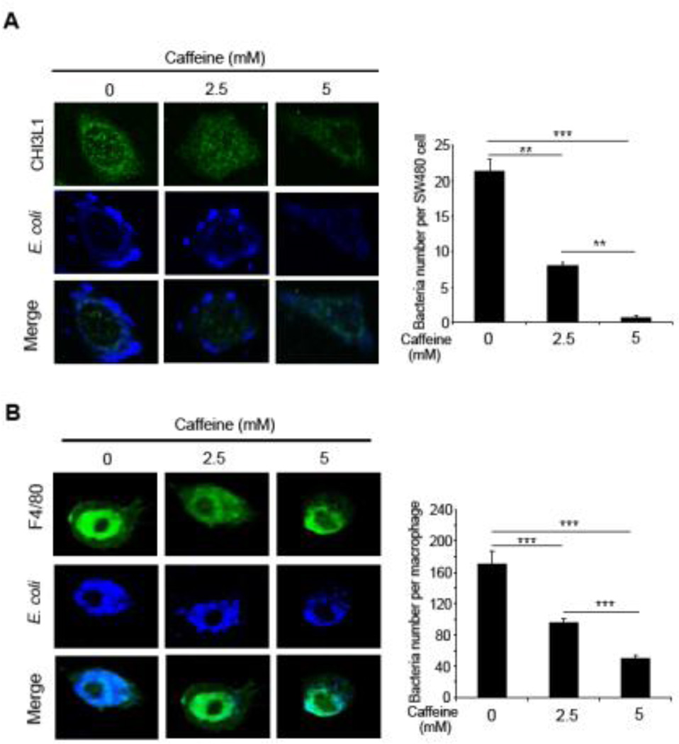
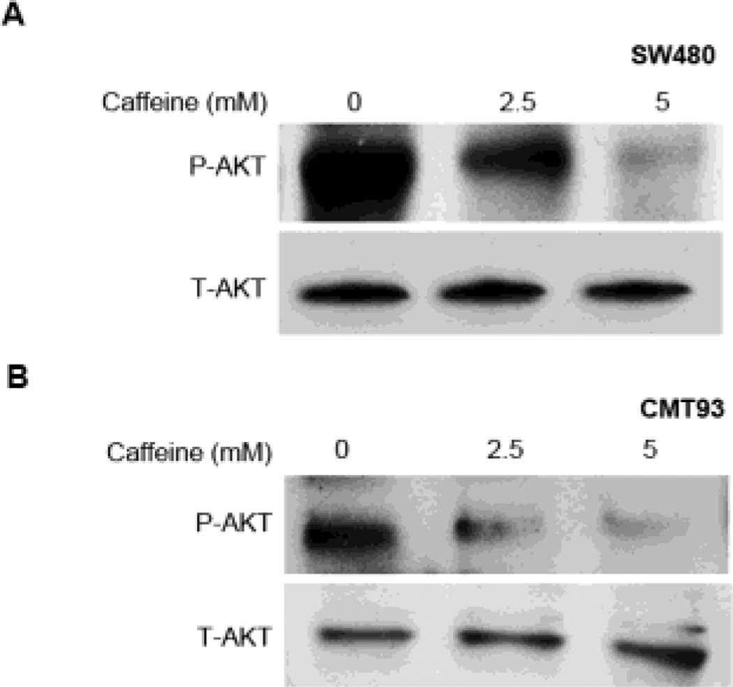
Results: In vitro, caffeine treatment in IEC lines down-regulated CHI3L1 mRNA expression, which resulted in the reduction of bacterial invasion in a caffeine dose-dependent manner. In vivo, mice treated with caffeine displayed a delayed response towards DSS-induced colitis, characterized by lower body weight loss, clinical and histological scores. Bacterial translocation into other organs and pro-inflammatory cytokines production were also reduced in the caffeine-treated mice with DSS-induced colitis. Caffeine treatment also resulted in the loss of CHI3L1-associated AKT signaling pathway activation both in vitro and in vivo.

Conclusion: Development of acute colitis is reduced upon caffeine treatment. The mechanism involves the down-regulation of CHI3L1 expression and its associated bacterial interaction effect. Therefore, caffeine is proposed as a safe and economical candidate for successful IBD management.


Figures

Figure 1
Figure 1. Caffeine suppresses chitinase mRNA expression in IECs
(A) SW480 cells were treated with 0, 2.5 or 5 mM caffeine for 48 hours incubation and detected for CHI3L1, AMCase and CHIT1 mRNA expression levels using RT-PCR (left) and Q-PCR (right). (B) Caffeine was first pre-incubated with bacteria-derived chitinase (BAC: 0.1 and 5 units) for 48 hours before adding into the culture medium of SW480 cells, followed by detection of CHI3L1 mRNA expression using RT-PCR (above) and Q-PCR (below). *P<0.05, **P<0.01, ***P<0.001, NS: no significant difference.
Figure 2
Figure 2. Caffeine blocked AIEC invasion into IECs and macrophages
(A, B) SW480 cells (A) or mouse-derived peritoneal macrophages (B) were pre-treated with 0, 2.5 or 5 mM caffeine for 1 hour and inoculated with AIEC at MOI of 10 (for SW480) or 20 (for macrophages) for 1 hour at 37°C, followed by gentamicin (100 µg/ml) treatment for additional 1 hour to exclude extracellular bacteria. SW480 cells and macrophages were costained with anti-E coli LPS antibody and anti-CHI3L1 (Xpress®) or anti-F4/80 antibody respectively, followed by confocal microscopy analysis. Intracellular bacteria were quantified and shown. **P<0.01, and ***P<0.001
Figure 3
Figure 3. Caffeine suppresses AKT signaling pathway activation in IECs
(A, B) SW480 (human) and CMT93 (mouse) IEC lines were treated with 0, 2.5 or 5 mM caffeine. Protein lysate were resolved using SDS-PAGE and detected for phosphorylated- or total-AKT using the respective specific antibodies.
Figure 4
Figure 4. Caffeine prevents onset of DSS-induced colitis in vivo
(A, B) Mice were pre-treated with 2.5 mM caffeine for 7 days, followed by simultaneous treatment with 3.5% DSS for additional 5 days and finally with caffeine treatment only for the next 6 days before sacrificing. Percent body weight and clinical scores from the treated and untreated mice groups are shown. (C, D) Colonic mRNA expression of CHI3L1, AMCase and CHIT1 were determined using RT-PCR (C) and Q-PCR (D). Data are average of two independent experiments (n=8 in each group). *P<0.05, **P<0.01, ***P<0.001, NS: no significant difference.
Figure 5
Figure 5. Caffeine treatment reduces severity of DSS-induced early colitis
(A, B) Colonic H&E staining (A) and histological scores (B) of caffeine treated and untreated DSS-treated mice are shown. 10×, objective. (C) Colonic sections were immuno-stained with anti-F4/80, -CD4 or -CD11b antibody and the number of positive cells per mm2 area were quantified. (D) Bacterial CFU from spleen, MLN, liver, cecum and colon homogenized extracts were quantified and shown. (E) Spleen, MLN, and colon cytokine production (TNFα IFNγ, IL-10, IL-4 and IL-17F) from the caffeine treated or untreated mice were determined using ELISA. Data are average of two independent experiments (n=8 in each group). *P<0.05, **P<0.01, ***P<0.001, NS: no significant difference.
Figure 6
Figure 6. Colonic Akt activation is suppressed by caffeine treatment
(A) Colonic samples from mice with or without caffeine treatment were lysed and 30 µg/lane of protein lysate was subjected to immunoblot with anti-phospho (P)- or anti-total (T)- Akt or MAPK p42/p44 antibodies. The data are representative of three independent experiments. (B) Densitometry analysis of phosphorylation signals normalized to total-Akt (closed bars) or -MAPK p42/p44 (open bars) signals are shown. Data are average of two independent experiments (n=3 in each group). *P<0.05, NS: no significant difference.

References

    1. Xavier RJ, Podolsky DK. Unravelling the pathogenesis of inflammatory bowel disease. Nature. 2007;448(7152):427–434. - PubMed
    1. Hamilton MJ, Snapper SB, Blumberg RS. Update on biologic pathways in inflammatory bowel disease and their therapeutic relevance. J Gastroenterol. 2012;47(1):1–8. - PMC - PubMed
    1. Franke A, McGovern DP, Barrett JC, Wang K, Radford-Smith GL, Ahmad T, et al. Genome-wide meta-analysis increases to 71 the number of confirmed Crohn's disease susceptibility loci. Nat Genet. 2010;42(12):1118–1125. - PMC - PubMed
    1. Franke A, Balschun T, Karlsen TH, Hedderich J, May S, Lu T, et al. Replication of signals from recent studies of Crohn's disease identifies previously unknown disease loci for ulcerative colitis. Nat Genet. 2008;40(6):713–715. - PubMed
    1. Anderson CA, Boucher G, Lees CW, Franke A, D'Amato M, Taylor KD, et al. Metaanalysis identifies 29 additional ulcerative colitis risk loci, increasing the number of confirmed associations to 47. Nat Genet. 2011;43(3):246–252. - PMC - PubMed

Publication types

MeSH terms

LinkOut - more resources