. 2013 Sep 20;288(38):27232-27242.
doi: 10.1074/jbc.M113.481184. Epub 2013 Aug 6.

Loss of epigenetic Kruppel-like factor 4 histone deacetylase (KLF-4-HDAC)-mediated transcriptional suppression is crucial in increasing vascular endothelial growth factor (VEGF) expression in breast cancer

Affiliations

Loss of epigenetic Kruppel-like factor 4 histone deacetylase (KLF-4-HDAC)-mediated transcriptional suppression is crucial in increasing vascular endothelial growth factor (VEGF) expression in breast cancer

Alpana Ray et al. J Biol Chem. .

Abstract

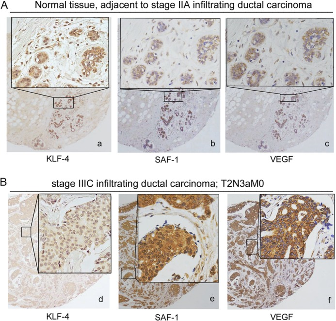
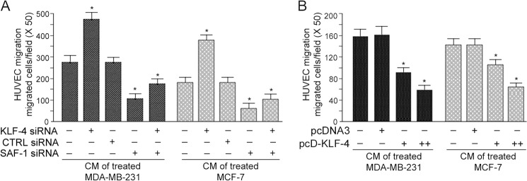
Vascular endothelial growth factor (VEGF) is recognized as an important angiogenic factor that promotes angiogenesis in a series of pathological conditions, including cancer, inflammation, and ischemic disorders. We have recently shown that the inflammatory transcription factor SAF-1 is, at least in part, responsible for the marked increase of VEGF levels in breast cancer. Here, we show that SAF-1-mediated induction of VEGF is repressed by KLF-4 transcription factor. KLF-4 is abundantly present in normal breast epithelial cells, but its level is considerably reduced in breast cancer cells and clinical cancer tissues. In the human VEGF promoter, SAF-1- and KLF-4-binding elements are overlapping, whereas SAF-1 induces and KLF-4 suppresses VEGF expression. Ectopic overexpression of KLF-4 and RNAi-mediated inhibition of endogenous KLF-4 supported the role of KLF-4 as a transcriptional repressor of VEGF and an inhibitor of angiogenesis in breast cancer cells. We show that KLF-4 recruits histone deacetylases (HDACs) -2 and -3 at the VEGF promoter. Chronological ChIP assays demonstrated the occupancy of KLF-4, HDAC2, and HDAC3 in the VEGF promoter in normal MCF-10A cells but not in MDA-MB-231 cancer cells. Co-transfection of KLF-4 and HDAC expression plasmids in breast cancer cells results in synergistic repression of VEGF expression and inhibition of angiogenic potential of these carcinoma cells. Together these results identify a new mechanism of VEGF up-regulation in cancer that involves concomitant loss of KLF-4-HDAC-mediated transcriptional repression and active recruitment of SAF-1-mediated transcriptional activation.

Keywords: Angiogenesis; Breast Cancer; DNA-Protein Interaction; Gene Regulation; Transcription Factors.


Figures

FIGURE 1.
FIGURE 1.
SAF-1-induced VEGF expression is higher in breast cancer cells. A, MCF-10A, MCF-7, MDA-MB-231, and MDA-MB-468 cells were co-transfected with equal amounts (0.5 μg) of 1.2VEGF-CAT or pBLCAT3 plasmid and increasing concentrations of pcDFLAG-SAF-1 plasmid (0.5 and 1.0 μg), as indicated. Twenty four hours after transfection, CAT activity was determined using an equivalent amount of cell extracts. Relative CAT activity was determined by comparing the activities of transfected plasmids with that of pBLCAT3 and correcting for transfection efficiency (β-gal). Results represent an average of three separate experiments. *, p < 0.05. Inset, schematic of human VEGF promoter. B, Western blot assay of transfected cells for detection of SAF-1 expression in the same samples as shown in A. Equal protein amount of cell extracts (50 μg) was fractionated in a 5/11% SDS-polyacrylamide gel, transferred to PVDF membrane, and immunoblotted with an anti-FLAG antibody. Blots were developed with enhanced chemiluminescence reagent. The membrane was stripped and reprobed with β-actin antibody to confirm equal loading. C, total RNA isolated from untreated and pcDSAF-1 (1.0 μg)-transfected cells, as indicated, was subjected to quantitative RT-PCR analysis with primers specific for VEGF. The results were normalized to the level of GAPDH in each sample and represent an average of three separate experiments. *, p < 0.05. D, Western blot analysis of cell extracts (70 μg of protein) from untreated and pcDSAF-1 (1.0 μg)-transfected cells, as used in C, with anti-VEGF antibody. The membrane was stripped and reprobed with β-actin antibody to confirm equal loading. Histograms summarize the Western blot results.
FIGURE 2.
FIGURE 2.
KLF-4 interacts with VEGF promoter. A, DNA sequences of VEGF from nucleotide position −119 to +5 contain the transcription start site, indicated by an arrow, and DNA-binding elements KLF-4 and SAF-1. B, nuclear extracts (10 μg of protein), as indicated, were incubated with 32P-labeled VEGF DNA containing sequences from −135 to +29. Resulting DNA·protein complexes (a, b, and c) were fractionated in a 6% nondenaturing polyacrylamide gel. In some assays, antibodies to KLF-4, Sp1 (Santa Cruz Biotechnology), SAF-1, or normal IgG were included during a preincubation reaction. Migration positions of KLF-4-, SAF-1-, and Sp1-specific complexes are indicated. Supershift of the DNA·protein complex is indicated by ss. C, total RNA was subjected to quantitative RT-PCR analysis with primers specific for KLF-4. The results were normalized to the level of GAPDH in each sample. D, cell extracts (50 μg of protein) were fractionated in a 5/11% SDS-polyacrylamide gel, transferred to PVDF membrane, and immunoblotted with anti-KLF-4 antibody for Western blot (WB) analysis. The membrane was stripped and reprobed with β-actin antibody as a loading control.
FIGURE 3.
FIGURE 3.
Overexpression of KLF-4 reduces VEGF expression in breast cancer cells. A, MDA-MB-231 cells were co-transfected with 1.2 VEGF CAT reporter plasmid (0.5 μg) and pcD-SAF-1 (0.5 μg) and pcD-KLF-4 (0.5, 0.75, 1.0, and 1.5 μg) expression plasmids, as indicated. Relative CAT activity was determined as described in Fig. 1A. The results represent an average of three independent experiments (*, p < 0.05). Inset shows protein expression (columns g′–k′) from KLF-4 and SAF-1 plasmids in transfected cells (columns g–k), which was assessed by Western blot (WB) analysis. The membrane was stripped and reprobed with β-actin antibody to confirm equal loading. B, VEGF mRNA level, after overexpression of KLF-4 in pcD-KLF-4 plasmid-transfected (0.5 and 1.0 μg) MDA-MB-231 cells, was determined by quantitative real time PCR analysis using the cDNA templates and the specific primers for GAPDH and VEGF. KLF-4 protein level in transfected cells was analyzed by Western blot (WB) analysis. The membrane was stripped and reprobed with β-actin antibody to confirm equal loading. C, MCF-7 and MDA-MB-231 cells were transfected with 1.2 VEGF CAT reporter plasmids (0.5 μg). In some transfection reactions, 200 nmol of KLF-4 siRNA or scrambled siRNA (CTRL siRNA) of the same length were used. Relative CAT activity was determined as described in Fig. 1A. Results represent an average of three separate experiments (**, p < 0.05). D and E, VEGF mRNA levels in MCF-7 and MDA-MB-231 cells, respectively, after transfection of 200 nm of KLF-4 siRNA, scrambled siRNA (CTRL siRNA), and SAF-1 siRNA, as indicated. Quantitative real time PCR assays were performed using the cDNA templates and the specific primers for GAPDH and VEGF. VEGF mRNA levels in KLF-4 siRNA-transfected cells was further normalized with control siRNA-transfected cells. The results shown are representative of three independent experiments (**, p < 0.02; ***, p < 0.05). F, MCF-10A cells were transfected with 1.2 VEGF CAT reporter plasmid (0.5 μg). In some transfection reactions, 200 nm KLF-4 siRNA or scrambled RNA (CTRL siRNA) oligonucleotide of the same length and increasing concentrations (0.5, 0.75, and 1.0 μg) of SAF-1 expression plasmid, pcD-SAF-1, were added. Relative CAT activity was determined as described in Fig. 1A. Results represent an average of three separate experiments (*, p < 0.05).
FIGURE 4.
FIGURE 4.
KLF-4 expression in clinical breast cancer and adjacent normal tissues. A, immunohistochemical analysis of KLF-4 (panel a), SAF-1 (panel b), and VEGF (panel c) in serial sections of normal breast tissues adjacent to cancer. B, immunohistochemical analysis of KLF-4 (panel d), SAF-1 (panel e), and VEGF (panel f) in serial sections of clinical breast cancer tissues. The insets represent higher magnification of the boxed area. A total of 60 breast tissue samples (30 cancer and 30 adjacent normal) were examined, and some representative samples are shown here. Pathological evaluation of the samples and the analysis of KLF-4, SAF-1 and VEGF expression are summarized in Table 1.
FIGURE 5.
FIGURE 5.
Inhibition of KLF-4 increases and overexpression of KLF-4 reduces cell migration potential. A, MDA-MB-231 and MCF-7 cells were transfected with KLF-4 siRNA, control siRNA (CTRL siRNA), or SAF-1 siRNA, as indicated. CM from these transfected cells were collected 24 h later. Culture media of HUVECs were fortified with the different CM preparations, and migrated cells were counted. Results represent mean ± S.E. of three independent experiments. *, p < 0.05. B, MDA-MB-231 and MCF-7 cells were transfected with increasing concentrations of pcDNA3 or pcD-KLF-4 plasmid DNA (0.0, 1.0, and 2.0 μg). Following transfection, CM were collected. Culture media of HUVECs were fortified with the different CM preparations, as indicated, and the migrated cells, following 24 h incubation, were counted. Results represent mean ± S.E. of three independent experiments. *, p < 0.05.
FIGURE 6.
FIGURE 6.
KLF-4 recruits HDAC2 and -3 to VEGF promoter. A, MCF-10A cells were cross-linked with formaldehyde, and chromatin isolated from these cells was subjected to ChIP-reChIP analysis. ChIP was first performed by immunoprecipitating with anti-KLF-4, anti-HDAC2, anti-HDAC3, or CTRL IgG, as indicated. The eluent of each immunocomplex was further immunoprecipitated using anti-HDAC2, anti-HDAC3, or CTRL IgG, as indicated. The precipitated chromatin DNA or input DNA was used for PCR amplification using VEGF-specific primers. B, MDA-MB-231 cells were subjected to ChIP-reChIP analysis by following the method as described in A. C, MDA-MB-231 cells were co-transfected with pBLCAT3 (0.5 μg) or 1.2VEGF CAT reporter (0.5 μg) and a combination of pcD-KLF-4 (0.25 μg), pcD-HDAC2 (0.5 μg), and pcD-HDAC3 (0.5 μg) DNAs, as indicated. Relative CAT activity was determined, and the results represent an average of three separate experiments. *, p < 0.05; **, p < 0.05.
FIGURE 7.
FIGURE 7.
Model illustrating the role of KLF-4, HDACs, and SAF-1 in regulating VEGF expression. In normal breast cells, KLF-4 is abundantly present, which, upon association with HDAC2 and -3, interacts with VEGF promoter. The interaction of the KLF-4·HDAC complex with VEGF is subject to very little competition from the minimally active SAF-1, resulting in transcriptional suppression of VEGF. In breast cancer cells, low abundance of KLF-4 and high abundance of SAF-1, allows predominant SAF-1 interaction with VEGF promoter and SAF-1-mediated increase of VEGF transcription.

Similar articles

Cited by

References

    1. Folkman J., Merler E., Abernathy C., Williams G. (1971) Isolation of a tumor factor responsible for angiogenesis. J. Exp. Med. 133, 275–288 - PMC - PubMed
    1. Engels K., Fox S. B., Whitehouse R. M., Gatter K. C., Harris A. L. (1997) Distinct angiogenic patterns are associated with high-grade in situ ductal carcinomas of the breast. J. Pathol. 181, 207–212 - PubMed
    1. Ferrara N. (1995) The role of vascular endothelial growth factor in pathological angiogenesis. Breast Cancer Res. Treat. 36, 127–137 - PubMed
    1. Ferrara N. (2002) Role of vascular endothelial growth factor in physiologic and pathologic angiogenesis: therapeutic implications. Semin. Oncol. 29, 10–14 - PubMed
    1. Yoshiji H., Gomez D. E., Shibuya M., Thorgeirsson U. P. (1996) Expression of vascular endothelial growth factor, its receptor, and other angiogenic factors in human breast cancer. Cancer Res. 56, 2013–2016 - PubMed

Publication types

MeSH terms