. 2013 Oct;23(10):2269-81.
doi: 10.1093/cercor/bht195. Epub 2013 Aug 7.

The neurobiology of thought: the groundbreaking discoveries of Patricia Goldman-Rakic 1937-2003

Affiliations

The neurobiology of thought: the groundbreaking discoveries of Patricia Goldman-Rakic 1937-2003

Amy F T Arnsten. Cereb Cortex. 2013 Oct.

Abstract

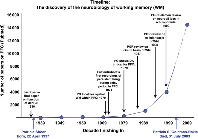
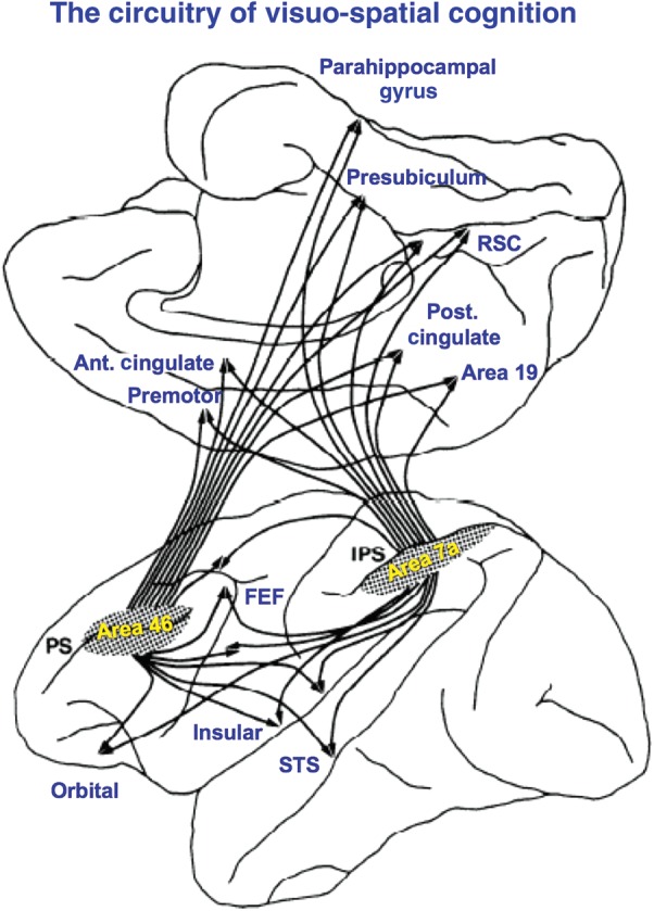
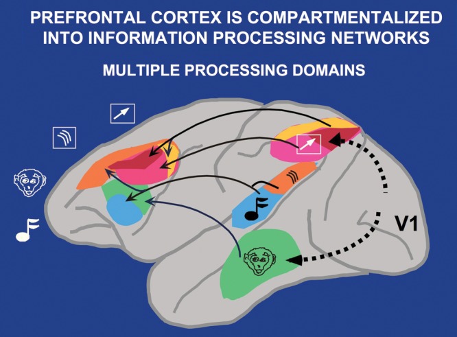
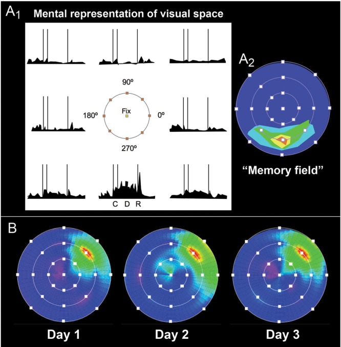
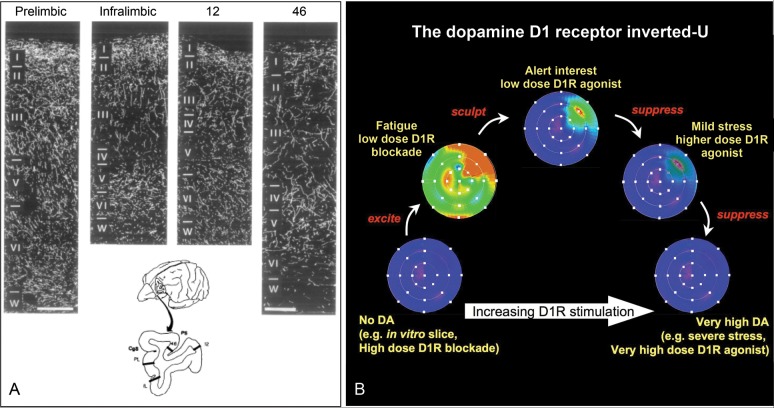
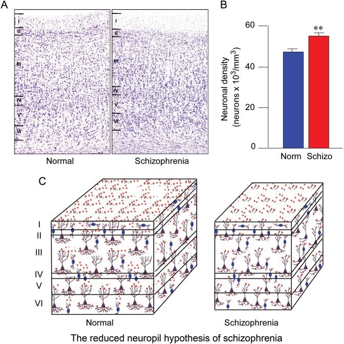
Patricia S. Goldman-Rakic (1937-2003) transformed the study of the prefrontal cortex (PFC) and the neural basis of mental representation, the basic building block of abstract thought. Her pioneering research first identified the dorsolateral PFC (dlPFC) region essential for spatial working memory, and the extensive circuits of spatial cognition. She discovered the cellular basis of working memory, illuminating the dlPFC microcircuitry underlying spatially tuned, persistent firing, whereby precise information can be held "in mind": persistent firing arises from recurrent excitation within glutamatergic pyramidal cell circuits in deep layer III, while tuning arises from GABAergic lateral inhibition. She was the first to discover that dopamine is essential for dlPFC function, particularly through D1 receptor actions. She applied a host of technical approaches, providing a new paradigm for scientific inquiry. Goldman-Rakic's work has allowed the perplexing complexities of mental illness to begun to be understood at the cellular level, including atrophy of the dlPFC microcircuits subserving mental representation. She correctly predicted that impairments in dlPFC working memory activity would contribute to thought disorder, a cardinal symptom of schizophrenia. Ten years following her death, we look back to see how she inspired an entire field, fundamentally changing our view of cognition and cognitive disorders.

Keywords: dopamine; mental representation; prefrontal cortex; schizophrenia; working memory.


Figures

Figure 1.
Figure 1.
Timeline of the discoveries of the PFC role in working memory (WM) and the key contributions of Goldman-Rakic. The graph shows the number of papers cited on PubMed using the search term “prefrontal cortex” for each decade ending in the year noted. Key publications by Goldman-Rakic and other early pioneers are indicated.
Figure 2.
Figure 2.
The cortical circuitry for spatial cognition, based on the work of Goldman-Rakic and Selemon. Note that both the dlPFC (area 46) and parietal cortex have many shared connections to subcortical structures that are not shown in this illustration, as well as “nonshared” connections that are not included in this diagram. Figure used with the permission of L. Selemon. Ant. Cingulate: anterior cingulate; FEF: frontal eye fields; IPS: intraparietal sulcus; Post. Cingulate: posterior cingulate; PS: principal sulcus; RSC: retrosplenial cortex; STS: superior temporal sulcus.
Figure 3.
Figure 3.
The parallel cortical circuits for space versus features in the visual and auditory domains. Parallel visual pathways for the processing of visual space and visual features emerge from the primary visual cortex, area V1. These pathways remain in parallel as they project into the PFC. Similar parallel projections were observed for the auditory spatial and feature streams. The visuospatial circuit is shown in pink/red/yellow; the auditory spatial circuit in orange; the visual feature circuit is shown in green, and the auditory feature circuit is shown in blue. Figure from a Goldman-Rakic presentation for Yale undergraduates with the permission of P. Rakic. Note that projections from the PFC back to the sensory cortex are not illustrated in this figure, but likely play an important role in top-down regulation of attention and sensory processing.
Figure 4.
Figure 4.
The physiology of mental representation in dlPFC. (A1) A schematic representation of a neuron with spatially tuned, persistent firing during the oculomotor delayed-response task. The possible spatial locations for cues are shown in the center of this figure, with the fixation point indicated in yellow. This neuron has persistent firing for the memory of a cue at 270°, but has less persistent firing for the memory of nearby locations, and actually inhibits firing during the delay period following cues distant to the neuron's “preferred location.” Goldman-Rakic considered this the cellular representation of visual space, the fundamental building block of mental representation. C: cue period; D: delay period; R: signal for saccadic response. (A2) The neuronal firing patterns of the neuron depicted in (A1) shown as a “memory field,” where dark blue represents low levels of firing during the delay period and brighter colors signify progressively higher firing rates. This method provides a more intuitive process for depicting the strength and precision of a neuron's spatial tuning. (B) The same neuron recorded on 3 separate days shows stable spatial tuning for 45°, as would be needed for the mental representation of visual space. This figure is from a Goldman-Rakic presentation for Yale undergraduates with the permission of P. Rakic; the data are from O’Scalaidhe and Goldman-Rakic, unpublished.
Figure 5.
Figure 5.
The dlPFC microcircuits underlying mental representation. (A) Microinjections of an anatomical tracer (purple) into layer IIIc of the primate dlPFC-labeled horizontal connections (gold) consistent with recurrent excitation between pyramidal cells. (B) Goldman-Rakic's schematic depiction of the primate layer IIIc microcircuits that provide the cellular basis for mental representation. Pyramidal cells are depicted by triangles; they excite each other through glutamatergic synapses on spines (white circles). GABAergic interneurons providing lateral inhibition are shown in blue. Note that although connections between pyramidal cells are depicted on the apical dendrites for the sake of clarity, they are likely most concentrated on the basal dendrites. Figures from a Goldman-Rakic presentation for Yale undergraduates with the permission of P. Rakic.
Figure 6.
Figure 6.
An example of the reciprocal relationship between pyramidal cells and GABAergic interneurons in the dlPFC. (A1) Photograph of a GABAergic basket cell (within blue rectangle) and a pyramidal cell (within orange rectangle) in the primate dlPFC. (A2) A schematic diagram of the likely connections between these neurons, whereby the basket cell (blue, fast-spiking with thin waveform) inhibits the pyramidal cell (orange, regular-spiking with longer waveform) through connections on the soma (shown) and proximal primary dendrites (not shown). (B1) The preferred direction of the presumed GABAergic interneuron during the delay period. (B2) The firing pattern of a fast-spiking, presumed GABAergic interneuron during the initial fixation (gray), cue presentation (purple), delay period (green), and response epochs (blue). (C1) The preferred direction of the regular-spiking, presumed pyramidal cell during the delay period. Note that it is opposite to the preferred direction of the interneuron. (C2) The firing pattern of the presumed pyramidal cell during the initial fixation (gray), cue presentation (purple), delay period (green), and response epochs (blue). Note that the firing of the pyramidal cell increases as the firing of the GABAergic interneuron decreases. Figures from a Goldman-Rakic presentation for Yale undergraduates with the permission of P. Rakic.
Figure 7.
Figure 7.
Representation of the visual field in principal sulcal dlPFC. (A) An image of the rostral principal sulcus that Goldman-Rakic used to illustrate the concept of progressive recordings down the sulcus as indicated by the white arrow. The autoradiographic image of the dlPFC is from an earlier anatomical study with Nauta showing columns of labeling from connections with the contralateral dlPFC. The actual recordings were performed in a more caudal region of the principal sulcal cortex. (B) Recordings by O’Scalaidhe and Goldman-Rakic (unpublished) down the length of the caudal principal sulcal cortex show progressive changes in the preferred direction of each neuron, thus providing comprehensive representation of the entire, contralateral visual field. Figures from a Goldman-Rakic presentation for Yale undergraduates with the permission of P. Rakic.
Figure 8.
Figure 8.
The key role of dopamine in the primate dlPFC. (A) The dopaminergic innervation of the primate PFC, including the dlPFC area 46, as visualized using an antibody directed against dopamine. Note the relatively sparse labeling in the dlPFC, a region that critically depends on dopamine actions. (From Williams and Goldman-Rakic 1993.) (B) A schematic illustration of the dopamine D1 receptor inverted-U influence on the pattern of Delay cell firing in the dlPFC. The memory fields of dlPFC neurons are shown under conditions of increasing levels of D1 receptor stimulation. Either very low or very high levels of D1 receptor stimulation markedly reduce delay-related firing. Low levels of D1 receptor stimulation are associated with noisy neuronal representations of visual space, while optimal levels reduce noise and enhance spatial tuning. The high levels of D1 receptor stimulation during stress exposure would reduce delay-related firing for all directions. Brighter colors indicate higher firing rates during the delay period. This figure is a schematic illustration of the physiological data presented in Williams and Goldman-Rakic (1995); Vijayraghavan et al. (2007); and Arnsten et al. (2009) and is consistent with the behavioral data from Arnsten et al. (1994); Murphy et al. (1996); Zahrt et al. (1997); and Arnsten and Goldman-Rakic (1998).
Figure 9.
Figure 9.
Reduced neuropil in the dlPFC in the brains of patients with schizophrenia. (A) Examples of Nissl-stained coronal sections of the dlPFC from a normal control subject and a subject with schizophrenia. (B) Neuronal density measured across all cortical layers is greater in the dlPFC in patients with schizophrenia. (C) Schematic illustration of greater cell packing in schizophrenia, that is, the same number of neurons is present in a smaller volume, suggesting that the intervening space containing neuropil is diminished. Reduced neuropil in the cortex of patients with schizophrenia suggests that impoverished connectivity of the dlPFC is a neuropathologic correlate of the disease. Figure generously provided by L. Selemon.

References

    1. Abi-Dargham A, Mawlawi O, Lombardo I, Gil R, Martinez D, Huang Y, Hwang DR, Keilp J, Kochan L, Van Heertum R, et al. Prefrontal dopamine D1 receptors and working memory in schizophrenia. J Neurosci. 2002;22:3708–3719. - PMC - PubMed
    1. Abi-Dargham A, Xu X, Thompson JL, Gil R, Kegeles LS, Urban NB, Narendran R, Hwang DR, Laruelle M, Slifstein M. Increased prefrontal cortical D1 receptors in drug naive patients with schizophrenia: a PET study with [11C]NNC112. J Psychopharmacol. 2012;26:794–805. - PubMed
    1. Akil M, Pierri JN, Whitehead RE, Edgar CL, Mohila C, Sampson AR, Lewis DA. Lamina-specific alterations in the dopamine innervation of the prefrontal cortex in schizophrenic subjects. Am J Psychiatry. 1999;156:1580–1589. - PubMed
    1. Arnsten AFT. The biology of feeling frazzled. Science. 1998;280:1711–1712. - PubMed
    1. Arnsten AFT, Cai JX, Murphy BL, Goldman-Rakic PS. Dopamine D1 receptor mechanisms in the cognitive performance of young adult and aged monkeys. Psychopharmacology. 1994;116:143–151. - PubMed

Personal name as subject