. 2013 Aug 7;5(197):197ra103.
doi: 10.1126/scitranslmed.3006034.

Identification of a Titin-derived HLA-A1-presented peptide as a cross-reactive target for engineered MAGE A3-directed T cells

Affiliations

Identification of a Titin-derived HLA-A1-presented peptide as a cross-reactive target for engineered MAGE A3-directed T cells

Brian J Cameron et al. Sci Transl Med. .

Abstract

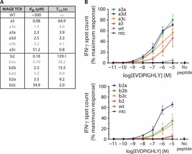
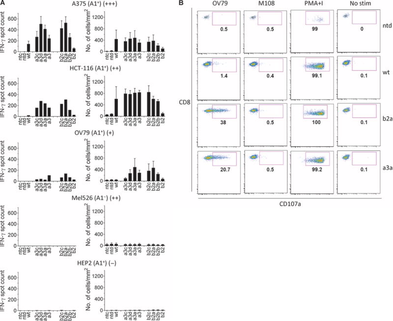
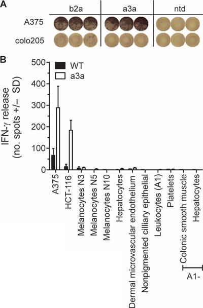
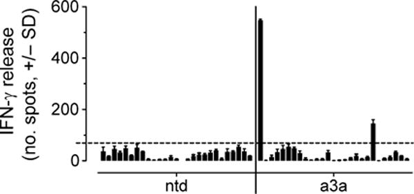
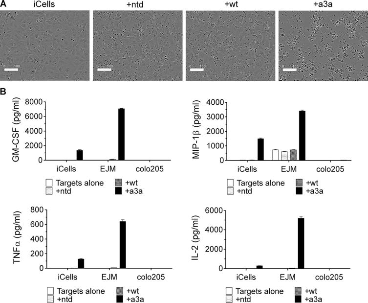
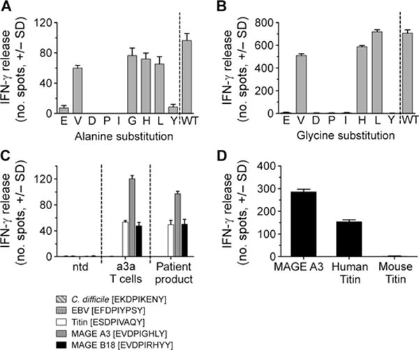
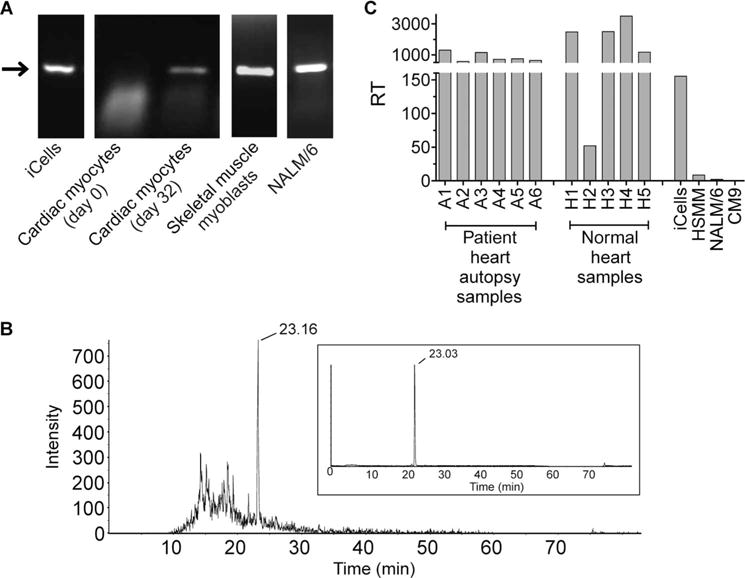
MAGE A3, which belongs to the family of cancer-testis antigens, is an attractive target for adoptive therapy given its reactivation in various tumors and limited expression in normal tissues. We developed an affinity-enhanced T cell receptor (TCR) directed to a human leukocyte antigen (HLA)-A*01-restricted MAGE A3 antigen (EVDPIGHLY) for use in adoptive therapy. Extensive preclinical investigations revealed no off-target antigen recognition concerns; nonetheless, administration to patients of T cells expressing the affinity-enhanced MAGE A3 TCR resulted in a serious adverse event (SAE) and fatal toxicity against cardiac tissue. We present a description of the preclinical in vitro functional analysis of the MAGE A3 TCR, which failed to reveal any evidence of off-target activity, and a full analysis of the post-SAE in vitro investigations, which reveal cross-recognition of an off-target peptide. Using an amino acid scanning approach, a peptide from the muscle protein Titin (ESDPIVAQY) was identified as an alternative target for the MAGE A3 TCR and the most likely cause of in vivo toxicity. These results demonstrate that affinity-enhanced TCRs have considerable effector functions in vivo and highlight the potential safety concerns for TCR-engineered T cells. Strategies such as peptide scanning and the use of more complex cell cultures are recommended in preclinical studies to mitigate the risk of off-target toxicity in future clinical investigations.


Conflict of interest statement

Competing interests: B.J.C., J.D., J.V.H., V.K., F.C.B., F.G., G.B., A.V., A.S.P., A.L., N.J.H., and B.K.J. are employees of Immunocore Ltd. A.B.G., J.E.B., K.J.A., A.D.B., N.J.P., D.D.W., G.B.-S., and B.K.J. are employees of Adaptimmune Ltd. A patent has been filed on the sequence and utility of the TCRs used in this study (WO12/03913). The remaining authors declare no competing interests.

Figures

Fig. 1
Fig. 1. Panel of MAGE A3 affinity-enhanced TCRs
(A) Biophysical parameters describing the interaction between 12 MAGE A3 affinity-enhanced TCRs and the HLA-A*01–restricted MAGE A3 peptide antigen. Affinity-enhanced TCRs were produced by phage display, and affinity and half-life were calculated from SPR measurements. (B) Sensitivity of TCR-engineered T cells to antigen. The TCRs from the panel were used to produce TCR-engineered T cells via lentivirus-mediated transduction. Sensitivity was assessed by IFN-γ release, using HEP2 cells HLA-A*01+ pulsed with a titration of MAGE A3 peptide, as targets. Control measurements were carried out in the absence of peptide. For clarity, data are shown for 8 of the 12 TCRs. TCRs with α chain mutations are shown in the top panel and those with β chain mutations in the lower panel. Experiments were carried out in triplicate. Error bars indicate SEM. wt, wild type; ntc, absence of T cells.
Fig. 2
Fig. 2. Functional analysis of TCR-engineered T cells
(A) Activity and specificity of engineered T cells expressing affinity-enhanced TCRs against various tumor cell lines and normal human hepatocytes (HEP2). The cell line is indicated above each graph along with HLA-A*01 status and an indication of MAGE A3 expression (shown in brackets as +/++/+++). IFN-γ release (left column) was determined by ELISpot, and cytotoxicity (right column) was determined using the IncuCyte platform (no. of apoptotic cells). Controls were carried out using nontransduced cells (ntd) and in the absence of T cells (ntc). Experiments were carried out in triplicate. Error bars indicate SEM. (B) Degranulation. T cell lines carrying the a3a or b2a affinity-enhanced TCRs were assessed for surface accumulation of CD107a in the presence of MAGE A3+ OV79 cells and MAGE A3 M108. Nontransduced T cells (ntd) and T cell carrying the wt TCR were included for comparison. Data were obtained using flow cytometry. Samples were initially gated on lymphocytes followed by gating on CD8+ and CD4+ events and finally a gate on Vβ5.1. Control measurements were carried out using PMA and ionomycin (PMA+I) and unstimulated T cells (No stim).
Fig. 3
Fig. 3. Specificity testing of a3a-engineered T cells
(A) Activation of b2a- but not a3a-engineered T cells by colo205 cells. Images shown represent raw IFN-γ ELISpot data. The MAGE A3+ A375 cell line is included for comparison. (B) A panel of primary cells derived from normal tissues was used to assess nonspecific activation of a3a-engineered T cells. A375 and HCT-116 tumor cell lines were used as positive controls. The nonmutated wt TCR was included for comparison. T cell activation was determined by IFN-γ release. Data represent the mean of three independent measurements ± SEM.
Fig. 4
Fig. 4. Alloreactivity panel
Representative data obtained from a3a-engineered T cells prepared from healthy donors. Nontransduced T cells (ntd) were included for comparison. The panel was compiled from various cell lines as detailed in Materials and Methods and encompassed more than 95% of the HLA serotype population. From left to right, the cell lines are as follows: EJM (positive control), colo205 (negative control), LMB 5835, GGT 4991, IZA 3089, MHX 1761, AAS 125, FAQ 3528, MBX 2763, AHT 192, LSR 5702, CVI 6184, MWX 3891, MSV 4803, NCI-H345, MDA-MB-231, TISI, IM-9, and nine EBV-transformed B cells obtained from independent donors. The dotted horizontal line indicates a background level. Data shown were derived from three independent measurements and represent means ± SEM.
Fig. 5
Fig. 5. Activation of wt and a3a-engineered T cells by iCell cardiomyocytes
(A) iCells were killed by a3a-engineered T cells but not by T cells transduced with the wt TCR. The IncuCyte platform was used to directly visualize iCells in the presence of a3a-engineered T cells. Phase-contrast images were obtained after 24 hours. Samples containing no T cells or nontransduced T cells (ntd) were used as negative controls. Scale bars, 100 μm. (B) Cytokine production. A Luminex assay was used to assess cytokine production from wt and a3a-engineered T cells in the presence of iCells. Data for granulocyte-macrophage colony-stimulating factor (GM-CSF), macrophage inflammatory protein–1β (MIP-1β), tumor necrosis factor–α (TNFα), and interleukin-2 (IL-2) are shown. EJM and colo205 tumor cell lines were included for comparison and are positive and negative for MAGE A3, respectively. Control measurements were made on targets alone and target cells plus nontransduced cells. Experiments were carried out in triplicate. Error bars indicate SEM.
Fig. 6
Fig. 6. Alternative specificity for a3a-engineered T cells
(A) Each residue in the MAGE A3 peptide sequence was sequentially replaced with alanine. Alanine-substituted peptides were pulsed onto HLA-A*01 HEP2 target cells, and IFN-γ release from a3a-engineered T cells was assessed. The residue replaced by alanine is indicated under each bar. (B) The same procedure was followed for glycine-substituted peptides, except substitution of the native glycine residue at position 6 was omitted. (C) Activation of a3a-engineered T cells by motif-containing peptides. Peptides were produced synthetically and pulsed onto HLA-A*01 HEP2 target cells. Activation of a3a-engineered T cell patient product and a3a-engineered T cells prepared from a healthy donor was determined by IFN-γ release. Nontransduced T cells (ntd) (from a healthy donor) were used as a control. (D) Activation of a3a-engineered T cells by mouse Titin. IFN-γ release was used to compare activation of a3a-engineered T cells against HEP2 cells pulsed with mouse and human Titin peptide. MAGE A3–pulsed cells are also included for comparison. All data shown were obtained from three independent measurements and represent means ± SEM.
Fig. 7
Fig. 7. Biological relevance of Titin
(A) Identification of Titin-expressing cells. RT-PCR was used to confirm Titin protein expression in the cell lines indicated. Cardiac myocytes were additionally assessed after being maintained at confluence for 32 days. The arrow indicates Titin (molecular weight of 327 bp). (B) Natural processing and presentation of Titin peptide. Peptides eluted from the surface of NALM/6 cells were analyzed by LC-MS/MS. Retention time of the Titin peptide is indicated. Comparison with a synthetic Titin peptide is shown inset. (C) Titin expression in heart tissue. Heart tissues obtained during patient autopsy and from tissue banks were assessed for relative expression of Titin, alongside the Titin-positive cell lines used in cellular assays (iCells, electrically active cardiomyocytes; CM9, normal cardiomyocytes; HSMM, human skeletal muscle myoblasts; NALM/6, B cell lymphoma cell line). RT indicates relative transcript number. Data shown were obtained from three independent measurements and represent means ± SEM.

References

    1. Restifo NP, Dudley ME, Rosenberg SA. Adoptive immunotherapy for cancer: Harnessing the T cell response. Nat Rev Immunol. 2012;12:269–281. - PMC - PubMed
    1. Morgan RA, Yang JC, Kitano M, Dudley ME, Laurencot CM, Rosenberg SA. Case report of a serious adverse event following the administration of T cells transduced with a chimeric antigen receptor recognizing. ERBB2 Mol Ther. 2010;18:843–851. - PMC - PubMed
    1. Parkhurst MR, Yang JC, Langan RC, Dudley ME, Nathan DA, Feldman SA, Davis JL, Morgan RA, Merino MJ, Sherry RM, Hughes MS, Kammula US, Phan GQ, Lim RM, Wank SA, Restifo NP, Robbins PF, Laurencot CM, Rosenberg SA. T cells targeting carcinoembryonic antigen can mediate regression of metastatic colorectal cancer but induce severe transient colitis. Mol Ther. 2011;19:620–626. - PMC - PubMed
    1. Robbins PF, Morgan RA, Feldman SA, Yang JC, Sherry RM, Dudley ME, Wunderlich JR, Nahvi AV, Helman LJ, Mackall CL, Kammula US, Hughes MS, Restifo NP, Raffeld M, Lee CC, Levy CL, Li YF, El-Gamil M, Schwarz SL, Laurencot C, Rosenberg SA. Tumor regression in patients with metastatic synovial cell sarcoma and melanoma using genetically engineered lymphocytes reactive with NY-ESO-1. J Clin Oncol. 2011;29:917–924. - PMC - PubMed
    1. Kalos M, Rapoport AP, Stadtmauer EA, Vogl DT, Weiss BM, Binder-Scholl GK, Smethurst DP, Kulikovskaya I, Gupta M, Macatee TL, Finklestein JM, Brewer JE, Bennett AD, Gerry AB, Pumphrey NJ, Tayton-Martin HK, Veloso E, Zheng Z, Badros AZ, Yanovich S, Akpek G, McConville H, Philip S, Levine BL, Jakobsen BK, June CH. Prolonged T cell persistence, homing to marrow and selective targeting of antigen positive tumor in multiple myeloma patients following adoptive transfer of T cells genetically engineered to express an affinity-enhanced T cell receptor against the cancer testis antigens NY-ESO-1 and Lage-1. Blood (ASH Annu Meet Abstr) 2012;120:755.

Publication types

MeSH terms