. 2013 Sep;2(9):641-54.
doi: 10.5966/sctm.2013-0017. Epub 2013 Aug 7.

Sustained knockdown of a disease-causing gene in patient-specific induced pluripotent stem cells using lentiviral vector-based gene therapy

Affiliations

Sustained knockdown of a disease-causing gene in patient-specific induced pluripotent stem cells using lentiviral vector-based gene therapy

Reto Eggenschwiler et al. Stem Cells Transl Med. 2013 Sep.

Abstract

Patient-specific induced pluripotent stem cells (iPSCs) hold great promise for studies on disease-related developmental processes and may serve as an autologous cell source for future treatment of many hereditary diseases. New genetic engineering tools such as zinc finger nucleases and transcription activator-like effector nuclease allow targeted correction of monogenetic disorders but are very cumbersome to establish. Aiming at studies on the knockdown of a disease-causing gene, lentiviral vector-mediated expression of short hairpin RNAs (shRNAs) is a valuable option, but it is limited by silencing of the knockdown construct upon epigenetic remodeling during differentiation. Here, we propose an approach for the expression of a therapeutic shRNA in disease-specific iPSCs using third-generation lentiviral vectors. Targeting severe α-1-antitrypsin (A1AT) deficiency, we overexpressed a human microRNA 30 (miR30)-styled shRNA directed against the PiZ variant of A1AT, which is known to cause chronic liver damage in affected patients. This knockdown cassette is traceable from clonal iPSC lines to differentiated hepatic progeny via an enhanced green fluorescence protein reporter expressed from the same RNA-polymerase II promoter. Importantly, the cytomegalovirus i/e enhancer chicken β actin (CAG) promoter-driven expression of this construct is sustained without transgene silencing during hepatic differentiation in vitro and in vivo. At low lentiviral copy numbers per genome we confirmed a functional relevant reduction (-66%) of intracellular PiZ protein in hepatic cells after differentiation of patient-specific iPSCs. In conclusion, we have demonstrated that lentiviral vector-mediated expression of shRNAs can be efficiently used to knock down and functionally evaluate disease-related genes in patient-specific iPSCs.

Keywords: Gene therapy; Hepatocyte differentiation; Lentiviral vector; Liver; Pluripotent stem cells; RNAi.


Figures

Figure 1.
Figure 1.
Characterization of murine PiZ induced pluripotent stem cells (iPSCs). (A): Quantitative reverse transcription-polymerase chain reaction expression analysis for endogenous pluripotency markers Nanog, Sox2, and Oct4 compared with a standard OG2 ES cell line. hOCT4 from the OKS-dT reprogramming construct was mostly silenced. (B): Immunostaining of PiZ iPSC colonies for Sox2, Oct4, and Nanog (top row), DAPI staining (middle row), and merged fluorescences with phase contrast (bottom row). (C): Fluorescence-activated cell sorting (FACS) analysis after SSEA-1 PE staining and isotype control. (D): Array comparative genomic hybridization showed an intact 40XY karoytype with a small (∼200 kbp) monoallelic deletion on chromosome 5 after reprogramming. (E): FACS analysis for dTomato in day 37 differentiated hepatic cells transduced with Alb-dTom lentivirus at day 17. Untransduced cells served as negative control. Abbreviations: Alb, albumin; DAPI, 4′,6-diamidino-2-phenylindole; ES, embryonic stem; hOCT4, human OCT4; SSEA, stage-specific embryonic antigen.
Figure 2.
Figure 2.
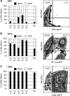
Comparison of different promoters for stable transgene expression during hepatic differentiation. (A, B): Murine PiZ induced pluripotent stem cells (iPSCs) were transduced with lentiviral vector-expressing eGFP under ubiquitous mammalian EFS and EF1α promoters. Clonal cell lines (cln 1–5) and a sorted bulk population (bulk) were established for each construct. Black bars show the percentage of eGFP-expressing cells in the dTomato-positive fraction after hepatic differentiation and transduction with lentiviral Alb-dTom. Right: Fluorescence-activated cell sorting analysis of corresponding bulk populations. (C): Analysis of clonal cell lines and a bulk population generated by lentiviral transduction of PiZ iPSCs with CAG-eGFP showed only reduced transgene silencing after differentiation. Abbreviations: cln, clone; EFS, EF1α-short; eGFP, enhanced green fluorescence protein; GFP, green fluorescence protein.
Figure 3.
Figure 3.
Knockdown in murine PiZ induced pluripotent stem cells (iPSCs) and in vitro analysis. (A): miR30-styled shRNA was inserted in between the eGFP and the PRE site on a third-generation lentiviral vector with an internal CAG promoter. (B): Fluorescence-activated cell sorting analysis for eGFP-shRNA-expressing cells in the Alb-dTom-positive fraction at day 37 of hepatic differentiation. Bulk populations were established by transduction of pluripotent PiZ iPSCs with lentiviral shRNA constructs (scr-sh mPi bulk and PiZ-sh mPi bulk), and Alb-dTom transduction was on day 37. (C): Vector copy number analysis for lentiviral integrations in established clonal cell lines. Parental line mPi showed two integrations from the OKS-dT reprogramming construct. Additional integrations from the eGFP-shRNA lentiviral vectors are shown in green. (D): Left: Quantitative reverse transcription-polymerase chain reaction analysis for expression of Ttr, Ck18, Fah, and hA1AT at day 37 of hepatic differentiation. All three clonal cell lines and the bulk population transduced with PiZ-shRNA showed significantly (p < .05, n = 3) reduced expression of hA1AT compared with scr-shRNA clones and bulk. Values were normalized to albumin. Right: Significance and fold change were calculated for the group of PiZ-shRNA-treated samples compared with the group of scr-shRNA-treated samples. hA1AT was significantly differentially expressed in the two groups, whereas Ttr, Ck18, and Fah were not. Abbreviations: Alb, albumin; Ck18, cytokeratin 18; cln, clone; dGag, deleted group specific antigen; eGFP, enhanced green fluorescence protein; Fah, fumarylacetoacetate-hydrolase; hA1AT, human α-1-antitrypsin; PBS, primer binding site; PPT, polypurine-tract; PRE, post-transcriptional regulatory element; RRE, Rev-responsive element; RSV, Rous sarcoma virus promoter/enhancer; RU5, redundant and unique 5′ region; scr, scramble; shRNA, short hairpin RNA; SIN, self-inactivating cassette; Ttr, transthyretin.
Figure 4.
Figure 4.
Analysis of PiZ knockdown in vivo. (A): Embryonic day (E) 13.5 chimeric fetuses generated from eGFP-short hairpin RNA (shRNA) transduced induced pluripotent stem cells (iPSCs). Fetal mice showed ubiquitous and strong eGFP expression, indicating high contribution of iPSCs to chimeras and sustained transgene expression (left and middle). Right: Enlarged fluorescence image of manually isolated E13.5 fetal liver. (B): Fluorescence-activated cell sorting (FACS) for the CD45−/eGFP+ fraction containing fetal hepatoblasts. (C): Quantitative reverse transcription-polymerase chain reaction analysis for Afp, Ck18, Ttr, and hA1AT in E13.5 CD45−/eGFP+ cells. The scr-shRNA-treated cell line and bulk population are shown in light blue and turquoise, respectively, and the PiZ-shRNA-treated cell line and bulk population are shown in light pink and dark pink, respectively. Cells from at least six fetal livers were pooled for each sample. PiZ-shRNA-treated cells showed reduced hA1AT expression compared with scr-shRNA-treated cells, whereas Afp, Ck18, and Ttr were at comparable levels in all samples. Values were normalized to albumin. (D, E): Western blot (D) and enzyme-linked immunosorbent assay (ELISA) (E) for human A1AT in lysates of E15.5 chimeric livers. Percentages of chimerism of respective samples are indicated in parentheses. The PiZ-shRNA sample showed a strong reduction in both assays Western blot and ELISA compared with scr-shRNA samples. Vinculin served as loading control in Western blot. (F): Immunohistochemical analysis of cryosections from E15.5 chimeric livers. Sections were stained using an antibody specific for human A1AT and analyzed using fluorescence microscopy. Pictures were taken with two seconds exposure time for Alexa 568 at a ×40 magnification. eGFP-positive cells stained strongly positive for hA1AT in the scr-shRNA-treated sample and weakly positive in the PiZ-shRNA-treated sample. Abbreviations: Alb, albumin; Ck18, cytokeratin 18; DAPI, 4′,6-diamidino-2-phenylindole; eGFP, enhanced green fluorescence protein; hA1AT, human α-1-antitrypsin; scr, scramble; sh, short hairpin RNA; Ttr, transthyretin; vinc, vinculin.
Figure 5.
Figure 5.
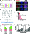
Generation and transduction of patient-specific induced pluripotent stem cells (iPSCs). (A): Quantitative reverse transcription-polymerase chain reaction analysis for pluripotency markers NANOG, SOX2, and OCT4 in the PiZZ iPSC clone hPi compared with the standard H9 human ES cell line. (B): Immunostaining of hPi colonies for nuclear OCT4, SOX2, and NANOG (top row); DAPI staining (middle row); and merged fluorescences with phase contrast (bottom row). (C): G banding shows a stable 46XX karyotype after reprogramming. (D): Single-nucleotide polymorphism sequencing for the PiZ G→A point mutation after reprogramming. (E): Fluorescence-activated cell sorting (FACS) analysis for Alb-dTom-positive cells in day 18 differentiated clone hPi. Cells were transduced with Alb-dTom at day 4 of differentiation, and untransduced cells served as control. (F): Vector copy number analysis for lentiviral integrations in clonal cell lines derived by transduction of hPi with lentiviral shRNAs. Parental line hPi has one integration from the reprogramming construct. Additional integrations from the eGFP-shRNA lentiviral vectors are shown in green. (G): FACS analysis for eGFP-shRNA-expressing cells in the Alb-dTom-positive fraction at day 18 of hepatic differentiation. Bulk populations were established by transduction of pluripotent PiZZ iPSCs with lentiviral shRNA constructs and Alb-dTom transduction was on day 4. Abbreviations: DAPI, 4′,6-diamidino-2-phenylindole; eGFP, enhanced green fluorescence protein; scr, scramble; shRNA, short hairpin RNA.
Figure 6.
Figure 6.
Knockdown of PiZ human α-1-antitrypsin (hA1AT) in patient-specific induced pluripotent stem cells (iPSCs) after differentiation. (A): Left: Quantitative reverse transcription-polymerase chain reaction expression analysis for TTR, CK18, CYP1A1, and α-1-antitrypsin (A1AT) at day 18 of hepatic differentiation. All three clonal cell lines and the bulk population transduced with PiZ-short hairpin RNA (shRNA) showed significantly reduced expression of hA1AT compared with scr-shRNA clones and bulk. Values were normalized to albumin. Right: Significance and fold change were calculated for the group of PiZ-shRNA-treated samples compared with the group of scr-shRNA-treated samples. A1AT was significantly differentially expressed between the two groups, whereas CK18 and CYP1A1 were not. (B): Left: Western blot for A1AT in day 18 differentiated PiZZ iPSC and scr-shRNA and PiZ-shRNA transduced bulk populations. The upper band (56 kDa) shows fully glycosylated A1AT, and the lower band (52 kDa) shows partially glycosylated A1AT. Right: Densities measured for both bands separately were added together and divided by density of vinculin, and then divided by the average of untreated samples. The PiZ-shRNA-treated bulk population showed a significant 66% reduction of A1AT. Lysates from HepG2 and HEK293T cells served as positive and negative controls, respectively. (C, D): Enzyme-linked immunosorbent assay (ELISA) for secreted A1AT (C) and albumin (D) in supernatants collected from day 18 differentiated iPSCs. The PiZ-shRNA-treated bulk population showed significantly decreased secretion of A1AT compared with scr-shRNA-treated cells, whereas secretion whereas secretion of albumin was at a comparable level. (E): Ethoxyresorufin-O-deethylase (EROD) assay for CYP1A1 activity. Values were at comparable levels among different iPSC samples. All values for ELISAs and EROD were normalized to 10,000 cells and 24 hours, and supernatant collected from HepG2 hepatoma cell line served as a positive control. (F): Immunocytochemical analysis for polymeric PiZ A1AT in cell clusters of day 18 differentiated iPSCs. Cells were stained using an antibody specific for polymeric human A1AT and analyzed using fluorescence microscopy. Pictures were taken with 2 seconds of exposure time for Alexa 568 at a magnification of ×40. Clusters stained strongly positive for polymeric hA1AT in scr-shRNA-treated cells and weakly positive in PiZ-shRNA-treated cells. Statistical analysis was performed using the t test. *, p < .05; **, p < .01. Abbreviations: ALB, albumin; CK18, cytokeratin 18; DAPI, 4′,6-diamidino-2-phenylindole; ns, not significant; scr, scramble; sh, short hairpin RNA; TTR, transthyretin.

References

    1. Sakuma T, Barry MA, Ikeda Y. Lentiviral vectors: Basic to translational. Biochem J. 2012;443:603–618. - PubMed
    1. Papapetrou EP, Lee G, Malani N, et al. Genomic safe harbors permit high beta-globin transgene expression in thalassemia induced pluripotent stem cells. Nat Biotechnol. 2011;29:73–78. - PMC - PubMed
    1. Frimpong K, Spector SA. Cotransduction of nondividing cells using lentiviral vectors. Gene Ther. 2000;7:1562–1569. - PubMed
    1. Yu X, Zhan X, D'Costa J, et al. Lentiviral vectors with two independent internal promoters transfer high-level expression of multiple transgenes to human hematopoietic stem-progenitor cells. Mol Ther. 2003;7:827–838. - PubMed
    1. Kim D, Kim CH, Moon JI, et al. Generation of human induced pluripotent stem cells by direct delivery of reprogramming proteins. Cell Stem Cell. 2009;4:472–476. - PMC - PubMed

Publication types

MeSH terms