. 2013 Sep 1;191(5):2581-8.
doi: 10.4049/jimmunol.1300748. Epub 2013 Aug 7.

The RodA hydrophobin on Aspergillus fumigatus spores masks dectin-1- and dectin-2-dependent responses and enhances fungal survival in vivo

Affiliations

The RodA hydrophobin on Aspergillus fumigatus spores masks dectin-1- and dectin-2-dependent responses and enhances fungal survival in vivo

Steven de Jesus Carrion et al. J Immunol. .

Abstract

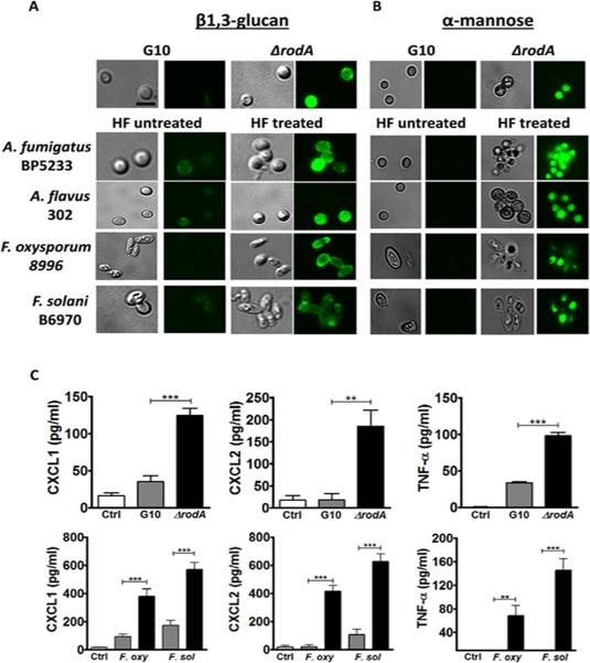
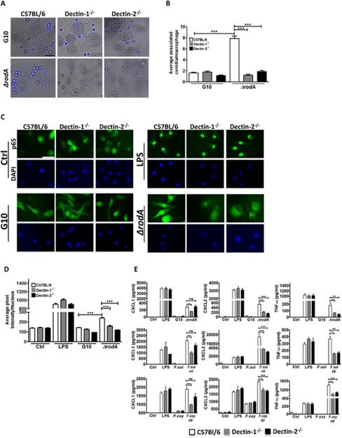
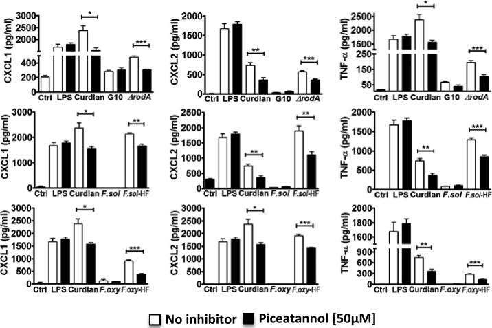
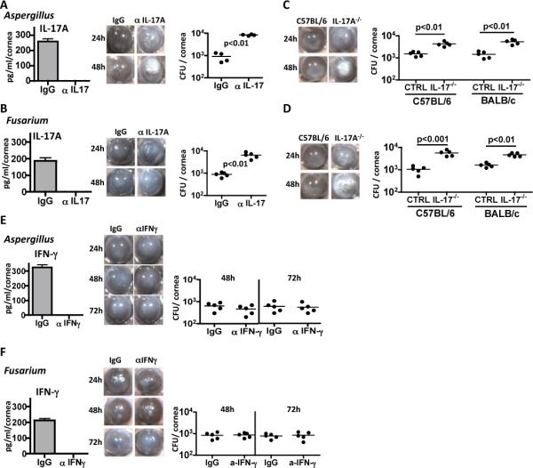
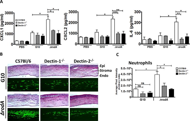
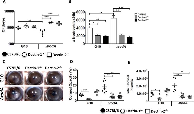
Aspergillus and Fusarium species are important causes of fungal infections worldwide. Airborne spores (conidia) of these filamentous fungi express a surface protein that confers hydrophobicity (hydrophobin) and covers cell wall components that would otherwise induce a host immune cell response. Using a mutant Aspergillus fumigatus strain (ΔrodA) that does not express the RodA hydrophobin, and Aspergillus and Fusarium conidia from clinical isolates that were treated with hydrofluoric acid (which removes the A. fumigatus RodA protein), we observed increased surface exposure of β1,3-glucan and α-mannose on Aspergillus and Fusarium conidia. We also found that ΔrodA and hydrofluoric acid-treated conidia stimulate significantly higher NF-κB p65 nuclear translocation and cytokine production by macrophages from C57BL/6, but not from Dectin-1(-/-) or Dectin-2(-/-) mice. Using a murine model of A. fumigatus corneal infection, we showed that ΔrodA conidia induced significantly higher cytokine production, neutrophil infiltration, and more rapid fungal clearance from C57BL/6 corneas compared with the parent G10 strain, which was dependent on Dectin-1 and Dectin-2. Together, these findings identify the hydrophobin RodA as a virulence factor that masks Dectin-1 and Dectin-2 recognition of conidia, resulting in impaired neutrophil recruitment to the cornea and increased fungal survival and clinical disease.


Figures

Figure 1
Figure 1. Removal of RodA protein exposes surface β1,3-glucan and α-mannose in Aspergillus and Fusarium conidia, leading to increased cytokine production by macrophages
A,B) Dormant conidia from Aspergillus and Fusarium clinical isolates were treated with hydrofluoric acid (HF) to remove the rodlet layer. A. HF-treated and ΔrodA conidia were fixed with PFA, and β1,3-glucan was detected using a dectin-1-Fc fusion protein and a FITC-conjugated goat anti-mouse IgG secondary antibody. B. Concanavalin A (ConA) was used to detect α-mannose, and DyLight 488 Streptavidin was used for detection. Original magnification is x100, and size bar is 2 μm. Cells were visualized by DIC and fluorescent microscopy using oil immersion C. C57Bl/6 bone marrow macrophages were incubated 6h with ΔrodA or HF treated conidia at a ratio of 1:50 (MOI=50), and CXCL1, CXCL2 and TNF-α secretion was measured by ELISA. These experiments were repeated at least twice with similar results.
Figure 2
Figure 2. The role of Dectin-1 and Dectin-2 in macrophage activation by ΔrodA and HF treated conidia
A. C57Bl/6, Dectin-1−/−, and Dectin-2−/− bone marrow macrophages were plated on coverslips and incubated 1h with ΔrodA or G10 conidia. Original magnification is x100, and the size bar is 10 μm. B. Mean+/-SD associated conidia per macrophage C. p65 translocation to the nucleus of C57Bl/6, Dectin-1−/−, and Dectin-2−/− bone marrow derived macrophages after 1h incubation with ΔrodA conidia. Macrophages were fixed, permeabilized, and stained with anti-p65 primary antibody and Alexafluor-488 tagged anti-rabbit secondary antibody. Cells were visualized by fluorescent microscopy using oil immersion (size bar is 10 μm). D. Nuclear translocation was quantified by image analysis using Metamorph software. E. CXCL1, CXCL2 and TNF-α production by C57Bl/6, Dectin-1−/−, and Dectin-2−/− bone marrow derived macrophages after 6h incubation with ΔrodA or HF treated conidia at a ratio of 1:50 (MOI=50). Cytokine production was measured by ELISA. P values are: *<0.05, **<0.001, ***<0.0001. Experiments were performed at least twice with similar results.
Figure 3
Figure 3. Role of spleen tyrosine kinase (Syk) pathway in macrophage cytokine production
C57Bl/6 macrophages were incubated 6h with ΔrodA or HF treated conidia at a ratio of 1:50 (MOI=50) in the presence of a Syk inhibitor (Piceatannol). CXCL1, CXCL2 and TNF-α were measured by ELISA. P values are *<0.05, **<0.001, ***<0.0001. Experiments were performed twice with similar results.
Figure 4
Figure 4. Virulence of A. fumigatus ΔrodA and G10 strains in infected corneas
5×10⁴ live G10 and ΔrodA conidia were injected into the corneal stroma of C57Bl/6 mice, and corneal opacity, neutrophil infiltration and CFUs were examined. A. Representative corneas at 24h, 48h and 72h post infection with strains G10 or ΔrodA, B, C. Percent and integrated corneal opacification scores quantified by image analysis (Figure S2). D, E. Total number of neutrophils and macrophages in the cornea at 24h post infection. Quantification was done by flow cytometry F. Colony forming units (CFU) from infected eyes with G10 and ΔrodA strains at indicated time points. P value is *<0.05, **<0.001, ***<0.0001. B, C, F: data points represent individual corneas or eyes; D, E: Mean -/-SD of five corneas per group. Experiments were repeated twice with similar results.
Figure 5
Figure 5. The role of Dectin-1 and Dectin-2 in the early host response following corneal infection with ΔrodA and G10 conidia
A. Cytokine production in the cornea at 6h post-infection. C57Bl/6, Dectin-1−/−, and Dectin-2−/− mice were injected intrastromally with 5×10⁴ G10 or ΔrodA conidia as before, and after 6h, corneas were dissected and homogenized, and CXCL1/KC, MIP-2, and IL-6 were measured by ELISA. B,C. Histological sections (5 μm) were stained with PASH, or immunostained with NIMP-R14 to detect neutrophils. B. Representative sections near the peripheral cornea at the site of neutrophil infiltration. Image bar represents 40X magnification, size bar is 20 μm. C. Quantification of NIMPR-14 staining by image analysis (Metamorph) shows average pixel intensity of the entire corneal section (mean+/-SD of five mice per group). P value is *<0.05, **<0.001, ***<0.0001. Experiments were repeated twice with similar results.
Figure 6
Figure 6. The role of Dectin-1 and Dectin-2 in corneal infection with A. fumigatus ΔrodA
C57Bl/6, Dectin-1−/−, and Dectin-2−/− mice were infected intrastromally with ΔrodA or G10 conidia, and CFU, corneal opacity and neutrophil infiltration were assessed after 24h. A. CFU in eye homogenate (data points represent individual eyes). B, C. Percent and total corneal opacification was quantified by image analysis (data points represent individual eyes). D. Representative corneas at 24h post infection. E. Total neutrophils in infected corneas. Corneas were dissected and digested with collagenase. Cells were incubated with NIMP-R14 and total neutrophils were quantified by flow cytometry. Data are mean+/-SD of five mice per group. P values are *<0.05, **<0.001, ***<0.0001, and these experiments were repeated twice with similar results.

References

    1. Oliveira M, Ribeiro H, Delgado JL, Abreu I. The effects of meteorological factors on airborne fungal spore concentration in two areas differing in urbanisation level. Int J Biometeorol. 2009;53:61–73. - PubMed
    1. Mahieu LM, De Dooy JJ, Van Laer FA, Jansens H, Ieven MM. A prospective study on factors influencing aspergillus spore load in the air during renovation works in a neonatal intensive care unit. J Hosp Infect. 2000;45:191–197. - PubMed
    1. Latge JP. Tasting the fungal cell wall. Cell Microbiol. 2010;12:863–872. - PubMed
    1. Linder MB, Szilvay GR, Nakari-Setala T, Penttila ME. Hydrophobins: the protein-amphiphiles of filamentous fungi. Fems Microbiology Reviews. 2005;29:877–896. - PubMed
    1. Aimanianda V, Bayry J, Bozza S, Kniemeyer O, Perruccio K, Elluru SR, Clavaud C, Paris S, Brakhage AA, Kaveri SV, Romani L, Latge JP. Surface hydrophobin prevents immune recognition of airborne fungal spores. Nature. 2009;460:1117–1121. - PubMed

Publication types