. 2013 Oct;87(20):10918-35.
doi: 10.1128/JVI.01123-13. Epub 2013 Aug 7.

Isolation, identification, and characterization of novel arenaviruses, the etiological agents of boid inclusion body disease

Affiliations

Isolation, identification, and characterization of novel arenaviruses, the etiological agents of boid inclusion body disease

Udo Hetzel et al. J Virol. 2013 Oct.

Abstract

Boid inclusion body disease (BIBD) is a progressive, usually fatal disease of constrictor snakes, characterized by cytoplasmic inclusion bodies (IB) in a wide range of cell types. To identify the causative agent of the disease, we established cell cultures from BIBD-positive and -negative boa constrictors. The IB phenotype was maintained in cultured cells of affected animals, and supernatants from these cultures caused the phenotype in cultures originating from BIBD-negative snakes. Viruses were purified from the supernatants by ultracentrifugation and subsequently identified as arenaviruses. Purified virus also induced the IB phenotype in naive cells, which fulfilled Koch's postulates in vitro. One isolate, tentatively designated University of Helsinki virus (UHV), was studied in depth. Sequencing confirmed that UHV is a novel arenavirus species that is distinct from other known arenaviruses including those recently identified in snakes with BIBD. The morphology of UHV was established by cryoelectron tomography and subtomographic averaging, revealing the trimeric arenavirus spike structure at 3.2-nm resolution. Immunofluorescence, immunohistochemistry, and immunoblotting with a polyclonal rabbit antiserum against UHV and reverse transcription-PCR (RT-PCR) revealed the presence of genetically diverse arenaviruses in a large cohort of snakes with BIBD, confirming the causative role of arenaviruses. Some snakes were also found to carry arenavirus antibodies. Furthermore, mammalian cells (Vero E6) were productively infected with UHV, demonstrating the potential of arenaviruses to cross species barriers. In conclusion, we propose the newly identified lineage of arenaviruses associated with BIBD as a novel taxonomic entity, boid inclusion body disease-associated arenaviruses (BIBDAV), in the family Arenaviridae.


Figures
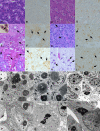
Fig 1
Fig 1
Histological features and immunocytochemistry (IHC) for arenavirus antigen in tissues of BIBD-negative (A to D) and BIBD-positive (E to L) snakes. For all tissues, an HE stain is shown on the left, and the IHC staining (polyclonal anti-UHV plus DAB) is on the right. Magnification, ×400. (A and B) Exocrine pancreas of BIBD-negative B. constrictor. There is no evidence of cytoplasmic inclusion bodies (A) and no reaction is seen in the IHC stain (B). (C and D) Liver of BIBD-negative P. reticulatus. There is no evidence of cytoplasmic IB in hepatocytes. The brown staining in panel D is due to a nonspecific reaction in intravascular heterophils and Kupffer cells. (E and F) Liver of BIBD-positive B. constrictor. Hepatocytes and bile duct epithelial cells exhibit cytoplasmic IB (E) that express arenavirus antigen (F). Arrows, IB. The inset in panel E shows erythrocytes in blood smear of the same animal, with typical IB (arrow) (May-Grünwald-Giemsa stain; magnification, ×800). (G and H) Exocrine pancreas of BIBD-positive B. constrictor. Abundant cytoplasmic IB of variable sizes (arrows) that express arenavirus antigen (H) are present in exocrine pancreatic acinar cells. (I and J) Kidney of BIBD-positive B. constrictor. Tubular epithelial cells exhibit abundant IHC-positive IB of variable sizes (arrows). (K and L) Brain/cerebral cortex of BIBD-positive C. annulatus. Neurons exhibit IHC-positive IB of variable sizes (arrows). (M to R) Ultrastructural characteristics of BIBD. TEM images of tissues from BIBD-positive snakes are shown in panels M to P. Liver of B. constrictor with irregularly delineated, electron-dense IB (asterisk) in the cytoplasm of an hepatocyte is shown in panel M. Arrow, mitochondrion. Magnification, ×10,000. Panel N shows exocrine pancreas of a B. constrictor with glandular epithelial cells with medium-sized cytoplasmic IB (asterisks). Arrow, rough endoplasmic reticulum; N, nucleus. Magnification, ×5,000. Panel O shows kidney of a B. constrictor with large cytoplasmic IB (asterisks) in glomerular mesangial cells. N, nucleus. Magnification, ×3,500. Panel P shows the brain of C. hortulanus with IB in neurons. Magnification, ×8,000. TEM images of BIBD-positive boid cell cultures are shown in panels Q and R. Cultured boid kidney cells, experimentally infected with supernatant of the BIBD-positive bone marrow cell line of snake 1 (B. constrictor) at day 8 postinoculation, are shown in panel Q. Two cells are shown, one with a cytoplasmic IB (asterisk). N, nucleus of second cell. Magnification, ×5,000. Cultured boid kidney cells, experimentally infected with supernatant of the BIBD-positive kidney cell line of snake 3 (B. constrictor) at day 8 postinoculation, are shown in panel R. Two cells, each with one cytoplasmic IB (asterisks), are shown. N: nucleus. Magnification, ×10,000.
Fig 2
Fig 2
In vitro studies in permanent boid tissue cultures (>30 passages). (A to C) Morphology of BIBD-negative (naive; A) and BIBD-positive (B and C) tissue cultures. Native cells are shown at a magnification of ×200. BIBD-negative cell culture (A) was derived from the kidney of a histologically BIBD-negative boa constrictor (snake 26). BIBD-positive cell cultures from histologically BIBD-positive snakes were derived from heart (snake 1; B) and bone marrow (snake 4; C). (D to K) Morphological features and immunocytochemistry (IHC) for arenavirus antigen in PFA-fixed, paraffin-embedded cell pellets of a naive boid kidney cell culture from snake 26 at day 8 after mock (D and E) or virus (F to K) inoculation. Purified virus was derived from supernatants of experimentally infected kidney cell lines by ultracentrifugation in density gradient. For all specimens, an HE stain is shown on the left, and the IHC staining (polyclonal anti-UHV plus DAB) is on the right. Magnification, ×400. Mock-infected cells are without evidence of cytoplasmic IB formation (D) and show no positive immune reaction (E). (F and G) Cells after inoculation with UHV, isolated from a BIBD-positive boa constrictor (snake 1). (H and I) Cells after inoculation with virus from infected kidney cell line from snake 2. (J and K) Cells after inoculation with virus purified from BIBD-positive B. constrictor (snake 5) (Table 1). Individual cells exhibit one or several cytoplasmic IB (F, H, and J) of variable sizes (arrows) that express arenavirus antigen (arrows in G, I, and K).
Fig 3
Fig 3
Fractionation of BIBD-positive and -negative cells and cell culture supernatants. (A) Isolation of 68-kDa protein from BIBD-positive culture of boid cells. BIBD-negative (snake 26) and BIBD-positive (snake 1) boid cells were lysed with detergent, debris was pelleted, the supernatant was collected (cell sup. 1), and the remaining pellet was washed once with lysis buffer. The resulting supernatants (cell sup. 2) and pellets were separated by SDS-PAGE and visualized by Coomassie staining. Inf, infected. (B) Cell culture supernatant. Material was pelleted through a sucrose cushion from mock and BIBD-positive boid cells. Question marks indicate unknown proteins. The proteins were visualized by Coomassie staining. (C) Transmission electron microscopy with negative staining of viruses analyzed in panel B. (D) Purification of virus in sucrose density gradient from mock-infected and BIBD-positive boid cells. The protein contents of fractions (f) collected from the bottom (f1) were analyzed by SDS-PAGE and visualized by Coomassie staining. (E) Negative staining of viruses purified in density gradient (fractions f8 and f9 from panel D). Mark, molecular mass marker.
Fig 4
Fig 4
Isolation and characterization of nucleic acids from virions purified by density gradient ultracentrifugation. (A) Nucleic acids isolated from density gradient fractions of BIBD-negative and -positive cell culture supernatants (pool of fractions 8 and 9 for both) were separated by agarose gel electrophoresis and visualized by ethidium bromide staining. Mark, lane with DNA ladder. The two segments of nucleic acid present in the fractions of the BIBD-positive cell culture supernatant are labeled L (large) and S (small). (B) Digestion of the nucleic acid isolated from BIBD-positive cell culture supernatant. The isolated nucleic acid (the same material as described for panel A) was treated with DNase I, RNase A, or left untreated, separated by agarose gel electrophoresis, and visualized by ethidium bromide staining. Mark, lane with DNA ladder. (C) Nucleic acids as analyzed by RNA gel electrophoresis.
Fig 5
Fig 5
(A) Cryo-EM image showing UHV virions embedded in vitrified water. Scale bar, 100 nm. (B) Central section through low-pass-filtered tomographic reconstruction of type 1 virion. White arrowheads, typical views of the spike; black arrowhead, secondary layer of density associated with the presence of spikes. (Inset) Section through subtomographic average of the surface of the type 1 virion. (C) Section through a low-pass-filtered tomographic reconstruction of type 2 virion. The inset shows a section through a subtomographic average of the surface of the type 2 virion. Scale bar, 10 nm (inset). Panels A to C are in the same scale. Insets in panels B and C are in the same scale. (D) Surface representation of the subtomographic reconstruction of the spike without application of symmetry. (E and F) Subtomographic reconstruction of the 3-fold averaged spike shown from the side (E) and tilted toward the viewer (F). Each separate domain corresponds to one GPC. In panels D to F, the surface representations are drawn at a density threshold of 1 standard deviation above the mean. For panels D and E, the gray scale sections through the spike at the three levels indicated on the side view show the three separate domains of the trimer in the tips and center, which merge together in the stalk next to the membrane. Protein density is in black. Scale bar, 10 nm (E). Panels D and E are in the same scale. (G) Section through tomographic reconstruction of a broken virion. Scale bar, 100 nm. The inset shows a magnified surface representation from a selected area of the virion tomogram. The solid surface is drawn at a density threshold of 2 standard deviations, and the mesh is at 1 standard deviation.
Fig 6
Fig 6
Evolutionary relationships of arenaviruses. (A) Phylogenetic tree based on full-length RdRp amino acid sequences of arenaviruses. (B) Coding strategy of structural proteins in UHV genome. IGR, intergenic region; GPC, glycoprotein precursor; NP, nucleoprotein; RdRp, RNA-dependent RNA polymerase; Z, Z protein. The arrows in the segments represent the orientations in which the respective structural proteins are encoded. Segments are not drawn to scale. (C) Phylogenetic tree based on core polymerase domains of representative members of the segmented negative-stranded RNA viruses. (D) Phylogenetic tree based on partial nucleotide sequences of the L segment to show the sequence diversity of arenaviruses in snakes. The curly lines in panel C denote different virus families, and the different shades in panel D denote phylogroups of BIBDAV. All three trees are maximum clade credibility trees with an arbitrary root, shown with mean branch lengths (substitutions per site). The Bayesian posterior probability values are given at the nodes, except for the tree in panel A, in which all Bayesian posterior probability values were 1.
Fig 7
Fig 7
Production and characterization of polyclonal rabbit antisera against UHV. (A) Density gradient purification of virus from supernatant of boid kidney cells (snake 26) infected with supernatant from a BIBD-positive bone marrow cell culture (snake 1). Fractions collected from the bottom were separated by SDS-PAGE, and proteins were visualized by Coomassie staining. (B) SDS-PAGE separation of the virus preparation used for immunization with Coomassie staining. (C) Immunoblot using rabbit antiserum against purified UHV. The preparation was subjected to immunoblotting with preimmune, first-bleed, and final-bleed sera of two immunized rabbits (21923 and 21924). (D) Purified virus (used as antigen) subjected to immunoblotting with mouse anti-LCMV, mouse anti-JUNV, human anti-BHV, rabbit anti-MACV, and rabbit anti-UHV. (E) Cell pellet (from the experiment shown in Fig. 3A) of BIBD-negative (center lane) and -positive (right lane) boid cell culture, subjected to immunoblotting with preimmune and anti-UHV (first bleed) serum. (F) Cell pellet (from the experiment shown in Fig. 3A) of BIBD-negative (center lane) and -positive (right lane) boid cell culture, subjected to immunoblotting with anti-MACV (rabbit), anti-MACV (mouse), and anti-BHV (human) antibodies. HMAF, hyperimmune mouse ascites fluid. The left-hand lanes in all gels represent the molecular mass markers. α, anti.
Fig 8
Fig 8
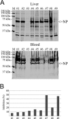
Viral antigens and antibodies in BIBD-positive snakes. (A) Liver and blood samples collected from nine snakes were solubilized in LSB, separated by SDS-PAGE, transferred onto nitrocellulose, and subjected to immunoblotting with anti-UHV serum (final bleed of animal 21923). (B) Competitive ELISA for the detection of arenavirus antibodies in the plasma of boid snakes. Samples are identified as follows: 1, snake 8; 2, snake 9;; 3, snake 35; 4, snake 5; 5, snake 6; 6, snake 7; 7, snake 10; 8, snake 11; and 9, snake 28 (Table 1).
Fig 9
Fig 9
(A) Histological and IHC analysis of PFA-fixed Vero E6 cell pellets at day 12 postinfection with UHV: HE-stained uninfected control cells without evidence of IB (frame a); HE-stained infected cells with numerous variably sized, typical cytoplasmic IB (arrows) (frame b); IHC with anti-UHV antiserum (IgG fraction of final bleed 21923, negatively purified) on infected cells, showing strong viral antigen expression (frame c). Magnification, ×400. (B) Immunofluorescence staining (anti-UHV antiserum) of Vero E6 cells infected with UHV at 5, 6, and 8 dpi. (C) Boid kidney cells infected with UHV (2 dpi), stained with rabbit anti-UHV serum. (D) Immunoblot of Vero E6 cell lysates with anti-UHV serum. Lane M, marker; lane 1, Vero E6 cells infected with supernatant of BIBD-negative boid cells (at 16 dpi); lane 2, Vero E6 cells infected with UHV (at 16 dpi); lane 3, Vero E6 cells infected with supernatant collected from Vero E6 cells infected with UHV at 16 dpi (at 12 dpi).

Comment in

References

    1. Vidal N, Hedges SB. 2009. The molecular evolutionary tree of lizards, snakes, and amphisbaenians. C. R. Biol. 332:129–139. - PubMed
    1. Schumacher J, Jacobson ER, Homer BL, Gaskin JM. 1994. Inclusion body disease in boid snakes. J. Zoo Wildl. Med. 25:511–524.
    1. Carlisle-Nowak MS, Sullivan N, Carrigan M, Knight C, Ryan C, Jacobson ER. 1998. Inclusion body disease in two captive Australian pythons (Morelia spilota variegata and Morelia spilota spilota). Aust. Vet. J. 76:98–100. - PubMed
    1. Raymond JT, Garner MM, Nordhausen RW, Jacobson ER. 2001. A disease resembling inclusion body disease of boid snakes in captive palm vipers (Bothriechis marchi). J. Vet. Diagn. Invest. 13:82–86. - PubMed
    1. Fleming GJ, Heard DJ, Jacobson ER. 2003. Cytoplasmic inclusions in corn snakes, Elaphe guttata, resembling inclusion body disease of boid snakes. J. Herpetol. Med. Surg. 13:18–22.

Publication types

LinkOut - more resources