. 2009;5(6):1174-1187.
doi: 10.1039/B812146J.

Bacterial Swarming: A Model System for Studying Dynamic Self-assembly

Affiliations

Bacterial Swarming: A Model System for Studying Dynamic Self-assembly

Matthew F Copeland et al. Soft Matter. 2009.

Abstract

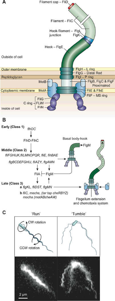
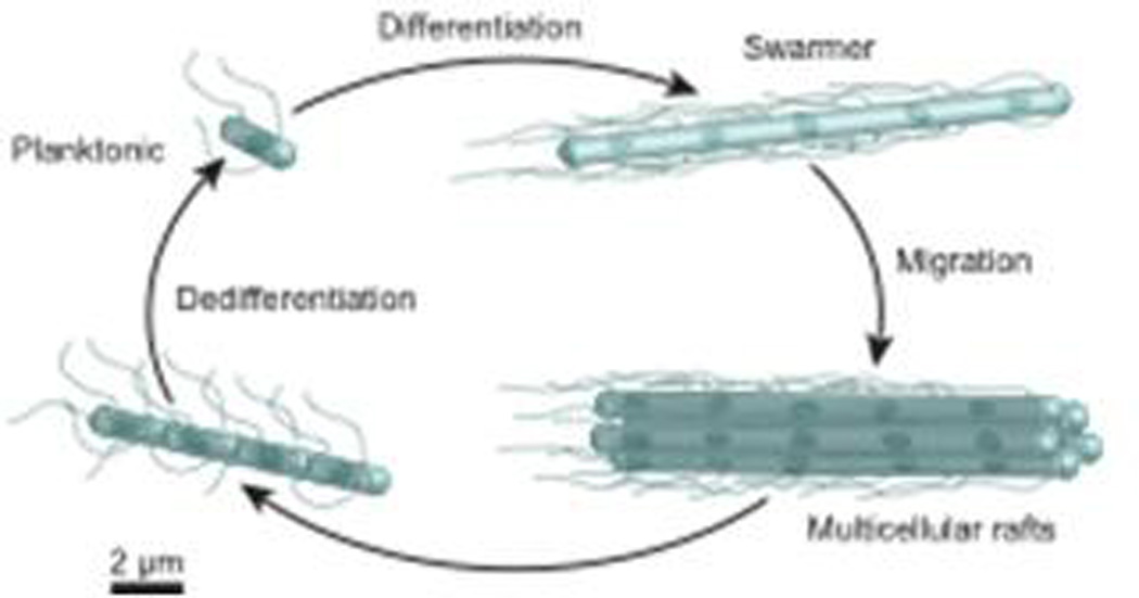
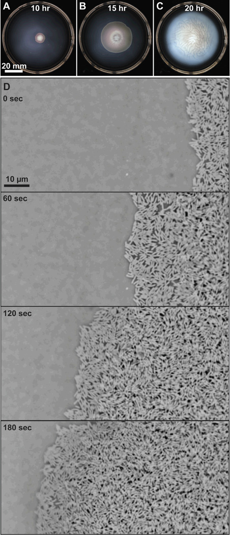
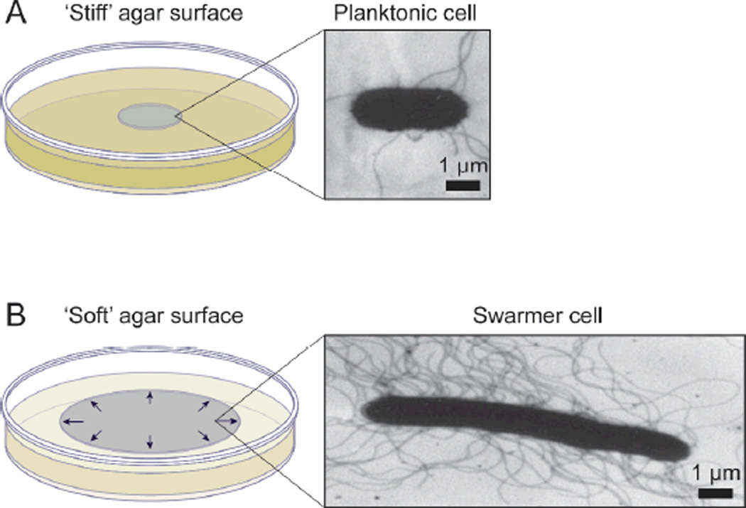
Bacterial swarming is an example of dynamic self-assembly in microbiology in which the collective interaction of a population of bacterial cells leads to emergent behavior. Swarming occurs when cells interact with surfaces, reprogram their physiology and behavior, and adapt to changes in their environment by coordinating their growth and motility with other cells in the colony. This review summarizes the salient biological and biophysical features of this system and describes our current understanding of swarming motility. We have organized this review into four sections: 1) The biophysics and mechanisms of bacterial motility in fluids and its relevance to swarming. 2) The role of cell/molecule, cell/surface, and cell/cell interactions during swarming. 3) The changes in physiology and behavior that accompany swarming motility. 4) A concluding discussion of several interesting, unanswered questions that is particularly relevant to soft matter scientists.


Figures

Figure 1
Figure 1
Structure, expression, assembly, and function of bacterial flagella. A) A cartoon depicting the components of flagella discussed in this review; some elements have intentionally been removed for clarity. B) A diagram of the hierarchy of the expression of flagellar genes in E. coli C) Fluorescence microscopy images of swimming E. coli cells and corresponding cartoons depicting a cell ‘running’ and ‘tumbling’. The cartoon on the top left depicts a cell with bundled flagella ‘running’; the image below shows a live cell in this configuration. When the cell ‘runs’ the bundle of flagella rotate CCW (as seen from behind the cell) and the cell body rotates CW. The cartoon on the top right depicts a cell ‘tumbling’ in which the flagella are splayed outward; the image immediately below shows a live cell in this configuration. Images are reprinted or modified with permission from A) Nature Publishing Group, Copyright 2008, B) American Association for the Advancement of Science, Copyright 2001, C) American Society for Microbiology, Copyright, 2000.
Figure 2
Figure 2
A cartoon depicting the general ‘life cycle’ of motile cells of bacteria as they swarm on surfaces. The length of the flagella (in relation to the length of cells) and the number of flagella per swarmer cell has been reduced for clarity. The blue spot depicts the bacterial chromosome. The scale bar is an approximate estimate of dimensions.
Figure 3
Figure 3
A-C) Time lapse images depicting the macroscopic migration of a swarming colony of wild-type E. coli strain RP437 on the surface of 0.45% Eiken agar (w/v) infused with nutrient broth (1.0% peptone, 0.5% NaCl, 0.3% beef extract, and 0.5% glucose). The agar solidified overnight at 25 °C before the center of the gel was inoculated with 2 µl of a saturated overnight culture of bacteria. The plate was incubated at 30 °C. The expansion of the swarm colony over time is shown at 10, 15, and 20 hours after inoculation. D) A sequence of microscopy images that demonstrate the time-dependent spreading of swarmer cells across an agar surface. The panels show the progression of the edge of the swarm colony over a three-minute period. The media and inoculation procedure was identical to the conditions used for images A-C. The images were acquired using phase contrast microscopy and a CCD camera; the images were inverted to improve the contrast between the cells and background. The cells are moving from the right-hand side of the image to the left.
Figure 4
Figure 4
A) A schematic diagram depicting a colony of bacteria grown on the surface of a ‘stiff’ agar gel (e.g. 1.5%, w/v) and a characteristic planktonic cell isolated from the colony. The TEM image shows a planktonic cell of S. liquefaciens MG1. B) A swarming colony of bacteria grown on the surface of a ‘soft’ agar gel (e.g. 0.45%, w/v) and a TEM image of a swarmer cell of S. liquefaciens MG1 isolated from the outer edge of a swarm colony. The arrows depict the radial outward expansion of the colony on the surface. TEM images in A and B are reprinted with permission from the American Society for Microbiology, Copyright 1999.
Figure 5
Figure 5
Examples of surface-active molecules involved in swarming migration. A) The structure of colony migration factor (CMF) from P. mirabilis. The two images show the swarming migration of wild-type P. mirabilis (left) and a mutant defective in production of CMF on agar gels. B) The structure of serrawettin W2. The images show the rescue of swarming motility on agar gels of a S. liquefaciens MG1 mutant (swrI/swrA) that does not synthesize serrawettin W2. The concentration of serrawettin W2 added to each plate is indicated. C) The structure of surfactin. The swarm plate on the left displays the swarming migration of wild-type B. subtilis strain 3610. The swarming defective B. subtilis mutant (right) contains a mutation in the srfAA gene, which encodes a translation product important for surfactin synthesis. D) The structure of mono-rhamnolipid. The image shows the swarming migration of wild-type P. aeruginosa (top structure) and an rhlA mutant (bottom structure), which is unable to synthesize rhamnolipids, on agar gels. The black arrows on each agar plate depict the edge of the swarming colony. Images are reprinted with permission from (A) Blackwell Science Ltd, Copyright 1995, (B) American Society for Microbiology, Copyright 1998, (C) Blackwell Publishing Ltd, Copyright 2004, and (D) American Society for Microbiology, Copyright 2000.
Figure 6
Figure 6
Examples of cell-cell contact in swarming motility. A) An image of the edge of a swarming colony of P. mirabilis (300x). The swarmer cells are arranged into tightly packed rafts of cells. B) S. liquefaciens MG1 cells at the leading edge of a swarming colony migrate together. No scale was provided in the original publication for images A) and B). C-D) TEM images of a wild-type P. mirabilis swarmer cell (C) and a mutant of the ccmA gene in P. mirabilis (D). The image (inset) shows the swarming motility of colonies of wild-type P. mirabilis (left) and the ccmA mutant (right) on an agar gel. E-F) SEM images of vapor-fixed swarmer cells of wild-type P. mirabilis (E) and a mutant with reduced swarming motility (~85% reduction in surface migration) (F). Images are reprinted with permission from (A) American Society for Microbiology, Copyright 1972, (B) American Society for Microbiology, Copyright 1999, (C) and (D) American Society for Microbiology, Copyright 1999, and (E) and (F) American Society for Microbiology, Copyright 2004.

References

    1. Whitesides GM, Grzybowski B. 2002;vol. 295:2418–2421. - PubMed
    1. Dworkin M, Shapiro JA. Bacteria as multicellular organisms. New York: Oxford University Press; 1997.
    1. Shapiro JA. Annu Rev Microbiol. 1998;52:81–104. - PubMed
    1. Fraser GM, Hughes C. Curr Opin Microbiol. 1999;2:630–635. - PubMed
    1. Baba T, Ara T, Hasegawa M, Takai Y, Okumura Y, Baba M, Datsenko KA, Tomita M, Wanner BL, Mori H. Mol Syst Biol. 2006;2 2006 0008. - PMC - PubMed