Review
. 2013 Aug;40(8):080901.
doi: 10.1118/1.4811136.

MR-guided focused ultrasound surgery, present and future

Affiliations
Review

MR-guided focused ultrasound surgery, present and future

David Schlesinger et al. Med Phys. 2013 Aug.

Abstract

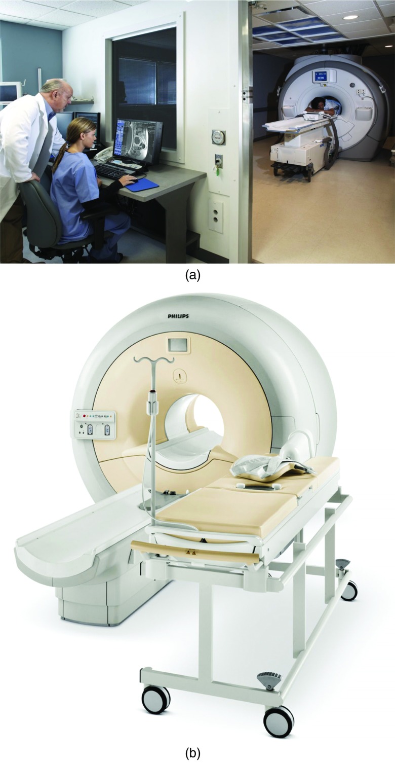
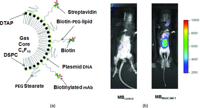
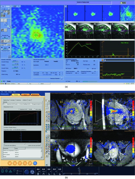
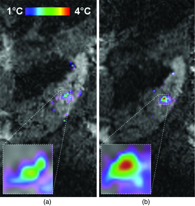
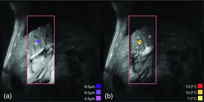
MR-guided focused ultrasound surgery (MRgFUS) is a quickly developing technology with potential applications across a spectrum of indications traditionally within the domain of radiation oncology. Especially for applications where focal treatment is the preferred technique (for example, radiosurgery), MRgFUS has the potential to be a disruptive technology that could shift traditional patterns of care. While currently cleared in the United States for the noninvasive treatment of uterine fibroids and bone metastases, a wide range of clinical trials are currently underway, and the number of publications describing advances in MRgFUS is increasing. However, for MRgFUS to make the transition from a research curiosity to a clinical standard of care, a variety of challenges, technical, financial, clinical, and practical, must be overcome. This installment of the Vision 20∕20 series examines the current status of MRgFUS, focusing on the hurdles the technology faces before it can cross over from a research technique to a standard fixture in the clinic. It then reviews current and near-term technical developments which may overcome these hurdles and allow MRgFUS to break through into clinical practice.


Figures

Figure 1
Figure 1
Examples of currently available commercial MR-guided focused ultrasound systems. (a) Insightec OR MR-guided system (image courtesy of InSightec Ltd.). (b) Philips Sonalleve MR-guided system (image courtesy of Philips Medical Systems).
Figure 2
Figure 2
MR-guided focused ultrasound for uterine fibroids. Images document the treatment of a fibroid patient treated in two MRgFUS sessions approximately 1 week apart. (a) Preprocedure: axial T1 spin-echo image of a patient with a large (∼542 cm3) uterine fibroid. Image shows some hetereogeneity of the fibroid, as well as some possible necrotic areas. (b) Postprocedure 1: axial, postcontrast, fat suppressed, fast spoiled gradient echo pulse sequence. Image shows large nonperfused volume in the center of the fibroid. Volume calculations estimate that 63% of fibroid volume was nonperfused. (c) Postprocedure 2: axial, postcontrast, fat suppressed, fast spoiled gradient echo pulse sequence. Image shows the remaining volume of the lesion is now nonperfused. Approximately 100% of the fibroid volume was ablated. [Images courtesy of the University of Virginia Department of Radiology].
Figure 3
Figure 3
Fluorescence microphotographs of two fragments (control and ultrasound-treated) of a swine artery after intravenous administration of DiI fluorescent-dye impregnated microbubbles (Ref. 167) (a) Fluorescence observed in the control fragment of the artery after microbubble administration. (b) Fluorescence observed in the ultrasound-treated fragment after microbubble administration and insonation with radiation-force ultrasound, followed by a “destruction” pulse to locally destroy the microbubbles. (c) Ultrasound image of the artery at the end of the applied ultrasound sequence with the locations of the excised control (a) and ultrasound-treated (b) fragments. [Figures courtesy of Abhay Patil, Philips Healthcare, and John Hossack, University of Virginia] (Ref. 167).
Figure 4
Figure 4
Contrast-enhanced T1-weighted MRI showing blood-brain barrier disruption induced in a brain volume in a macaque by focused ultrasound and microbubbles. The disruption was produced in a 1 cm3 volume using low-energy focused ultrasound pulses combined with a circulating microbubble ultrasound contrast agent. The sonications were applied transcranially using a clinical prototype MRI-guided focused ultrasound system (ExAblate 4000, InSightec). Note the lack of contrast enhancement in the ultrasound beam path. This noninvasive technique is being investigated to target the delivery of drugs that normally do not reach the brain due to the presence of the blood-brain barrier. [Image courtesy of Dr. Nathan McDannold, Brigham & Women's Hospital, Boston, MA.]
Figure 5
Figure 5
FUS/microbubble-mediated gene transcription (Ref. 197). (a) A positively charged microbubble is complexed with a luciferase-encoding plasmid, and carries an antibody against a marker for Crohn's disease. Control bubbles carry a nonspecific Immunoglobulin G (IgG) antibody. Bubbles are injected intravenously and left to circulate for 2 days, with the targeted bubbles accumulating in the target intestinal inflammation zone and attaching to the Crohn's disease marker on the vascular endothelium. After the circulating bubbles exit the bloodstream, ultrasound is performed. Two days later, luciferin is injected, and optical imaging of the induced bioluminescence is performed. (b) Control vs experimental results. The left-hand figure shows an animal injected with control bubbles. Right hand figure shows animal injected with targeted antibody bubbles. Note the accumulation bubbles in the targeted animal, demonstrating transfection. [Image courtesy of Alexander Klibanov, University of Virginia] (Ref. 197).
Figure 6
Figure 6
Examples of treatment planning systems for MRgFUS. (a) Treatment planning for the InSightec Exablate Neuro. Planning screens allow the operator to set treatment parameters, monitor beam paths per transducer, thermal lesion location, time/temperature graphs, and ultrasound frequency spectrum. [Image courtesy of the InSightec Ltd.] (b) Treatment planning for the Philips Sonalleve MRgFUS system. This system allows the operator to monitor real-time temperature rise at the target, as well as in near-field and far-field regions [Image courtesy of Philips Healthcare].
Figure 7
Figure 7
Color-coded temperature map overlaid on T2* weighted anatomical MR images of porcine kidneys demonstrating a real-time motion compensation technique (Ref. 272). (a) Heating deposition without motion compensation. (b) Heating deposition with motion compensation. Notice the increase in heating magnitude and sharper temperature falloff. [Images courtesy of Mario Ries, Ph.D., Laboratory for Functional and Molecular Imaging, Bordeaux, France].
Figure 8
Figure 8
MR-ARFI: (a) MR-ARFI and (b) MR-thermometry images acquired in the in vivo porcine liver. Both images are small FOV EPI acquisitions superimposed on a larger FOV gradient echo image acquired a few minutes before. After visualization of the displacement focus on MR-ARFI to verify the target location (Ref. 291), a steered HIFU ablation was performed with thermal monitoring, shown in the reduced FOV image on the right (Ref. 27). MR-ARFI images require only 3 J of energy, whereas a low temperature rise test ablation would require upwards of 800 J of energy. [Images of courtesy of Dr. Andrew B. Holbrook and Dr. Kim Butts Pauly, Stanford University].

References

    1. Cline H. E., Schenck J. F., Hynynen K., Watkins R. D., Souza S. P., and Jolesz F. A., “MR-guided focused ultrasound surgery,” J. Comput. Assist. Tomogr. 16, 956–965 (1992).10.1097/00004728-199211000-00024 - DOI - PubMed
    1. Hynynen K., Darkazanli A., Unger E., and Schenck J. F., “MRI-guided noninvasive ultrasound surgery,” Med. Phys. 20, 107–115 (1993).10.1118/1.597093 - DOI - PubMed
    1. Arthur R. M., Straube W. L., Trobaugh J. W., and Moros E. G., “Non-invasive estimation of hyperthermia temperatures with ultrasound,” Int. J. Hyperthermia 21, 589–600 (2005).10.1080/02656730500159103 - DOI - PubMed
    1. Vaezy S., Shi X., Martin R. W., Chi E., Nelson P. I., Bailey M. R., and Crum L. A., “Real-time visualization of high-intensity focused ultrasound treatment using ultrasound imaging,” Ultrasound Med. Biol. 27, 33–42 (2001).10.1016/S0301-5629(00)00279-9 - DOI - PubMed
    1. Maleke C. and Konofagou E. E., “An all-ultrasound-based system for real-time monitoring and sonication of temperature change and ablation,” Conf. Proc. IEEE Eng. Med. Biol. Soc. 1, 164–167 (2006).10.1109/IEMBS.2006.260845 - DOI - PubMed

Publication types

MeSH terms