. 2013 Dec;49(6):1120-6.
doi: 10.1165/rcmb.2013-0039OC.

Molecular magnetic resonance imaging of pulmonary fibrosis in mice

Affiliations

Molecular magnetic resonance imaging of pulmonary fibrosis in mice

Peter Caravan et al. Am J Respir Cell Mol Biol. 2013 Dec.

Abstract

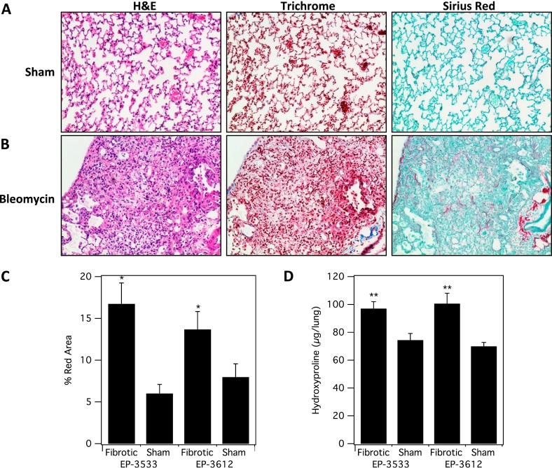
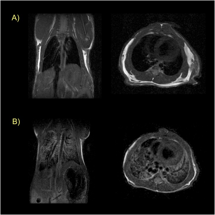
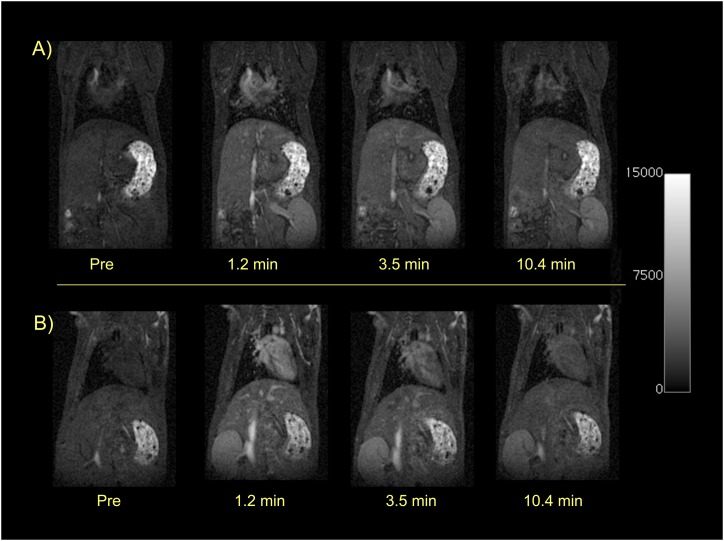
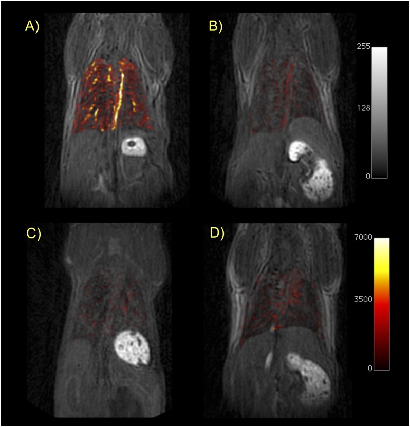
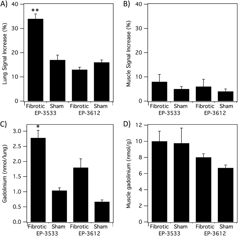
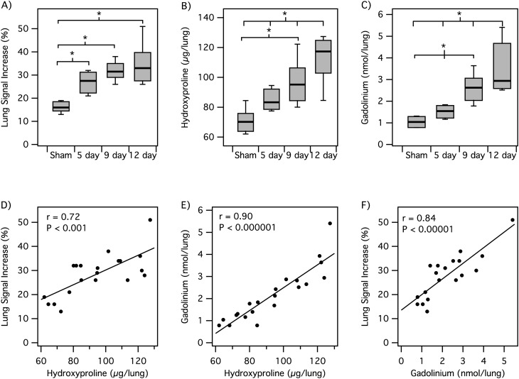
Idiopathic pulmonary fibrosis is a chronic, progressive, fibrosing interstitial pneumonia of unknown cause resulting in dyspnea and functional decline until death. There are currently no effective noninvasive tools to monitor disease progression and response to treatment. The objective of the present study was to determine whether molecular magnetic resonance imaging of the lung using a probe targeted to type I collagen could provide a direct, noninvasive method for assessment of pulmonary fibrosis in a mouse model. Pulmonary fibrosis was generated in mice by transtracheal instillation of bleomycin (BM). Six cohorts were imaged before and immediately after intravenous administration of molecular imaging probe: (1) BM plus collagen-targeted probe, EP-3533; (2) sham plus EP-3533; (3) BM plus nonbinding control probe, EP-3612; (4) sham plus EP-3612; (5) BM plus EP-3533 imaged early; and (6) BM plus EP-3533 imaged late. Signal-to-noise ratio (SNR) enhancement was quantified in the lungs and muscle. Lung tissue was subjected to pathologic scoring of fibrosis and analyzed for gadolinium and hydroxyproline. BM-treated mice had 35% higher lung collagen than sham mice (P < 0.0001). The SNR increase in the lungs of fibrotic mice after EP-3533 administration was twofold higher than in sham animals and twofold higher than in fibrotic or sham mice that received control probe, EP-3612 (P < 0.0001). The SNR increase in muscle was similar for all cohorts. For EP-3533, we observed a strong, positive, linear correlation between lung SNR increase and hydroxyproline levels (r = 0.72). Collagen-targeted probe EP-3533-enhanced magnetic resonance imaging specifically detects pulmonary fibrosis in a mouse model of disease.


Figures

<i>Figure 1.</i>
Figure 1.
Characterization of fibrosis and collagen deposition in the mouse bleomycin (BM) model. Representative images of lung tissue stained with hematoxylin and eosin (H&E), trichrome, and Sirius red for sham (A) and BM-treated mice (B). (C) Sirius red staining was quantified and was significantly higher in fibrotic (BM-treated) mice (*P < 0.001). (D) Hydroxyproline (Hyp) analysis of ex vivo harvested lung tissue from all four cohorts shows significantly higher Hyp/lung in the fibrotic (BM) -treated cohorts (**P < 0.0001).
<i>Figure 2.</i>
Figure 2.
Magnetic resonance (MR) characterization of pulmonary edema. (A) Coronal (left) and axial (right) T1-weighted rapid acquisition with refocused echo (RARE) images of a sham mouse showing absence of signal in the lungs. (B) Coronal (left) and axial (right) images at approximately the same location in a BM-treated mouse. Edema is obvious from the opacification of the lungs in this animal. All images taken before probe injection.
<i>Figure 3.</i>
Figure 3.
Dynamic T1-weighted MR imaging (MRI) showing rapid renal elimination of the probes. Sham mice before and immediately after intravenous injection of 10 μmol/kg EP-3533 (top panel) or EP-3612 (bottom panel). Both probes show immediate and similar enhancement of the heart, blood pool, and kidneys. By 10 minutes after injection, the signal in the heart and blood pool is approaching baseline, whereas the kidneys remain enhanced. These dynamic images show the rapid blood clearance and predominant renal elimination pathway that is similar for both EP-3533 and EP-3612.
<i>Figure 4.</i>
Figure 4.
Ultrashort echo time (UTE)-MRI of the lungs. Coronal T1-weighted UTE images of mice from each cohort in this study: (A) BM mouse + collagen-targeted EP-3533; (B) sham mouse + EP-3533; (C) BM + control probe EP-3612; (D) sham + EP-3612. False color overlay is the difference image obtained by subtracting the UTE image acquired before probe injection from the UTE image taken after probe injection. Mice with pulmonary fibrosis that receive the collagen-specific probe (A) show much higher pulmonary signal enhancement than sham mice or mice receiving the control probe. All images rendered at the same scale.
<i>Figure 5.</i>
Figure 5.
MRI and ex vivo analyses. (A) Lung signal-to-noise ratio (SNR) increase after probe injection. Fibrotic mice receiving EP-3533 had twofold higher SNR increase compared with sham mice or mice receiving EP-3612 (**P < 0.0001). (B) Muscle SNR shows no significant differences between fibrotic and sham animals for either probe. (C) Ex vivo gadolinium (Gd) analysis shows significantly higher Gd in lungs of fibrotic mice that received EP-3533 compared with sham mice or mice that received EP-3612 (*P < 0.05). (D) Ex vivo Gd concentration in muscle shows no significant differences between fibrotic and sham animals for either probe.
<i>Figure 6.</i>
Figure 6.
MRI of disease progression. (AC) Box plots showing that lung MR signal change after EP-3533 injection (A), lung Hyp (B), and lung Gd levels (C) all increase with time after BM inoculation (*P < 0.05). (DF) Scatter plots for animals imaged with EP-3533 showing positive linear correlations between lung MR signal increase and lung Hyp (D), lung Gd and lung Hyp levels (E), and lung MR signal increase and lung Gd (F).

References

    1. Bjoraker JA, Ryu JH, Edwin MK, Myers JL, Tazelaar HD, Schroeder DR, Offord KP. Prognostic significance of histopathologic subsets in idiopathic pulmonary fibrosis. Am J Respir Crit Care Med. 1998;157:199–203. - PubMed
    1. King TE, Jr, Schwarz MI, Brown K, Tooze JA, Colby TV, Waldron JA, Jr, Flint A, Thurlbeck W, Cherniack RM. Idiopathic pulmonary fibrosis: relationship between histopathologic features and mortality. Am J Respir Crit Care Med. 2001;164:1025–1032. - PubMed
    1. Raghu G, Weycker D, Edelsberg J, Bradford WZ, Oster G. Incidence and prevalence of idiopathic pulmonary fibrosis. Am J Respir Crit Care Med. 2006;174:810–816. - PubMed
    1. Raghu G, Collard HR, Egan JJ, Martinez FJ, Behr J, Brown KK, Colby TV, Cordier JF, Flaherty KR, Lasky JA, et al. An official ATS/ERS/JRS/ALAT statement: idiopathic pulmonary fibrosis: evidence-based guidelines for diagnosis and management. Am J Respir Crit Care Med. 2011;183:788–824. - PMC - PubMed
    1. Hunninghake GW, Zimmerman MB, Schwartz DA, King TE, Jr, Lynch J, Hegele R, Waldron J, Colby T, Muller N, Lynch D, et al. Utility of a lung biopsy for the diagnosis of idiopathic pulmonary fibrosis. Am J Respir Crit Care Med. 2001;164:193–196. - PubMed

Publication types