Review
. 2013 Nov;29(11):611-20.
doi: 10.1016/j.tig.2013.07.003. Epub 2013 Aug 6.

The zebrafish as a model for complex tissue regeneration

Affiliations
Review

The zebrafish as a model for complex tissue regeneration

Matthew Gemberling et al. Trends Genet. 2013 Nov.

Abstract

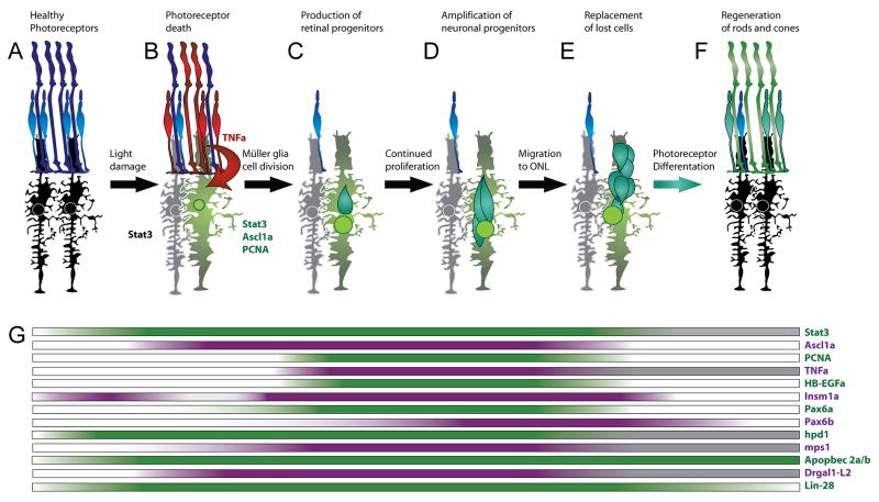
For centuries, philosophers and scientists have been fascinated by the principles and implications of regeneration in lower vertebrate species. Two features have made zebrafish an informative model system for determining mechanisms of regenerative events. First, they are highly regenerative, able to regrow amputated fins, as well as a lesioned brain, retina, spinal cord, heart, and other tissues. Second, they are amenable to both forward and reverse genetic approaches, with a research toolset regularly updated by an expanding community of zebrafish researchers. Zebrafish studies have helped identify new mechanistic underpinnings of regeneration in multiple tissues and, in some cases, have served as a guide for contemplating regenerative strategies in mammals. Here, we review the recent history of zebrafish as a genetic model system for understanding how and why tissue regeneration occurs.

Keywords: brain; fin; heart; regeneration; retina; spinal cord; stem cells; zebrafish.


Figures

Figure 1
Figure 1. Model for regeneration after partial resection of the cardiac ventricle
After injury, the RA-synthesizing enzyme, raldh2, is induced throughout the endocardium within a few hours of amputation (hpa) and later the epicardium, before these responses localize to the wound. By 7 days post amputation (dpa), gata4 regulatory sequences are activated throughout the cortical muscle layer of the ventricle. At this point, cardiomyocyte proliferation is stimulated, under the influences of hypoxia and signaling pathways as described in the main text, and epicardial cells have begun to integrate into the wound. By 14 dpa, vascularization of the regenerating muscle begins, aided by Fgf and Pdgf signaling. By 30 dpa, a new wall of cardiac muscle is typically formed, in large part by the progeny of early cardiomyocytes that activate gata4 sequences. At this point, the myocardium is vascularized and electrically coupled with the existing muscle.
Figure 2
Figure 2. Model for regeneration after light damage to photoreceptors
A. Prior to light damage, the retina consists of healthy photoreceptors (blue) and quiescent Müller glia (black). B. Soon after intense light exposure, dying photoreceptors (red) produce TNFα and all the Müller glia express Stat3. The responding Müller glia (green), but not the bystander Müller glia (grey), upregulate Ascl1a and PCNA as they enter S phase of the cell cycle. C. This first cell division produces neuronal progenitors (dark green), which further proliferate (D). E. The new retinal progenitor cells migrate to the site of damage. F. Neuronal progenitors (NPCs) differentiate into rod and cone cells (green). G. Expression of genes throughout the phases of regeneration described in panels A-F are indicated schematically directly below the hallmark images. Colored shading represents increased expression relative to undamaged retinas. Grey shading represents times not assessed for gene expression.

References

    1. Streisinger G, et al. Production of clones of homozygous diploid zebra fish (Brachydanio rerio) Nature. 1981;291(5813):293–6. - PubMed
    1. Streisinger G, et al. Segregation analyses and gene-centromere distances in zebrafish. Genetics. 1986;112(2):311–9. - PMC - PubMed
    1. Schier AF, Talbot WS. Molecular genetics of axis formation in zebrafish. Annu Rev Genet. 2005;39:561–613. - PubMed
    1. Liu J, Stainier DY. Zebrafish in the study of early cardiac development. Circ Res. 2012;110(6):870–4. - PMC - PubMed
    1. Wolman M, Granato M. Behavioral genetics in larval zebrafish: learning from the young. Dev Neurobiol. 2012;72(3):366–72. - PMC - PubMed

Publication types

LinkOut - more resources