

Actinomycosis in the mandible: CT and MR findings
- PMID: 23928143
- PMCID: PMC7965762
- DOI: 10.3174/ajnr.A3673
Actinomycosis in the mandible: CT and MR findings
Abstract
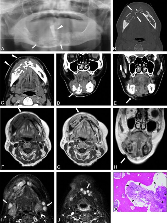
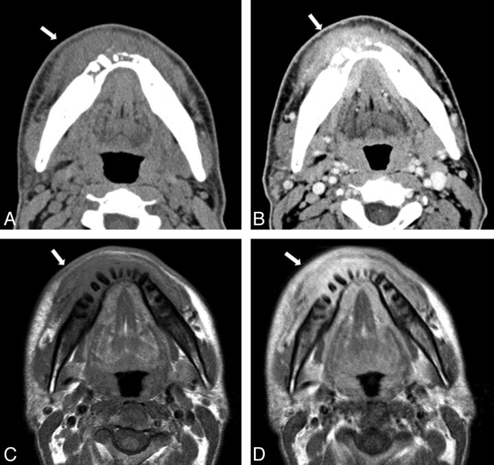
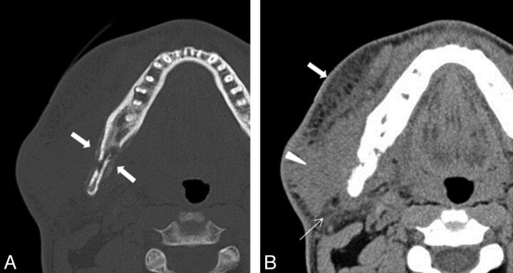
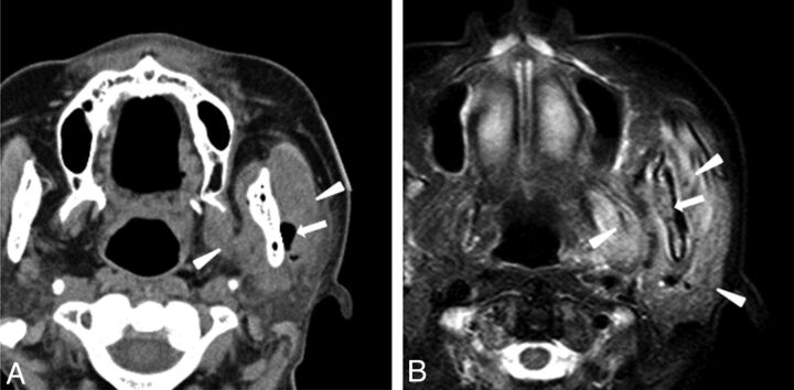
Mandibular actinomycosis is an uncommon disease. We retrospectively reviewed 6 patients with pathologically proven mandibular actinomycosis who underwent both CT and MR imaging to evaluate the characteristic imaging findings. CT results showed an irregularly marginated lesion with increased bone marrow attenuation, osteolysis, and involvement of the skin in all patients. Periosteal reaction and intralesional gas were seen in 4 patients. MR imaging results revealed low signal on T1-weighted and high signal on T2-weighted images of the mandible, and moderate heterogeneous enhancement was seen in all patients who received intravenous contrast. Cervical lymphadenopathy was not observed. Involvement of the masseter, lateral pterygoid, and medial pterygoid muscles was seen in 4 patients, whereas parotid gland and submandibular gland as well as parapharyngeal space involvement were seen in 3 patients. Familiarity with the imaging findings of mandibular actinomycosis may help to diagnosis this entity.
Figures
References
-
- Hansen T, Kunkel M, Springer E, et al. Actinomycosis of the jaws–histopathological study of 45 patients shows significant involvement in bisphosphonate-associated osteonecrosis and infected osteoradionecrosis. Virchows Arch 2007;451:1009–17 - PubMed
-
- Sa'do B, Yoshiura K, Yuasa K, et al. Multimodality imaging of cervicofacial actinomycosis. Oral Surg Oral Med Oral Pathol 1993;76:772–82 - PubMed
-
- Belmont MJ, Behar PM, Wax MK. Atypical presentations of actinomycosis. Head Neck 1999;21:264–68 - PubMed
MeSH terms
LinkOut - more resources
Full Text Sources
Other Literature Sources
Medical