

Immunomagnetic enrichment and flow cytometric characterization of mouse microglia
- PMID: 23928152
- PMCID: PMC3809889
- DOI: 10.1016/j.jneumeth.2013.07.017
Immunomagnetic enrichment and flow cytometric characterization of mouse microglia
Abstract
Background: The inflammatory response after a CNS injury is regulated by microglia/macrophages. Changes in the ratio of M1 classically activated pro-inflammatory cells versus M2 alternatively activated anti-inflammatory cells reveal the direction of the immune response. These cells are routinely identified and enumerated by morphology and cell-surface markers using immunohistochemistry.
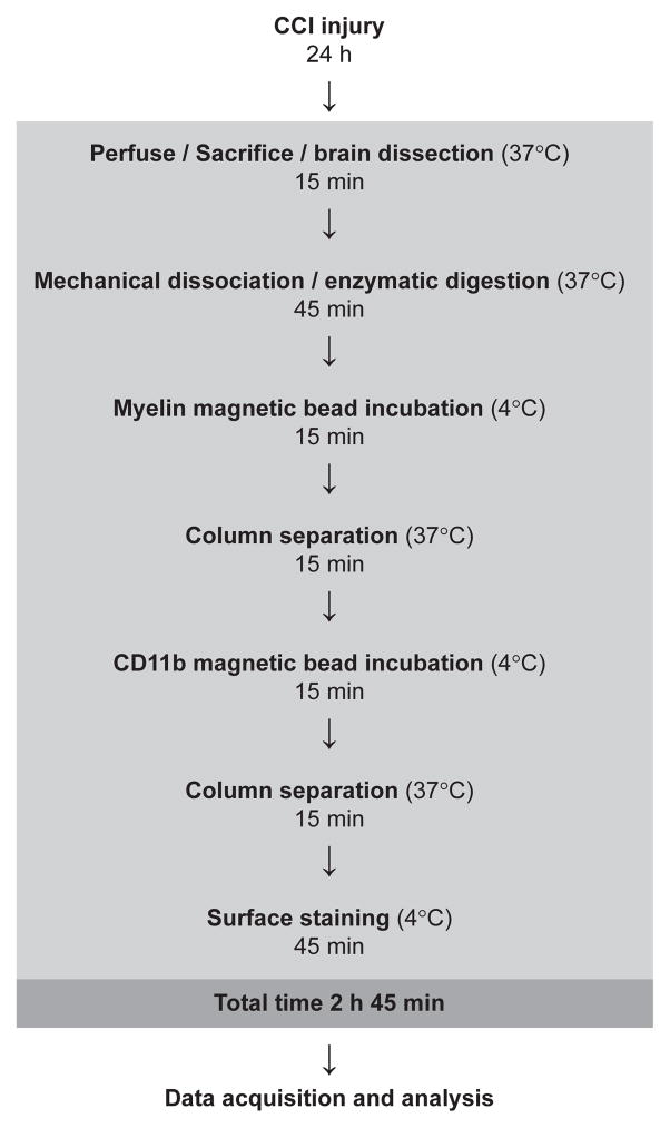
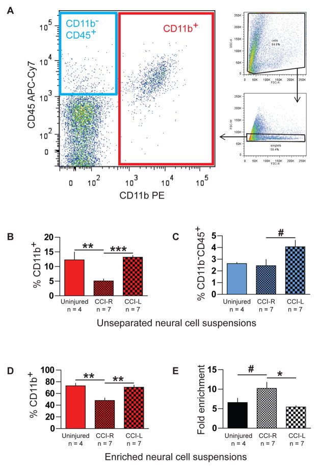
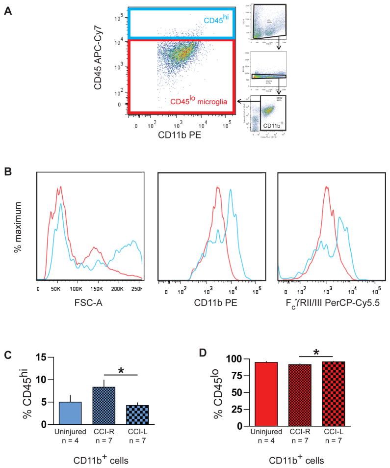
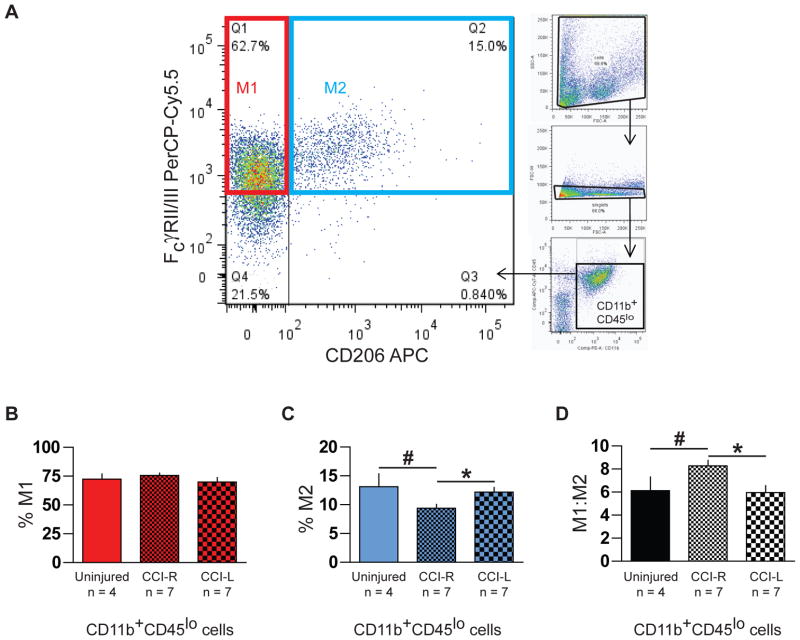
New method: We used a controlled cortical impact (CCI) mouse model for traumatic brain injury (TBI), then isolated microglia/macrophages from neural cell suspensions using magnetic beads conjugated to CD11b monoclonal antibody to obtain the entire myeloid population. Polarization states of CD11b(+)CD45(lo) microglia were evaluated by expression of M1 surface marker FcγRII/III and M2 surface marker CD206.
Results: After TBI, we observed an increase in M1:M2 ratio in the ipsilateral hemisphere when compared to the contralateral side, indicating that 24h after CCI, a shift in microglia polarization occurs localized to the hemisphere of injury. Comparison with existing method(s): The major impetus for developing and refining the methods was the need to accurately quantify microglial activation states without reliance on manual morphometric counting of serial immunohistochemistry slides. Flow cytometric analysis of enriched cell suspensions provides quantitative measurement of microglial polarization states complementary to existing methods, but for entire populations of cells.
Conclusions: In summary, we used immunomagnetic beads to isolate myeloid cells from injured brain, then stained surface antigens to flow cytometrically identify and categorize microglia as either classically activated M1 or alternatively activated M2, generating a ratio of M1:M2 cells which is useful in studying attempts to reduce or redirect neuroinflammation.
Keywords: Flow cytometry; Microglia; Neuroinflammation; Traumatic brain injury.
Copyright © 2013 Elsevier B.V. All rights reserved.
Figures
References
-
- David S, Kroner A. Repertoire of microglial and macrophage responses after spinal cord injury. Nat Rev Neurosci. 2011;12:388–399. - PubMed
-
- Ford AL, Goodsall AL, Hickey WF, et al. Normal adult ramified microglia separated from other central nervous system macrophages by flow cytometric sorting. Phenotypic differences defined and direct ex vivo antigen presentation to myelin basic protein-reactive CD4+ T cells compared. J Immunol. 1995;154:4309–4321. - PubMed
-
- Gordon S. Alternative activation of macrophages. Nat Rev Immunol. 2003;3:23–35. - PubMed
-
- Graeber MB. Changing face of microglia. Science. 2010;330:783–788. - PubMed
MeSH terms
Substances
Grants and funding
LinkOut - more resources
Full Text Sources
Other Literature Sources
Research Materials
Miscellaneous
