. 2013 Sep 27;288(39):28180-94.
doi: 10.1074/jbc.M113.491613. Epub 2013 Aug 8.

YidC protein, a molecular chaperone for LacY protein folding via the SecYEG protein machinery

Affiliations

YidC protein, a molecular chaperone for LacY protein folding via the SecYEG protein machinery

Lu Zhu et al. J Biol Chem. .

Abstract

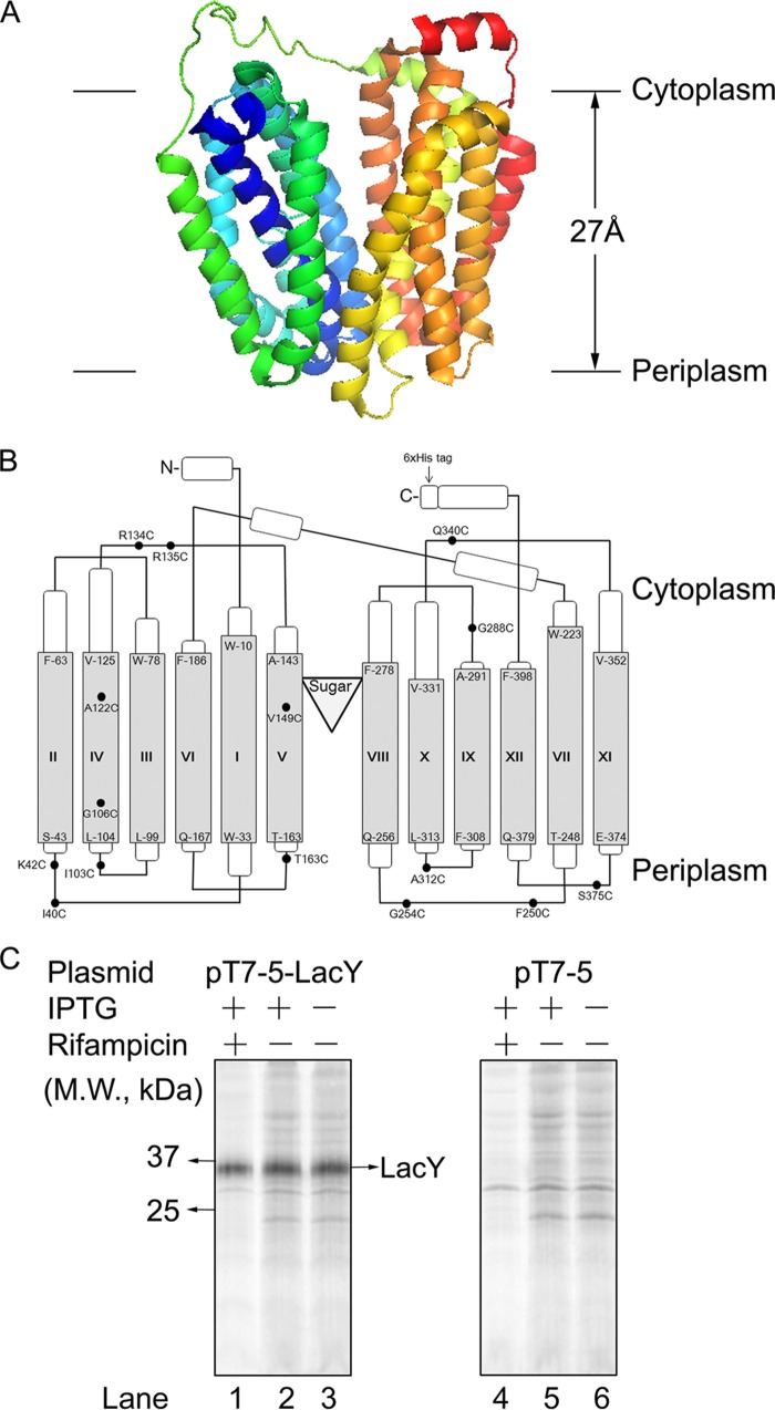
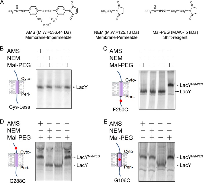
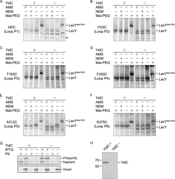
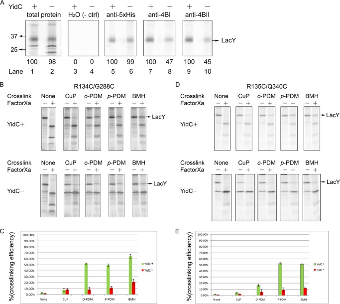
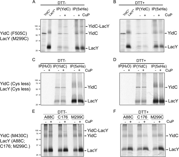
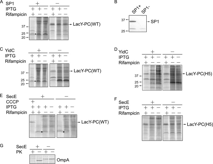
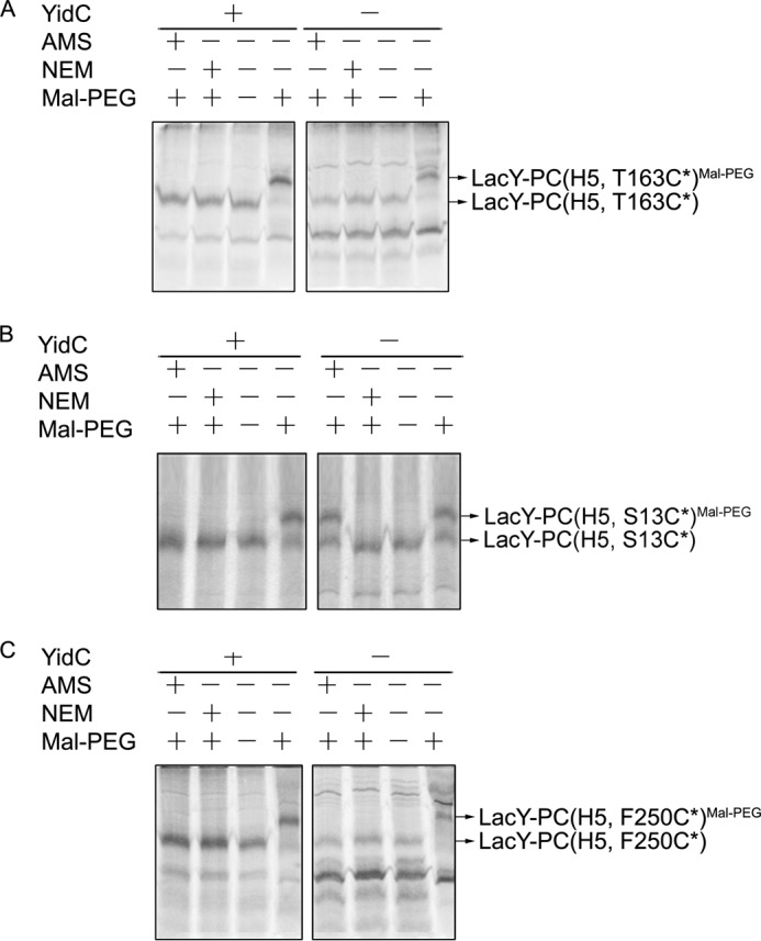
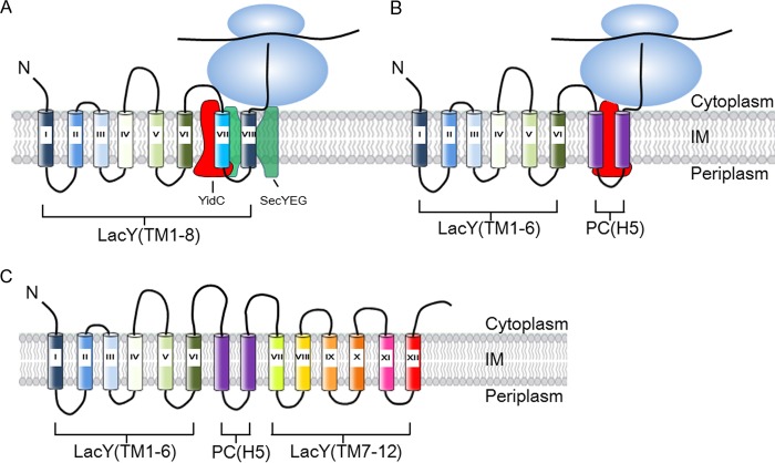
To understand how YidC and SecYEG function together in membrane protein topogenesis, insertion and folding of the lactose permease of Escherichia coli (LacY), a 12-transmembrane helix protein LacY that catalyzes symport of a galactoside and an H(+), was studied. Although both the SecYEG machinery and signal recognition particle are required for insertion of LacY into the membrane, YidC is not required for translocation of the six periplasmic loops in LacY. Rather, YidC acts as a chaperone, facilitating LacY folding. Upon YidC depletion, the conformation of LacY is perturbed, as judged by monoclonal antibody binding studies and by in vivo cross-linking between introduced Cys pairs. Disulfide cross-linking also demonstrates that YidC interacts with multiple transmembrane segments of LacY during membrane biogenesis. Moreover, YidC is strictly required for insertion of M13 procoat protein fused into the middle cytoplasmic loop of LacY. In contrast, the loops preceding and following the inserted procoat domain are dependent on SecYEG for insertion. These studies demonstrate close cooperation between the two complexes in membrane biogenesis and that YidC functions primarily as a foldase for LacY.

Keywords: Chaperone; Chaperonin; LacY; Membrane Biogenesis; Membrane Enzymes; Membrane Insertion; Membrane Proteins; Protein Folding; YidC.


Figures

FIGURE 1.
FIGURE 1.
Structure and expression of LacY. A, the structure of LacY in the inward conformation viewed across the membrane plane. The lactose homolog β-d-galactopyranosyl-1-thio-β-d-galactopyranoside is not shown in the structure. B, the membrane topology and secondary structure model of LacY on the basis of the x-ray structure. The loops depict the connectivity. The Cys residues used in the alkylation and cross-linking studies are indicated in the LacY schematic. We also show the position where PC(H5) is inserted in LacY in the LacY-PC chimera (arrow). C, selective [35S]Met-labeling of LacY in the membrane by rifampicin treatment. Membranes were isolated from JS7131 cells containing the pT7-5-LacY vector (left panel) or empty vector pT7-5 (right panel) that were [35S]Met-labeled before or after IPTG induction. Lanes 1 and 4 represent membranes that are derived from rifampicin-treated cells. M.W., molecular weight.
FIGURE 2.
FIGURE 2.
The gel shift assay to assess the topology of LacY. A, Cys-reactive reagents that were used to probe the topology of LacY. The gel shift assay for the Cys-less (B), F250C (periplasm (Peri)) (C), G288C (cytoplasm (Cyto)) (D), and G106C (transmembrane) LacY proteins (E) is shown. M.W., molecular weight. Rifampicin-treated radiolabeled cells expressing the indicated LacY mutants were treated with or without membrane-impermeable AMS or with membrane-permeable NEM. The cells were then washed with PBS buffer to remove the unreactive chemical reagent and pelleted. Membranes were isolated, and LacY was solubilized in detergent buffer. Where indicated, the samples were treated with Mal-PEG, which modifies unreacted Cys residues, and then analyzed by SDS-PAGE and phosphorimaging.
FIGURE 3.
FIGURE 3.
Membrane insertion of LacY is dependent on SRP and SecYEG. A, translocation of the fourth periplasmic loop of LacYF250C under SecE expression (+) and SecE depletion conditions (−). E. coli strain CM124 bearing pT7-5 encoding LacY F250C was grown with arabinose or glucose. Cells were treated with rifampicin and IPTG and labeled with [35S]Met, and the isolated membranes were analyzed by gel shift assay as described in Fig. 2. B, Western blot analysis confirms that SecY is depleted when SecE is depleted from the membrane by growth of CM124 under glucose conditions. N term, N-terminal. C, translocation of loop 4 (F250C) in LacY under Ffh expression (+) and Ffh depletion (−) conditions. E. coli WAM121 grown in the presence of arabinose or glucose was analyzed exactly as described in A. D, Western blot analysis showing that Ffh is depleted from the membrane by growth of WAM121 in glucose medium (see “Experimental Procedures” for details).
FIGURE 4.
FIGURE 4.
Translocation of the periplasmic loops of LacY does not require YidC. Translocation of the periplasmic loops of LacY under YidC expression (+) and YidC depletion (−) conditions. E. coli strain JS7131 bearing pT7-5 lacY I40C (A), I103C (B), T163C (C), F250C (D), A312C (E), and S375C (F) were grown, labeled with [35S]Met, and analyzed by the gel shift method as described in Fig. 3B. Note that there is a YidC depletion-induced band at the lower molecular weight position in A–F (arrowhead, see A). G, membrane insertion of procoat-Lep H5 (S13C) under YidC expression (+) and YidC depletion conditions (−). JS7131 containing pMS119 procoat-Lep H5 (S13C) was [35S]Met-labeled without rifampicin treatment and analyzed by protease accessibility assay. OmpA served as a positive control for proteolysis. Samples were analyzed by SDS-PAGE and phosphorimaging. H, a representative Western blot confirms that YidC is depleted by growth of JS7131 bearing the pGP1–2 and pT7-5-LacY plasmids in LB medium with glucose added for 5 h at 30 °C.
FIGURE 5.
FIGURE 5.
YidC is required for proper helix packing of LacY. A, the binding of monoclonal antibodies 4B1 and 4B11 to LacY is impaired under YidC depletion conditions. Expression of LacY under YidC expression (+) and YidC depletion (−) conditions was performed in JS7131 as described in Fig. 4A. Radiolabeled cells in either YidC (+) or YidC (−) conditions were split into five aliquots and treated equally. Radioactive bands were visualized using a Typhoon PhosphorImager. The total 35S-labeled proteins in the membrane before immunoprecipitation are shown in lanes 1 and 2. Mock treatment by immunoprecipitation against water was used as a negative control (ctrl, lanes 3 and 4). Penta-His antibody (Qiagen) recognizing the His-tagged LacY was used as a positive control (lanes 5 and 6). Immunoprecipitation using monoclonal antibodies against the 4B1 and 4B11 epitopes was used to assay the folding of LacY at periplasmic loop 4 (VII/VIII) (lanes 7 and 8) and cytoplasmic loops VIII/IX and X/XI (lanes 9 and 10), respectively. The induced band in the total protein in the membrane that appears slightly below the 25-kDa molecular mass upon YidC depletion is most likely the PspA. B–E, LacY folding is impaired under YidC depletion conditions, as determined by intramolecular cross-linking between Cys pairs of LacY. B, the E. coli strain JS7131 bearing pKR35-R134C/G288C was grown in the presence of arabinose (YidC+) and glucose (YidC−), radiolabeled using the rifampicin blocking technique as described in Fig. 4, and then membranes were isolated. Where indicated, the right-side-out vesicles were treated with BMH, p-PDM, o-PDM, and CuP. Thereafter, solubilized membrane protein samples were treated with Factor Xa protease (NEB Co.). Protein samples were then analyzed by SDS-PAGE and phosphorimaging. D, JS7131 cells harboring pKR35-R135C/Q340C were grown in arabinose and glucose conditions and radiolabeled using the rifampicin technique. The LacY R135C/Q340C in the membrane fraction derived from cells grown under YidC (+) and YidC (−) conditions was analyzed for cross-linking as described in B and subjected to SDS-PAGE and phosphorimaging. The Cys pairs were introduced into LacY L6XB, having a Factor Xa-biotin acceptor domain between TM6 and TM7 (75). The efficiency of LacY cross-linking under YidC (+) and YidC (−) conditions between the R134C/G288C (C) and R135C/Q340C pairs (E) was determined by quantification of the band density of the full-length LacY using ImageJ software. The error bars indicate the S.D. in triplicate measurements.
FIGURE 6.
FIGURE 6.
YidC contacts LacY during membrane protein biogenesis. Cross-linking between the single-Cys YidC and single-Cys LacY mutants was performed under conditions where the chromosomal YidC was depleted. A and B, to deplete the chromosomally encoded YidC, E. coli JS7131 harboring the YidC and LacY expression vectors pACYC184-YidC (F505C) and pLZ2-LacY (M299C) were grown in the presence of 0.2% (m/v) glucose at 30 °C. The cells were expressed using the rifampicin technique as described in Fig. 2. JS7131 cells were pulse-labeled with trans-[35S]Met for 5 min with or without treatment of 1 mm CuP for 10 min at 30 °C (see “Experimental Procedures”). The total membrane protein sample was immunoprecipitated (IP) with antibody to either YidC or Penta-His (recognizing the 6× His tag at the LacY C terminus). Protein samples with (B) or without (A) adding reducing agent (200 mm DTT), were analyzed using a 10% (m/v) SDS-PAGE and by phosphorimaging. The LacY (lane 2) or YidC (lane 1) protein controls were obtained as follows. E. coli BL21 cells bearing pET28b-YidC (WT) were pulse-labeled for 1 min with trans-[35S]Met, and total protein was immunoprecipitated using anti-YidC serum. E. coli T184 cells cotransformed with pGP1–2 and pT7-5-LacY were pulse-labeled for 5 min after treating with rifampicin, and total membrane proteins were immunoprecipitated using Penta-His antibody. C and D, mock cross-linking between YidC and LacY was performed with E. coli JS7131, with both of the expressed Cys-less proteins, using pACYC184-YidC (Cys-less) and pLZ2-LacY (Cys-less). In addition to YidC and Penta-His immunoprecipitation, a mock immunoprecipitation was performed with water. After CuP treatment, samples were treated with (D) or without (C) DTT. E and F, disulfide cross-linking between YidC (M430C) and LacY single-Cys mutants (A88C, C176, or M299C) was performed as described in A and B. Penta-His antiserum was used to pull down LacY with a 6× His tag.
FIGURE 7.
FIGURE 7.
YidC can function to insert the procoat domain fused within LacY. A, E. coli FTL85 bearing pLZ-t7 and pT7-5-LacY-PC (WT) was grown under SP1 expression (+) and SP1 depletion (−) conditions. As indicated, cells were treated with rifampicin and/or IPTG prior to radiolabeling. B, Western blot analysis confirms that SP1 is depleted by growth of FTL85 in LB medium plus 0.2% glucose at 30 °C for 5 h (SP1-). C, E. coli JS7131 bearing pT7-5-LacY-PC (WT) was grown under YidC expression (+) and YidC depletion conditions (−). Where indicated, cells were treated with rifampicin and/or IPTG prior to radiolabeling. D, JS7131 harboring pT7-5-LacY-PC (H5) was analyzed as described in panel C. For A and C, ★ depicts the SP1-cleaved fragments of LacY-PC. E. coli CM124 cells bearing pLZ2 encoding LacY-PC (WT) (E) or LacY-PC (H5) (F) were grown in LB medium with arabinose or glucose to express or deplete SecE, respectively, and then treated, where indicated, with rifampicin and IPTG prior to radiolabeling. A portion of CM124 cells grown under arabinose conditions was treated with 50 μm (final concentration) carbonyl cyanide m-chlorophenylhydrazone (CCCP) for 45 s after incubation with rifampicin/IPTG prior to pulse-labeling with trans-[35S]Met for 5 min (E, lane 1). G, A portion of CM124 cells bearing pLZ2-LacY-PC (WT) was grown under arabinose or glucose conditions, switched into fresh M9 minimal medium, and incubated at 30 °C for 30 min. The CM124 cells were pulse-labeled with [35S]Met without rifampicin treatment and analyzed by protease accessibility assay. Sec-dependent OmpA protein was immunoprecipitated using anti-OmpA serum, and the protein samples were analyzed by SDS-PAGE and phosphorimaging as described in Fig. 4G. PK, proteinase K.
FIGURE 8.
FIGURE 8.
Different YidC translocase requirements for insertion of the PC domain and the flanking hydrophilic loops of LacY-PC. LacY-PC (H5) with a Cys in the loop preceding the PC domain (T163C*) (A), in the PC domain at position 13 (PC S13C*) (B), or in a loop immediately after the PC domain (F250C*) (C) were analyzed for membrane insertion under YidC expression (+) and YidC depletion conditions (−). JS7131 cells bearing pGP1–2 and pT7-5 encoding the respective LacY-PC (H5) derivatives were grown in medium supplemented with arabinose or glucose. Cells were treated with rifampicin and IPTG, radiolabeled with trans-[35S]Met, and analyzed using cysteine modification reagents as described in Fig. 2. The isolated membrane protein samples were treated with Mal-PEG and analyzed by SDS-PAGE and phosphorimaging.
FIGURE 9.
FIGURE 9.
Proposed model for cooperative insertion of LacY-PC by SecYEG and YidC. A, the periplasmic loops of LacY are inserted by the SecYEG machinery, whereas YidC binds the TM segments and helps fold the protein. N, designates the N terminus. B, the procoat domain of LacY-PC is inserted by the YidC-only pathway, whereas all the LacY periplasmic loops are inserted by the SecYEG machinery. In this latter pathway, YidC presumably plays a role in the folding of LacY as well. It should be noted that it is not known for LacY when the TM segments are released from YidC and integrated into the lipid bilayer. IM, inner membrane. C, the topology of LacY-PC (H5) on the basis of this study. The PC (H5) region is depicted in purple.

Similar articles

Cited by

References

    1. Xie K., Dalbey R. E. (2008) Inserting proteins into the bacterial cytoplasmic membrane using the Sec and YidC translocases. Nat. Rev. Microbiol. 6, 234–244 - PubMed
    1. Driessen A. J., Nouwen N. (2008) Protein translocation across the bacterial cytoplasmic membrane. Annu. Rev. Biochem. 77, 643–667 - PubMed
    1. Dalbey R. E., Wang P., Kuhn A. (2011) Assembly of bacterial inner membrane proteins. Annu. Rev. Biochem. 80, 161–187 - PubMed
    1. Luirink J., Yu Z., Wagner S., de Gier J. W. (2012) Biogenesis of inner membrane proteins in Escherichia coli. Biochim. Biophys. Acta 1817, 965–976 - PubMed
    1. Stephenson K. (2005) Sec-dependent protein translocation across biological membranes. Evolutionary conservation of an essential protein transport pathway (review). Mol. Membr. Biol. 22, 17–28 - PubMed

Publication types

MeSH terms