

Evidence that orphanin FQ mediates progesterone negative feedback in the ewe
- PMID: 23928375
- PMCID: PMC3800756
- DOI: 10.1210/en.2013-1274
Evidence that orphanin FQ mediates progesterone negative feedback in the ewe
Abstract
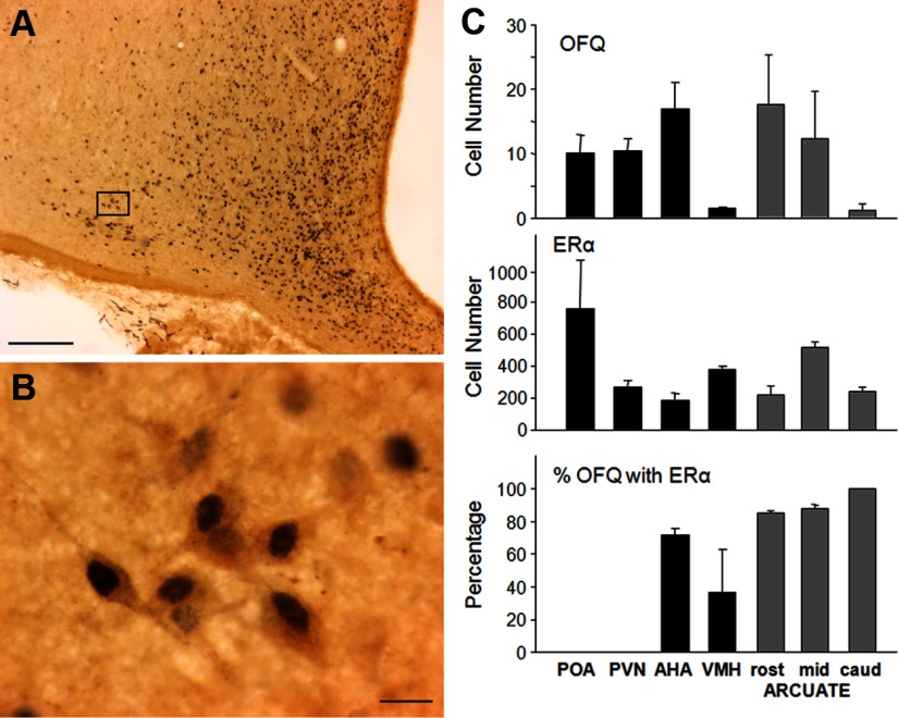
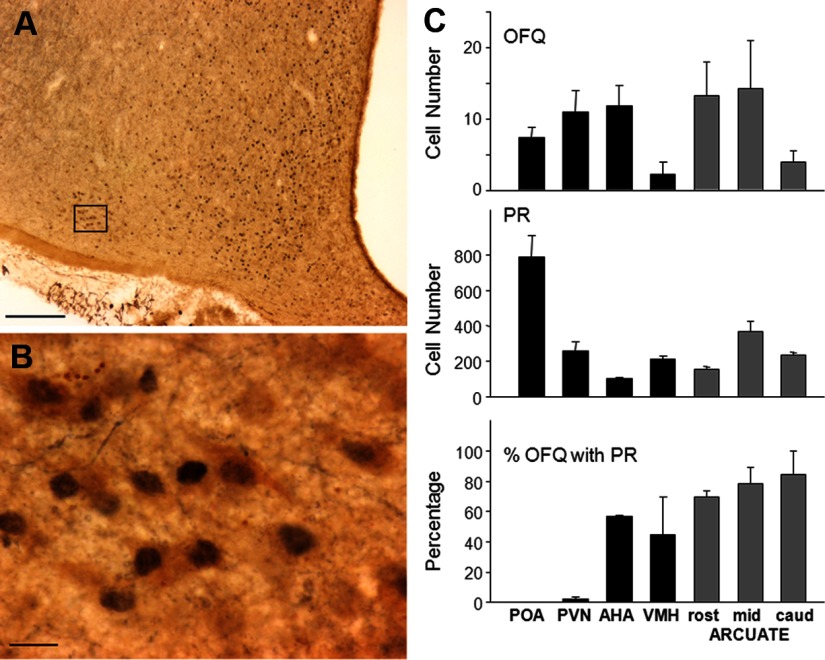
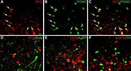
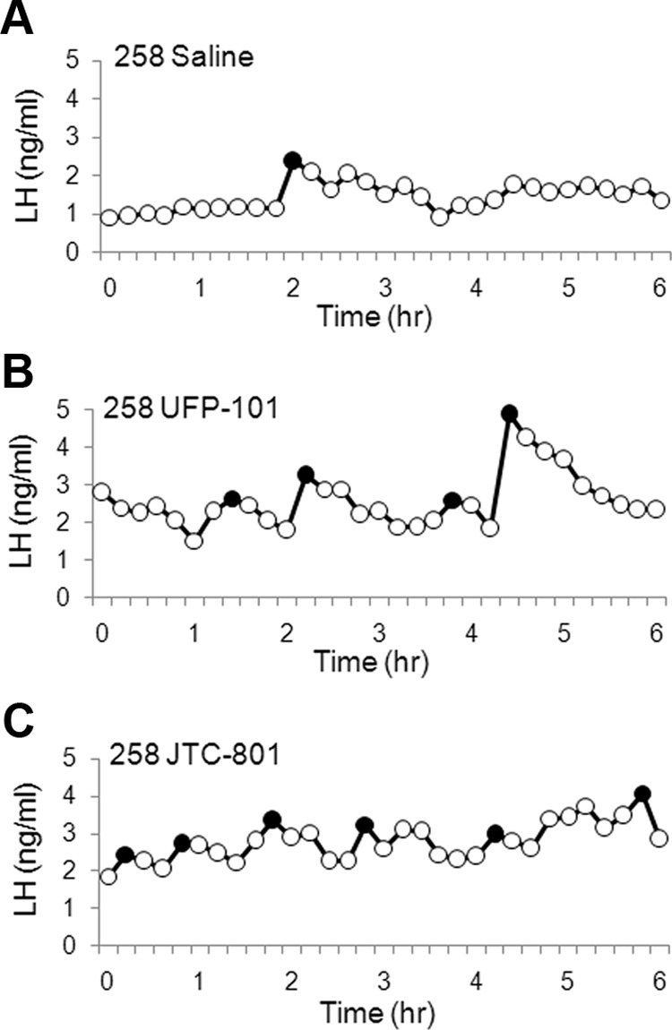
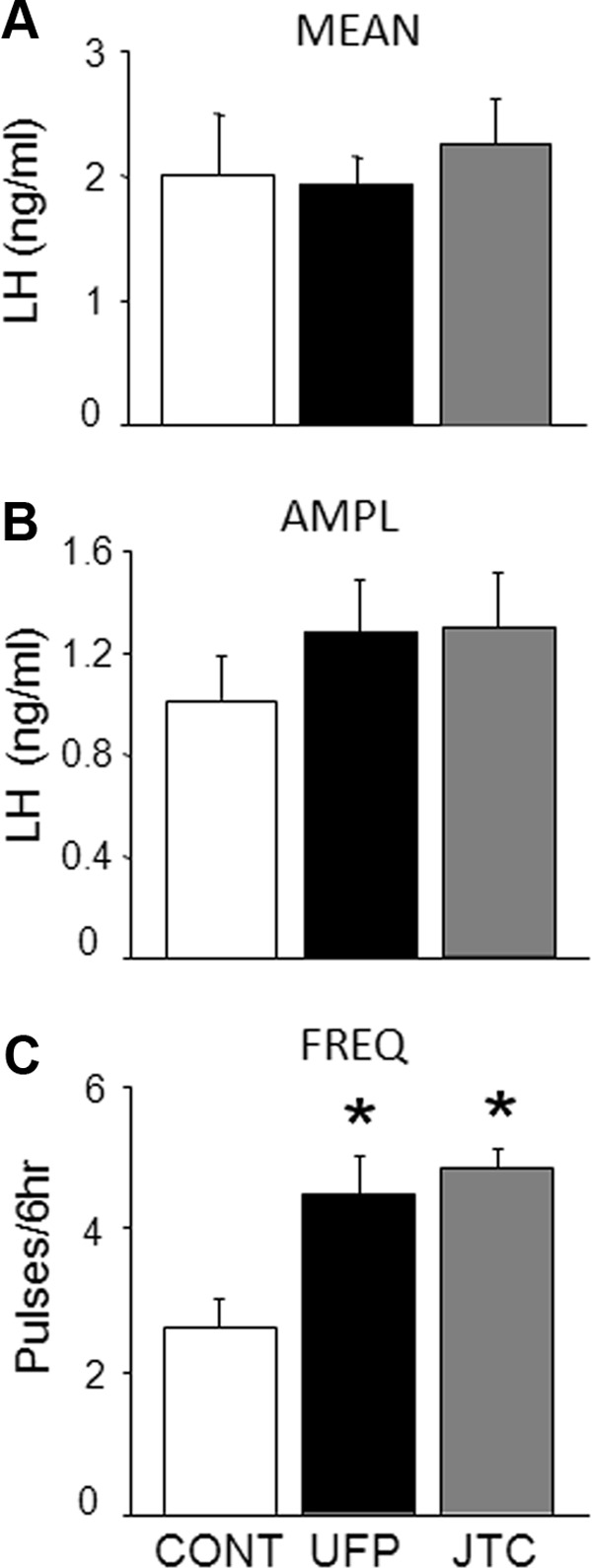
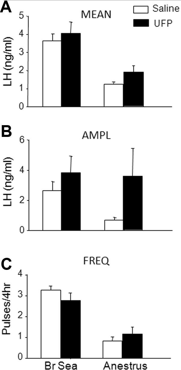
Orphanin FQ (OFQ), a member of the opioid family, is found in many areas of the hypothalamus and, when given centrally OFQ inhibits episodic LH secretion in rodents and sheep. Because GnRH neurons are devoid of the appropriate receptors to mediate steroid negative feedback directly, neurons that release OFQ may be involved. Using immunocytochemistry, we first determined that most OFQ neurons in the arcuate nucleus (ARC) and other hypothalamic regions of luteal phase ewes contained both estrogen receptor α and progesterone (P) receptor. Given a similar high degree of steroid receptor colocalization in other ARC subpopulations, we examined whether OFQ neurons of the ARC contained those other neuropeptides and neurotransmitters. OFQ did not colocalize with kisspeptin, tyrosine hydroxylase, or agouti-related peptide, but all ARC OFQ neurons coexpressed proopiomelanocortin. To test for a role for endogenous OFQ, we examined the effects of an OFQ receptor antagonist, [Nphe1,Arg14,Lys15]Nociceptin-NH2 (UFP-101) (30 nmol intracerebroventricular/h), on LH secretion in steroid-treated ewes in the breeding season and ovary-intact ewes in anestrus. Ovariectomized ewes with luteal phase concentrations of P and estradiol showed a significant increase in LH pulse frequency during infusion of UFP-101 (4.5 ± 0.5 pulses/6 h) compared with saline infusion (2.6 ± 0.4 pulses/6 h), whereas ewes implanted with only estradiol did not. Ovary-intact anestrous ewes displayed no significant differences in LH pulse amplitude or frequency during infusion of UFP-101. Therefore, we conclude that OFQ mediates, at least in part, the negative feedback action of P on GnRH/LH pulse frequency in sheep.
Figures
References
-
- Evans NP, Dahl GE, Glover BH, Karsch FJ. Central regulation of pulsatile gonadotropin-releasing hormone (GnRH) secretion by estradiol during the period leading up to the preovulatory GnRH surge in the ewe. Endocrinology. 1994;134:1806–1811 - PubMed
-
- Evans NP, Dahl GE, Mauger D, Karsch FJ. Estradiol induces both qualitative and quantitative changes in the pattern of gonadotropin-releasing hormone secretion during the presurge period in the ewe. Endocrinology. 1995;136:1603–1609 - PubMed
-
- Clarke IJ, Thomas GB, Yao B, Cummins JT. GnRH secretion throughout the ovine estrous cycle. Neuroendocrinology. 1987;46:82–88 - PubMed
-
- Karsch FJ, Cummins JT, Thomas GB, Clarke IJ. Steroid feedback inhibition of pulsatile secretion of gonadotropin-releasing hormone in the ewe. Biol Reprod. 1987;36:1207–1218 - PubMed
-
- Herbison AE, Robinson JE, Skinner DC. Distribution of estrogen receptor-immunoreactive cells in the preoptic area of the ewe: co-localization with glutamic acid decarboxylase but not luteinizing hormone-releasing hormone. Neuroendocrinology. 1993;57:751–759 - PubMed
Publication types
MeSH terms
Substances
Grants and funding
LinkOut - more resources
Full Text Sources
Other Literature Sources
