. 2014 Aug;73(8):1575-84.
doi: 10.1136/annrheumdis-2013-203561. Epub 2013 Aug 8.

ADAMTS-7 forms a positive feedback loop with TNF-α in the pathogenesis of osteoarthritis

Affiliations

ADAMTS-7 forms a positive feedback loop with TNF-α in the pathogenesis of osteoarthritis

Yongjie Lai et al. Ann Rheum Dis. 2014 Aug.

Abstract

Objective: To examine the expression of ADAMTS-7 during the progression of osteoarthritis (OA), defining its role in the pathogenesis of OA, and elucidating the molecular events involved.

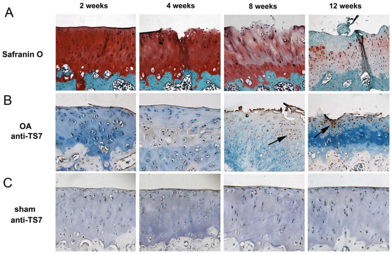
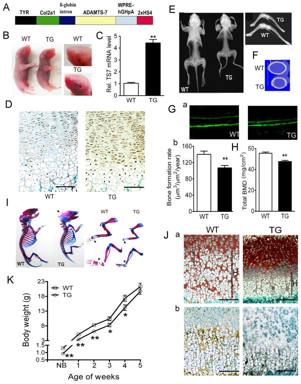
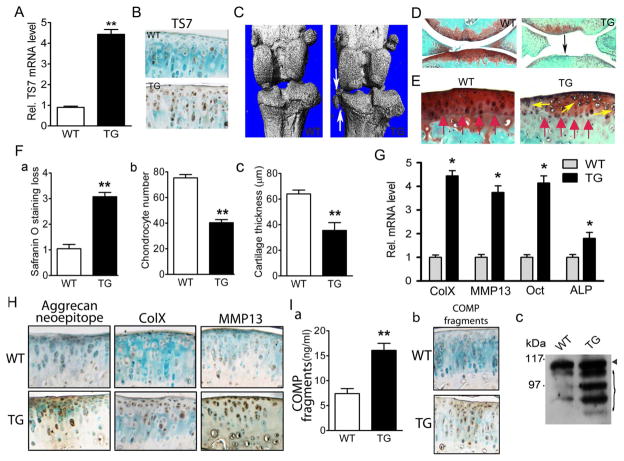
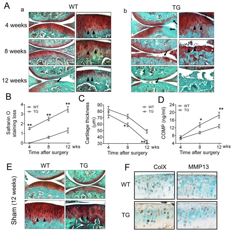
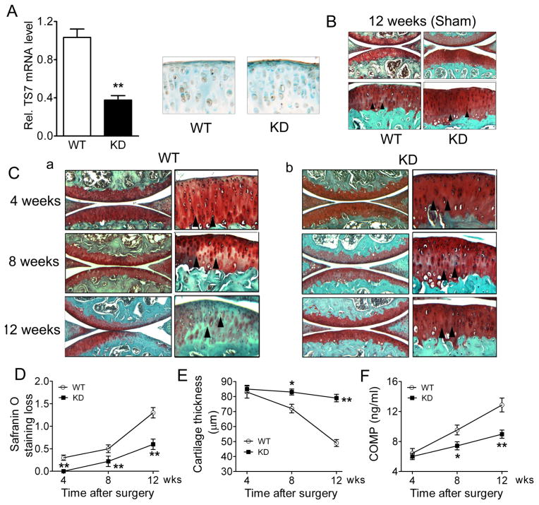
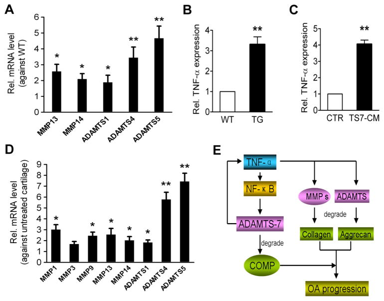
Methods: ADAMTS-7 expression in cartilage of a rat OA model was assayed using immunohistochemistry. Cartilage-specific ADAMTS-7 transgenic mice and ADAMTS-7 small interfering (si)RNA knockdown mice were generated and used to analyse OA progression in both spontaneous and surgically induced OA models. Cartilage degradation and OA was evaluated using Safranin-O staining, immunohistochemistry, ELISA and western blotting. In addition, mRNA expression of tumour necrosis factor (TNF)-α and metalloproteinases known to be involved in cartilage degeneration in OA was analysed. Furthermore, the transactivation of ADAMTS-7 by TNF-α and its downstream NF-κB signalling was measured using reporter gene assay.

Results: ADAMTS-7 expression was elevated during disease progression in the surgically induced rat OA model. Targeted overexpression of ADAMTS-7 in chondrocytes led to chondrodysplasia characterised by short-limbed dwarfism and a delay in endochondral ossification in 'young mice' and a spontaneous OA-like phenotype in 'aged' mice. In addition, overexpression of ADAMTS-7 led to exaggerated breakdown of cartilage and accelerated OA progression, while knockdown of ADAMTS-7 attenuated degradation of cartilage matrix and protected against OA development, in surgically induced OA models. ADAMTS-7 upregulated TNF-α and metalloproteinases associated with OA; in addition, TNF-α induced ADAMTS-7 through NF-κB signalling.

Conclusions: ADAMTS-7 and TNF-α form a positive feedback loop in the regulation of cartilage degradation and OA progression, making them potential molecular targets for prevention and treatment of joint degenerative diseases, including OA.

Keywords: Arthritis; Chondrocytes; Osteoarthritis; TNF-alpha.


Conflict of interest statement

Competing interests None.

Figures

Figure 1
Figure 1
ADAMTS-7 is upregulated in a surgically induced rat osteoarthritis (OA) model. (A) Safranin-O staining of femur articular cartilage in the operated knees. Samples from rat knee cartilage at different time points after surgery, as indicated, were harvested, and histopathological studies were performed. (B) Expression of ADAMTS-7 during OA development, assayed by immunohistochemistry. Femur cartilage sections were stained with anti-ADAMTS-7 serum (brown) and counterstained with methyl green. Arrows indicate the signal. (C) ADAMTS-7 expression in sham-operated knee femur cartilage, examined by immunohistochemistry with anti-ADAMTS-7 serum. Representative sections are shown. Original magnification ×40.
Figure 2
Figure 2
Targeted overexpression of ADAMTS-7 in chondrocytes leads to short-limbed dwarfism and a delay in endochondral ossification in young mice. (A) Diagram of Col2a1-ADAMTS-7 transgenic (TG) construct. Full-length human ADAMTS-7 cDNA was cloned into the 6.0 kb chondrocyte-specific promoter in a coat colour vector. TYR, tyrosinase minigene; WPRE, woodchuck hepatitis virus post-transcriptional regulatory element; HS4, chicken β-globin insulator. (B) Smaller size of newborn Col2a1-TG founders compared with wild-type (WT) littermate controls (left panel). High magnification of black eye in the transgenic mouse indicates the strong transgene expression (right panel). The arrow indicates the black eye in TG mouse. (C) Real-time RT-PCR analysis of ADAMTS-7 mRNA expression from newborn TG mice and WT control. Values are mean ±SEM from three independent founders per genotype. (D) Increased ADAMTS-7 expression in the newborn TG mouse growth plate detected by immunohistochemistry. (E) Representative pictures of whole body from WT and TG mice at 3 weeks old, assayed by X-ray. (F) The coronal section of middle femurs in 3-week-old WT and TG mice, assayed by micro-CT. (G) Calcein double labelling in 4-week-old mice. (a) The bone formation rate represented by the distance between two green labels (original magnification ×40); (b) bone formation rate was compared between WT and TG mice (n=5 in each group). (H) Dual-energy X-ray absorptiometry scan analysis of total bone mineral density (BMD) in WT and TG mice at the age of 3 weeks (n=6 in each group). (I) Skeletal preparation with Alcian blue/Alizarin red staining of newborn WT and TG mice. Representative pictures are shown as whole-mount view (left), and high magnification of forelimbs and hind limbs (right). Black arrows indicate the mineralisation site of the bone. (J) Delayed hypertrophy in TG mice. (a) Safranin-O staining of the tibial growth plates in 1-week-old mice; H, hypertrophic zone. (b) Hypertrophic chondrocytes-specific marker type X collagen was examined by immunohistochemistry in WT and TG mice. Brown signal indicates the location of collagen X in the matrix. (K) Body weight curve of WT and TG mice from newborn to 5 weeks old (n=6 for each group). Data are means ±SEM. *p<0.05, **p<0.01 between WT and TG mice. Bars=100 μm.
Figure 3
Figure 3
Eight-month-old ADAMTS-7 transgenic (TG) mice spontaneously developed a mild osteoarthritis (OA)-like phenotype. (A) Relative level of ADAMTS-7 transgene in 8-month-old adult mice, assayed by real-time reverse transcriptase (RT)-PCR. Normalised values were calibrated against the wild-type (WT), given the value of 1 (n=6 in each group). (B) Immunohistochemical analysis of ADAMTS-7 expression in articular cartilage of 8-month-old mice. (C) Micro-CT images of knee joints from WT and TG mice. Osteophyte formation is indicated by white arrows. (D) Safranin O staining of knee joint articular cartilage. Significant proteoglycan loss in TG mice is indicated (black arrow). (E) High-magnification pictures of Safranin-O staining showing tidemark shift (red arrows) and chondrocyte cluster formation (yellow arrows) in aged transgenic mice. (F) Comparison of OA severity between WT and TG mice in aging-associated model, as assessed by Safranin-O staining loss score (a), chondrocyte number (b) and articular cartilage layer thickness (c) in the tibia. n=6 for each group. (G) Transcript levels of mature chondrocyte marker genes for collagen X (ColX), matrix metalloproteinase 13 (MMP13), osteocalcin (Oct), alkaline phosphatase (ALP) from WT and TG articular cartilage were measured by real-time RT-PCR. The units are arbitrary, and the normalised values were calibrated against the control, here given the value of 1. Each real-time RT-PCR was performed in triplicate. (H) Immunostaining for aggrecan degradation neoepitope (left), ColX (middle) and MMP13 (right) in articular cartilage from WT and TG mice. (I) Analyses of COMP degradation fragments in WT and TG mice. (a) Serum levels of COMP fragments in WT and TG mice, assayed by COMP sandwich ELISA using a fragment-preferred COMP monoclonal antibody (n=6 for each group). (b) Higher levels of COMP fragments in TG cartilage, assayed by immunohistochemistry with a fragment-preferred COMP monoclonal antibody. (c) Femoral head articular cartilage from WT and TG mice was cultured for 3 days, and conditioned medium was precipitated by 100% trichloroacetic acid, then subjected to 10% reducing sodium dodecyl sulphate/polyacrylamide gel electrophoresis and detected by a polyclonal COMP antibody. The arrowhead indicates the intact COMP, and the bracket indicates the degraded COMP fragments. Values are normalised mean±SEM. *p<0.05, **p<0.01 versus WT mice. Original magnification ×100 in (B), (E), (H) and (I) and ×40 in (D).
Figure 4
Figure 4
Overexpression of ADAMTS-7 led to accelerated osteoarthritis (OA) progression in a surgically induced model. (A) Histological analysis of knee joints in wild-type (WT) (a) and transgenic (TG) (b) mice at various time points after surgery. The surgical model was induced in 8-week-old WT and TG mice by transecting the right knee medial meniscotibial ligament (WT, n=6; TG, n=8 for each time point). Representative low (left, original magnification ×40) and high (right, original magnification ×100) magnifications of articular cartilage are presented. Arrows indicate the Safranin-O staining loss, and arrowheads indicate the tidemark. (B) OA severity in the course of OA development, assessed by Safranin-O staining loss score. (C) OA severity in the course of OA development, assayed by cartilage thickness measurement. (D) Serum levels of COMP fragments in the OA progression, measured by ELISA. (E) Safranin-O staining of sham-operated knee joints 12 weeks after surgery. Representative low (top, ×40) and high (bottom, ×100) magnifications are presented. (F) Immunohistochemistry for collagen X (ColX) and matrix metalloproteinase (MMP13) in the WT and TG mouse articular cartilage 4 weeks after surgery (original magnification ×100). Values are normalised mean±SEM. *p<0.05, **p<0.01 versus WT mice.
Figure 5
Figure 5
ADAMTS-7 knockdown (KD) mice displayed attenuated osteoarthritis (OA) development in surgically-induced model. (A) The relative level of ADAMTS-7 in 8-week-old KD mice, assayed by real-time RT-PCR. Normalised values were calibrated against the wild-type (WT), given the value of 1. (B) Immunohistochemistry analysis for ADAMTS-7 expression in the articular cartilage from both 8-week-old WT and KD mice. (C) Safranin-O staining of sham-operated knee joints 12 weeks after surgery. Representative low (top, ×40) and high (bottom, ×100) magnifications are presented. (D) Histological analysis of knee joints in WT (a) and KD (b) mice at various time points, as indicated, after surgery. Low (left, original magnification ×40) and high (right, original magnification ×100) magnification of articular cartilage are presented. Arrowheads indicate tidemark. OA severity was quantified by Safranin-O staining loss score (E) and cartilage thickness measurement (F). (G) Serum levels of COMP fragments were measured by sandwich ELISA in the course of OA development. Values are the normalised mean±SEM, n=5 and n=8 for WT and KD, respectively. *p<0.05, **p<0.01 versus WT mice.
Figure 6
Figure 6
ADAMTS-7 upregulates tumour necrosis factor (TNF)-α and metalloproteinases in cartilage. (A) Levels of matrix metalloproteinase (MMP) 13, MMP14, ADAMTS-1, ADAMTS-4 and ADAMTS-5 mRNA in articular cartilage from 8-month-old wild-type (WT) and transgenic (TG) mice, assayed by real-time RT-PCR. (B) RNA level of TNF-α in articular cartilage from 8-month-old WT and TG mice, assayed by real-time RT-PCR. The normalised values were calibrated against the control, given the value of 1. (C) ADAMTS-7 upregulates TNF-α levels in cartilage explants. Femoral head cartilage from 2-month-old WT mice was harvested, and cartilage explants were cultured for 48 h with either Dulbecco’s modified Eagle’s medium only (CTR; n=5) or ADAMTS-7-conditioned medium (TS7-CM; n=6) collected from HEK-293 EBNA stable cell lines transfected with a construct encoding ADAMTS-7, followed by real-time RT-PCR. (D) Expression levels of MMP1, MMP3, MMP9, MMP13, MMP14, ADAMTS-1, ADAMTS-4 and ADAMTS-5 mRNA in cartilage explants stimulated by ADAMTS-7, assayed by real-time RT-PCR. Normalised values were calibrated against the untreated controls, given the value of 1. (E) A proposed model for the role and regulation of ADAMTS-7 in the development of osteoarthritis. Each real-time RT-PCR was carried out in triplicate. All the data are presented as mean±SEM, *p <0.05, **p<0.01.

References

    1. Herndon JH, Davidson SM, Apazidis A. Recent socioeconomic trends in orthopaedic practice. J Bone Joint Surg Am. 2001;83-A:1097–105. - PubMed
    1. Salzet M. Leech thrombin inhibitors. Curr Pharm Des. 2002;8:493–503. - PubMed
    1. Hurskainen TL, Hirohata S, Seldin MF, et al. ADAM-TS5, ADAM-TS6, and ADAM-TS7, novel members of a new family of zinc metalloproteases. General features and genomic distribution of the ADAM- TS family. J Biol Chem. 1999;274:25555–63. - PubMed
    1. Glasson SS, Askew R, Sheppard B, et al. Deletion of active ADAMTS5 prevents cartilage degradation in a murine model of osteoarthritis. Nature. 2005;434:644–48. - PubMed
    1. Stanton H, Rogerson FM, East CJ, et al. ADAMTS5 is the major aggrecanase in mouse cartilage in vivo and in vitro. Nature. 2005;434:648–52. - PubMed

Publication types

MeSH terms