. 2013 Sep;75(3):410-6.
doi: 10.1097/TA.0b013e31829617c6.

Autologous bone marrow mononuclear cells therapy attenuates activated microglial/macrophage response and improves spatial learning after traumatic brain injury

Affiliations

Autologous bone marrow mononuclear cells therapy attenuates activated microglial/macrophage response and improves spatial learning after traumatic brain injury

Supinder S Bedi et al. J Trauma Acute Care Surg. 2013 Sep.

Abstract

Background: Autologous bone marrow-derived mononuclear cells (AMNCs) have shown therapeutic promise for central nervous system insults such as stroke and traumatic brain injury (TBI). We hypothesized that intravenous injection of AMNC provides neuroprotection, which leads to cognitive improvement after TBI.

Methods: A controlled cortical impact (CCI) rodent TBI model was used to examine blood-brain barrier (BBB) permeability, neuronal and glial apoptosis, as well as cognitive behavior. Two groups of rats underwent CCI with AMNC treatment (CCI-autologous) or without AMNC treatment (CCI-alone), consisting of 2 million AMNC per kilogram body weight harvested from the tibia and intravenously injected 72 hours after injury. CCI-alone animals underwent sham harvests and received vehicle injections.

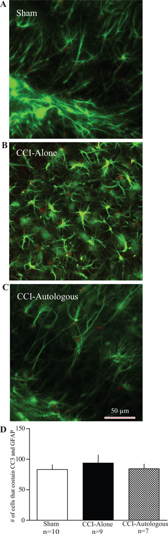
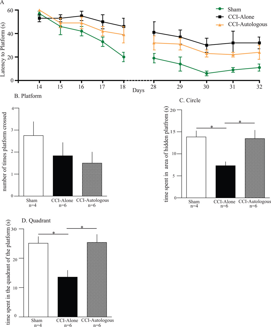
Results: Ninety-six hours after injury, AMNC significantly reduced the BBB permeability in injured animals, and there was an increase in apoptosis of proinflammatory activated microglia in the ipsilateral hippocampus. At 4 weeks after injury, we observed significant improvement in probe testing of CCI-Autologous group in comparison to CCI-Alone in the Morris Water Maze paradigm.

Conclusion: Our data demonstrate that the intravenous injection of AMNC after TBI leads to neuroprotection by preserving early BBB integrity, increasing activated microglial apoptosis and improving cognitive function.


Conflict of interest statement

Disclosure/Conflict of Interest

There are no known conflicts between the authors and the information presented in this paper.

Figures

Figure 1
Figure 1. Autologous cell therapy preserves the blood brain barrier
Blood brain barrier (BBB) permeability measured via Evan's blue extravasation. BBB permeability measurement (mean absorbance/weight of tissue (mg) from homogenized cortical tissue (Y axis) derived from both hemispheres. There was a significant decrease in BBB permeability after autologous treatment (CCI-Autologous) when compared to CCI-Alone in the injured right hemisphere. There was also a significant increase in BBB permeability in the CCI-Alone when compared to Sham in the injured right hemisphere. There were no significant differences between Sham and CCI-Autologous in the right hemisphere. Additionally there were no differences between all three groups in the left hemispheres.
Figure 2
Figure 2. Autologous cell therapy results in apoptosis of activated pro-inflammatory microglia mediated by cleaved caspase 3 (CC3)
Photomicrographs of microglia from hippocampal brain slices stained for cleaved caspase 3 (red) and microglia (green). A: Example of a sham brain slice. There is modest staining of CC3. In addition there are non-activated microglia (morphology). B: Example of a CCI-Alone brain slice. There are activated ameboid microglia but little overlap between the activated microglia and CC3. C: Example of CCI- Autologous brain slice. There is co-staining of CC3 and activated microglia. Scale bar 50 µm. D: Graph comparing the number of cells that co-stained with CC3 and microglia antibody. There is a significant increase in apoptotic (CC3+) and activated microglia in brain slices that received autologous cell treatment when compared to CCI-Alone. In addition, there was an increase in apoptotic, activated microglia between CCI-Alone and Sham.
Figure 3
Figure 3. Autologous cell therapy does not affect neuronal apoptosis
Photomicrographs of neurons from hippocampal brain slices stained for cleaved caspase 3 (red) and NeuN (green). A: Example of a sham brain slice. There is modest staining of CC3 with no overlap with NeuN. B: Example of a CCI-Alone brain slice. Like sham animals, there is very little overlap between CC3 and NeuN. C: Example of CCI - Autologous brain slice. There is very little overlap between CC3 and NeuN. Scale bar 50 µm D: Graph comparing the number of cells that co-stained with CC3 and NeuN. There is a modest, but not significant decrease in overlap between CC3 and NeuN in brain slices that received autologous cell treatment when compared to CCI-Alone.
Figure 4
Figure 4. Autologous cell therapy does not affect astrocytic apoptosis
Photomicrographs of astrocytes from hippocampal brain slices stained for cleaved caspase 3 (red) and GFAP (green). A: Example of a sham brain slice. There is modest staining of CC3 with no overlap with GFAP. B: Example of a CCI-Alone brain slice. Like sham animals, there is very little overlap between CC3 and GFAP. C: Example of CCI - Autologous brain slice. There is very little overlap between CC3 and GFAP. Scale bar 50 µm D: Graph comparing the number of cells that co-stained with CC3 and GFAP. There is a modest, but not significant decrease in overlap between CC3 and GFAP in hippocampal brain slices that received autologous cell treatment when compared to CCI-Alone.
Figure 5
Figure 5. Autologous cell therapy improves circle and quadrant time in the Morris Water Maze during probe trial
Probe trials completed on 3 groups of animals (Sham, CCI-Alone, and CCI-Autologous) after 5 days of Morris Water Maze training (4 weeks following cortical injury). A. 2 weeks and 4 weeks post-injury training of the MWM. There was no significant difference between CCI-Autologous and CCI-Alone after the training. B. There were no significant differences in the swim speed (velocity) of the animals among all three groups. C. There were no differences in the number the times the animal crossed the area where the platform use to be. D. There was a significant increase in the circle times for the CCI-Autologous group as compared to the CCI-Alone. E. There was a significant increase in the time spent in the quadrant where the platform was located previously for the CCI-Autologous group as compared to the CCI-Alone.

References

    1. Thurman DJ, Alverson C, Dunn KA, Guerrero J, Sniezek JE. Traumatic brain injury in the United States: A public health perspective. J Head Trauma Rehabil. 1999 Dec;14(6):602–615. - PubMed
    1. Faul M, Wald MM, Rutland-Brown W, Sullivent EE, Sattin RW. Using a cost-benefit analysis to estimate outcomes of a clinical treatment guideline: testing the Brain Trauma Foundation guidelines for the treatment of severe traumatic brain injury. J Trauma. 2007 Dec;63(6):1271–1278. - PubMed
    1. Savitz SI, Misra V, Kasam M, Juneja H, Cox CS, Jr, Alderman S, et al. Intravenous autologous bone marrow mononuclear cells for ischemic stroke. Ann Neurol. 2011 Jul;70(1):59–69. - PubMed
    1. Cox CS, Jr, Baumgartner JE, Harting MT, Worth LL, Walker PA, Shah SK, et al. Autologous bone marrow mononuclear cell therapy for severe traumatic brain injury in children. Neurosurgery. 2011 Mar;68(3):588–600. - PubMed
    1. Brenneman M, Sharma S, Harting M, Strong R, Cox CS, Jr, Aronowski J, et al. Autologous bone marrow mononuclear cells enhance recovery after acute ischemic stroke in young and middle-aged rats. J Cereb Blood Flow Metab. 2010 Jan;30(1):140–149. - PMC - PubMed

Publication types