. 2013 Nov;27(11):4596-605.
doi: 10.1096/fj.13-229799. Epub 2013 Aug 8.

Evidence for the contribution of muscle stem cells to nonhypertrophic skeletal muscle remodeling in humans

Affiliations

Evidence for the contribution of muscle stem cells to nonhypertrophic skeletal muscle remodeling in humans

Sophie Joanisse et al. FASEB J. 2013 Nov.

Abstract

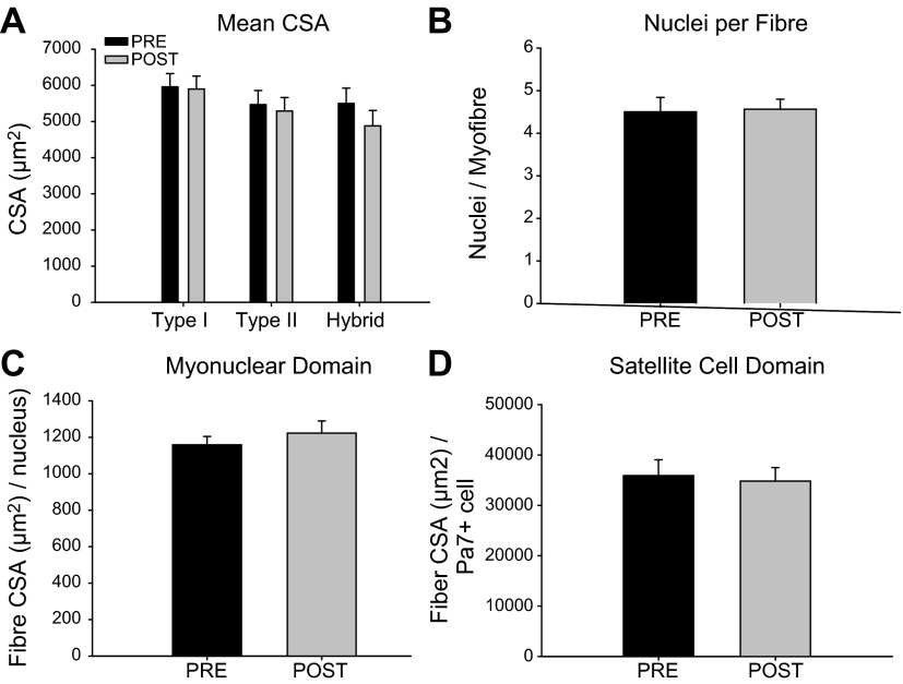
The purpose of this study was to explore the possible role of muscle stem cells, also referred to as satellite cells (SCs), in adaptation and remodeling following a nonhypertrophic stimulus in humans. Muscle biopsies were obtained from the vastus lateralis of previously untrained women (n=15; age: 27±8 yr, BMI: 29±6 kg/m(2)) before and after 6 wk of aerobic interval training. The fiber type-specific SC response to training was analyzed using immunofluorescent microscopy of muscle cross sections. Following training, the number of SCs associated with fibers expressing myosin heavy-chain type I and II isoforms (hybrid fibers) increased (pre: 0.062±0.035 SC/hybrid fiber; post: 0.38±0.063 SC/hybrid fiber; P<0.01). In addition, there was a greater number of MyoD(+)/Pax7(-) SCs, indicative of differentiating SCs, associated with hybrid fibers (0.18±0.096 MyoD(+)/Pax7(-) SC/hybrid fiber) compared to type I (0.015±0.00615 MyoD(+)/Pax7(-) SC/type I fiber) or II (0.012±0.00454 MyoD(+)/Pax7(-) SC/type II fiber) fibers (P<0.05). There was also a training-induced increase in the number of hybrid fibers containing centrally located nuclei (15.1%) compared to either type I (3.4%) or II fibers (3.6%) (P<0.01). These data are consistent with the hypothesis that SCs contribute to the remodeling of muscle fibers even in the absence of hypertrophy.

Keywords: Pax7; aerobic interval training; satellite cells.


Figures

Figure 1.
Figure 1.
Absence of hypertrophy following training. Fiber type-specific CSA, myonuclei, and SCs were quantified by visualization of myofiber borders (laminin and MHCI/II) nuclei (DAPI), and SCs (Pax7). A) Mean CSA of type I, II, and hybrid fibers pre- and post-training. B–D) Calculations of nuclei per fiber (B), myonuclear domain [fiber area (μm2) per nucleus; C] and SC domain [fiber area (μm2) per Pax7+ cell; D] pre- and post-training.
Figure 2.
Figure 2.
Fiber type-specific association of SCs pre- and post-training. A) Representative image of a Pax7/laminin/MHCI/MCHII/nuclei immunofluorescent stain on a muscle cross section. Yellow arrows denote Pax7+ cells associated with a hybrid fiber, white arrow denotes a Pax7+ cell associated to a type I fiber. a–d) Single-channel views of MHCI (green; a), MHCII and laminin (red; b), Pax7 (pink; c), and nuclei (blue, DAPI; d). Scale bar = 10 μm. B) SC response of type I, type II hybrid fiber-associated SCs pre- and post-training, expressed per myofiber. Bars represent means ± se.*P < 0.05 vs. type I fibers; **P < 0.01 vs. pretraining and type I and II fibers.
Figure 3.
Figure 3.
Fiber type-specific SC domain and centrally located nuclei following training. A) Representative image of an MHCI/MHCII/nuclei immunofluorescent stain on a muscle cross section. Yellow arrows denote hybrid fibers; white arrows denote centrally located nuclei within a hybrid fiber. a, b) Single-channel views of MHCI (green; a) and MHCII (red; b). Scale bar = 10 μm. B) Percentage of type I, II, and hybrid fibers with centrally located nuclei following training. C) Fiber type-specific SC domain following training, expressed as fiber area (μm2) per Pax7+ cell. Bars represent means ± se. **P < 0.01 vs. type I and II fibers.
Figure 4.
Figure 4.
Association of SCs to fibers expressing nMHC. A) Representative image of a Pax7/nMHC/laminin/nuclei immunofluorescent stain on a muscle cross section. Scale bar = 100 μm. B) Magnified view of boxed area in A. Arrows denote Pax7+ cells associated with a fiber expressing nMHC. Scale bar = 10 μm. a–d) Single-channel views of nMHC (green; a), Pax7 (red)/laminin (orange) [note that Pax7-positive cells, denoted by white arrows, lie beneath the basal lamina (orange) and are associated with a fiber expressing nMHC; b], Pax7 (red; c), and nuclei (blue, DAPI; d).
Figure 5.
Figure 5.
Fiber type-specific MyoD response of SCs following training. A) Representative image of a Pax7/MyoD/laminin/MCHII/nuclei immunofluorescent stain on a muscle cross section. Yellow arrow denotes a Pax7/MyoD+ cell associated with a hybrid fiber; white arrow denotes a Pax7+/MyoD cell associated with a hybrid fiber; green arrow denotes a Pax7+/MyoD cell associated with a type II fiber. a–d) Single-channel views of Pax7 (green; a), MHCII and laminin (red; b), MyoD (pink; c), and nuclei (blue, DAPI; d). Scale bar = 10 μm. B–D) Fiber-type specific association of quiescent (Pax7+/MyoD; B), activated (Pax7+/MyoD+; C), and differentiating (Pax7/MyoD+; D) SCs following training, expressed per myofiber. Bars represent means ± se. **P < 0.05 vs. type I and II fibers.

References

    1. Mauro A. (1961) Satellite cell of skeletal muscle fiber. J. Biophys. Biochem. Cytol. 9, 493–495 - PMC - PubMed
    1. Crameri R. M., Langberg H., Magnusson P., Jensen C. H., Schrøder H. D., Olesen J. L., Suetta C., Teisner B., Kjaer M. (2004) Changes in satellite cells in human skeletal muscle after a single bout of high-intensity exercise. J Physiol (Lond.) 558, 333–340 - PMC - PubMed
    1. Schultz E., Jaryszak D. L., Valliere C. R. (1985) Response of satellite cells to focal skeletal muscle injury. Muscle Nerve 8, 217–222 - PubMed
    1. McKay B. R., De Lisio M., Johnston A. P. W., O'Reilly C. E., Phillips S. M., Toth K. G., Tarnopolsky M. A., Parise G. (2009) Association of interleukin-6 signalling with the muscle stem cell response following muscle-lengthening contractions in humans. PLoS One 4, e6027. - PMC - PubMed
    1. Tajbakhsh S. (2003) Stem cells to tissue: molecular, cellular and anatomical heterogeneity in skeletal muscle. Curr. Opin. Genetics Dev. 13, 413–422 - PubMed