Review
. 2013 Jul;41 Suppl 4(0 4):S49-55.
doi: 10.1016/S0300-5712(13)70006-X.

How can sensitive dentine become hypersensitive and can it be reversed?

Affiliations
Review

How can sensitive dentine become hypersensitive and can it be reversed?

David H Pashley. J Dent. 2013 Jul.

Abstract

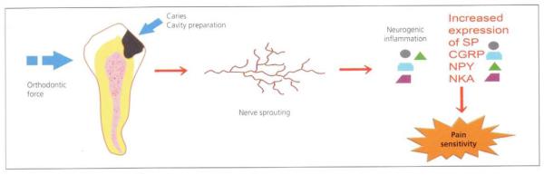
This paper reviews a number of studies in oral biology and endodontics that deal with the reactivity of the pulpo-dentine complex in response to mechanical and immunological stimuli. It can be hypothesized that these reactions could also apply to changes in dentine sensitivity following periodontal procedures. Some of these changes involve neurogenic inflammation of the pulp under exposed open tubules; this increases the rate of outward fluid flow through the tubules, making the overlying exposed dentine more sensitive. Other changes may be due to inflammation-related nerve sprouting of pulpal nerves, which can lead to innervation of more tubules than normal. Changes may also involve upregulation of new, more sensitive ion channels in the membranes of these nerves. The goal of the paper is to increase awareness of the complex issues involved in dentine sensitivity, so that future investigators may develop agents or techniques to stimulate mechanisms that mitigate dentine sensitivity, or to block mechanisms that aggravate the condition, for therapeutic effect.

Keywords: Dentine sensitivity; Pulpal reaction.


Figures

Fig. 1
Fig. 1. Scanning electron micrographs of epoxy replicas of vital human dentine surfaces
(A) Smear layer-covered dentine only allowed small amounts of dentinal fluid to permeate through smear layers in the 3–4 min required for the polyvinylsiloxane impression material to set. (B) Much more fluid was observed on the surface of acid-etched dentine free of smear layers. Reproduced from Pashley and Tay, with kind permission from Quintessence Publishing Co.
Fig. 2
Fig. 2. Smear layer-covered human coronal dentine
Smear layer (SL) debris slightly penetrates into the lumen of dentinal tubules to form a smear plug (SP). Reproduced from Pashley, with kind permission from the Finnish Dental Society.
Fig. 3
Fig. 3. Smear layer-covered human dentine
(A) The fractured edge reveals the presence of smear plugs (P) in tubules (T). (B) The smear layer-covered dentine has been exposed to bacterial plaque for 1 week in vivo. Note the loss of the smear layer and the appearance of open dentinal tubules. Reproduced from Brännström, with kind permission from Elsevier.
Fig. 4
Fig. 4. Histologic appearance of young dental pulp
(A) After evaporative air blast to exposed dentine surface in vivo. Note the loss of the odontoblastic cell bodies and the appearance of their nuclei in the adjacent dentinal tubules. The cell-free zone is seen just above the cell-rich (CR) zone. There are no inflammatory cells in this pulp. (B) After exposed coronal dentine was left open to oral fluids for 1 week. The pulp is heavily inflamed. This dentine was very hypersensitive. Note the absence of an odontoblast layer and the cell-free and CR zones, and the presence of acute inflammatory cells throughout the pulp. A large dilated venule can be seen in the bottom of the field. Reproduced from Brännström, with kind permission from Elsevier.
Fig. 5
Fig. 5. Changes in dentine permeability of dog dentine in vivo
Dentine permeability increased in teeth that had coronal pulp tissue removed just before preparing class V cavities on the buccal surface (dotted line, square symbols). Dentine permeability decreased rapidly in vital teeth over time (solid line with round filled symbols). Numbers in parentheses are numbers of individual teeth studied. Reproduced from Pashley et al., with kind permission from Elsevier.
Fig. 6
Fig. 6. Changes in dentine permeability in dog teeth with and without fibrinogen
Teeth from fibrinogen-depleted dogs (treated with snake venom 2 weeks before the permeability assessment) showed higher, more constant dentine permeability over 6 hours than did teeth from dogs with normal fibrinogen plasma levels. Lp, hydraulic conductivity Reproduced from Pashley et al., with kind permission from Elsevier.
Fig. 7
Fig. 7. Permeability of dentine treated with immunoglobulin G (IgG) in vitro
Dentinal permeability decreased during passage of 160 kDa IgG from the pulpal side of the dentine toward the occlusal side under a simulated normal pulpal pressure of 20 cm H2O. Reproduced from Hahn and Overton, with kind permission from Elsevier.
Fig. 8
Fig. 8. Schematic diagram illustration the consequences of pulpal irritation
Regardless of source, pulpal irritation induces pulpal inflammation that causes nerve sprouting. Increased numbers of pulpal nerves release increased amounts of substance P (SP), calcitonin-gene-related peptide (CGRP), neuropeptide Y (NPY) and neurokinin A (NKA). These neuropeptides sustain neurogenic inflammation that contributes to maintaining sensitive dentine in a hypersensitive state. Reproduced from Caviedes-Bucheli et al., with kind permission from Elsevier.
Fig. 9
Fig. 9. Confocal microscopy of pulpal nerves
Sodium channels (NaCh; red) and Caspr (green) immunoreactions within pulpal nerves in (A) a normal pulp and (B) a painful pulp. Caspr identifies nodes of Ranvier in myelinated nerves. There are more unmyelinated nerves in painful (i.e. inflamed) than normal pulps. Reproduced from Byers et al., with kind permission from Quintessence Publishing Co.

References

    1. Brännström M. Dentin and pulp in restorative dentistry. Wolfe Medical Publications Ltd; London: 1982.
    1. Brännström M. Etiology of dentin hypersensitivity. Proceedings of the Finnish Dental Society. 1992;88(Suppl 1):7–14. - PubMed
    1. Ciucchi B, Bouillaguet S, Holz J, Pashley DH. Dentinal fluid dynamics in human teeth, in vivo. Journal of Endodontics. 1995;21:191–4. - PubMed
    1. Pashley DH, Tay FR, Haywood VB, Collins MA, Drisko CL. Consensus-based recommendations for the diagnosis and management of dentin hypersensitivity. Compendium of Continuing Education in Dentistry. 2008;8(Special Issue):1–37.
    1. Adams D, Addy M, Absi EG. Abrasive and chemical effects of dentifrices. In: Embery G, Rolla G, editors. Clinical and biological aspects of dentifrices. Oxford University Press; Oxford: 1992. pp. 345–55.

Publication types

LinkOut - more resources