. 2013 Nov;6(6):1459-69.
doi: 10.1242/dmm.010447. Epub 2013 Aug 7.

Moderate and high amounts of tamoxifen in αMHC-MerCreMer mice induce a DNA damage response, leading to heart failure and death

Affiliations

Moderate and high amounts of tamoxifen in αMHC-MerCreMer mice induce a DNA damage response, leading to heart failure and death

Kevin Bersell et al. Dis Model Mech. 2013 Nov.

Abstract

Numerous mouse models have utilized Cre-loxP technology to modify gene expression. Adverse effects of Cre recombinase activity have been reported, including in the heart. However, the mechanisms associated with cardiac Cre toxicity are largely unknown. Here, we show that expression of Cre in cardiomyocytes induces a DNA damage response, resulting in cardiomyocyte apoptosis, cardiac fibrosis and cardiac dysfunction. In an effort to increase the recombination efficiency of a widely used tamoxifen-sensitive Cre transgene under control of the α-myosin-heavy-chain promoter (αMHC-MerCreMer), we observed myocardial dysfunction and decreased survival, which were dependent on the dose of tamoxifen injected. After excluding a Cre-independent contribution by tamoxifen, we found that Cre induced myocardial fibrosis, activation of pro-fibrotic genes and cardiomyocyte apoptosis. Examination of the molecular mechanisms showed activation of DNA damage response signaling and p53 stabilization in the absence of loxP sites, suggesting that Cre induced illegitimate DNA breaks. Cardiomyocyte apoptosis was also induced by expressing Cre using adenoviral transduction, indicating that the effect was not dependent on genomic integration of the transgene. Cre-mediated homologous recombination at loxP sites was dose-dependent and had a ceiling effect at ∼80% of cardiomyocytes showing recombination. By titrating the amount of tamoxifen to maximize recombination while minimizing animal lethality, we determined that 30 μg tamoxifen/g body weight/day injected on three consecutive days is the optimal condition for the αMHC-MerCreMer system to induce recombination in the Rosa26-lacZ strain. Our results further highlight the importance of experimental design, including the use of appropriate genetic controls for Cre expression.


Figures

Fig. 1.
Fig. 1.
Tamoxifen injections cause dose-dependent lethal heart failure in αMHC-MerCreMer mice. Tamoxifen was given in three doses as indicated in μg/g body weight. (A) Tamoxifen induces dose-dependent mortality within 1 week of injection. Mortality at doses of 60 and 90 μg/g body weight was statistically significantly different from no tamoxifen (P<0.02 by logrank test, asterisks). (B) Myocardial disorganization and accumulation of extracellular material in moribund mice that were euthanized. Scale bars: 100 μm. (C) Echocardiography performed 1 week after completion of tamoxifen treatment shows diminished myocardial function at 3×60 μg/g body weight. (D) Cre induction does not result in cardiac hypertrophy. (E,F) C57/BL6 mice received five doses of tamoxifen of 90 μg/g body weight. Cardiac structure or function, determined by echocardiography 2 weeks after injection of tamoxifen (E), and heart weight (F) were unchanged, indicating that tamoxifen alone was not toxic. (G) Echocardiography performed 4 weeks after completion of tamoxifen administration (3×60 μg/g body weight) shows diminished myocardial function. n≥4 animals per group (A,C–G), n=2 (G, oil); statistical significance determined with ANOVA (C,D,F) and t-test (E,G).
Fig. 2.
Fig. 2.
Tamoxifen injections induce fibrosis in αMHC-MerCreMer mice. Mice received tamoxifen at the indicated doses in μg/g body weight and frequencies. Fibrosis was visualized with AFOG staining. (A) Earliest evidence of fibrosis, 2 and 3 days after tamoxifen injections (3×60 μg/g body weight). (B) Representative cross-sections showing regional fibrosis 4 weeks after injection of 3×60 μg/g body weight tamoxifen. (C) Examples of fibrosis development 2 weeks after tamoxifen administration at the indicated doses and frequencies. (D) Quantification of multiple mice and sections shows that Cre leads to fibrosis after tamoxifen doses above 30 μg/g body weight. (E) Increased myocardial fibrosis 4 weeks after administration of 3×60 μg tamoxifen/g body weight. Scale bars: 100 μm (A,C) and 1 mm (B). n≥4 animals per group (C–E). (F,G) Quantitative RT-PCR reveals an increase of pro- and anti-fibrotic markers, normalized to GAPDH expression, in tamoxifen- compared with oil-injected animals 2 weeks after tamoxifen injection of 3×30 μg/g body weight (F, n=4) and 4 weeks after injection of 3×60 μg/g body weight (G, n=8): brain natriuretic peptide (BNP), mineralocorticoid receptor (MR), α-smooth muscle actin (α-SMA) and transforming growth factor-β1 (TGF-β1). Statistical significance tested by ANOVA (D) and t-test (E–G). *P<0.05.
Fig. 3.
Fig. 3.
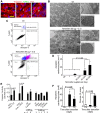
Tamoxifen injections induce cardiomyocyte apoptosis in αMHC-MerCreMer mice.αMHC-MerCreMer+/+ mice received oil or tamoxifen at the indicated doses (in μg/g body weight) and frequencies on consecutive days. (A) Representative TUNEL stainings after injection of 3×90 μg tamoxifen/g body weight show apoptotic cardiomyocyte nuclei surrounded by organized and disorganized sarcomeres. Scale bar: 25 μm. (B) Electron microscopy showed disorganization of mitochondria, sarcomere disarray, mitochondrial rupture and distorted mitochondrial matrix/cristae after injection of 3×60 μg tamoxifen/g body weight. (C) Flow cytometry of isolated cardiomyocytes stained with annexin V and 7-amino-actinomycin (7-AAD) showed viable cardiomyocytes with a small population of necrotic cardiomyocytes in oil-injected control mice. Cardiomyocytes from tamoxifen-treated mice were viable (annexin-V- and 7-AAD-negative), in early apoptosis (annexin-V-positive and 7-AAD-negative), in late apoptosis (annexin-V- and 7-AAD-positive) or in necrosis (annexin-V-negative and 7-AAD-positive). (D) Quantification at 2 weeks after tamoxifen injection shows increased cardiomyocyte apoptosis at doses above 3×30 μg/g body weight. (E) Quantitative analysis of activated caspase-3 activity following injection of tamoxifen shows no increased caspase activity. Positive controls were HeLa cells treated with staurosporine (ST) and liver from animals injected with doxorubicin (DOX). Negative controls were littermates injected with oil (B–E). (F) Comparing apoptosis and fibrosis between 1 and 14 days after 3×90 μg tamoxifen/g body weight shows that apoptosis precedes widespread fibrosis formation. n≥4 animals per group (D,F); number of animals indicated below the x-axis (E). Statistical significance tested by ANOVA.
Fig. 4.
Fig. 4.
Tamoxifen injections induce DNA damage response signaling in αMHC-MerCreMer mice.αMHC-MerCreMer+/+ mice received an injection of 60 μg/g body weight of tamoxifen on three consecutive days. Western blot analysis reveals an increase of γH2AX (A) and p53 (B) at various time points. Littermates injected with oil served as a negative control.
Fig. 5.
Fig. 5.
Transduction with Cre-expressing adenoviruses induces cardiomyocyte apoptosis and shows that transgene insertion is not required. Adult rat ventricular cardiomyocytes were transduced with adenoviruses encoding for lacZ or for Cre under control of the troponin T promoter (TNT-Cre) or CMV promoter (CMV-Cre). (A) Whereas necrotic cardiomyocytes (7-AAD-positive) were present in all wells, apoptotic cardiomyocytes (annexin-V-positive) were present in Cre-transduced plates only. (B) Quantification of annexin-V staining showed cardiomyocyte apoptosis associated with Cre-expressing adenoviruses.
Fig. 6.
Fig. 6.
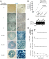
Site-specific recombination and toxicity occur at different levels of Cre recombinase activity in αMHC-MerCreMer mice, revealing a ‘therapeutic window’. Experiments were performed in αMHC-MerCreMer+/+; Rosa26R+/– mice. (A) Representative examples of micrographs after X-gal staining. Scale bars: 1 mm and 100 μm for left and right panels, respectively. (B) Dosing 3×30 μg tamoxifen/g body weight maximizes site-specific recombination while minimizing cumulative 14-day mortality. (C) The efficiency of creating genomic recombination was determined in αMHC-MerCreMer+/+; ErbB4flox/flox mice after injection of 3×30 μg tamoxifen/g body weight. Genomic PCR shows ∼50% recombination. The sizes of the specific PCR products from the floxed allele (547 bp) and from the recombined allele (Rec.; 507 bp) are indicated with arrows. Tam, tamoxifen. (D) Injecting 3×30 μg tamoxifen/g body weight (indicated by vertical arrows) does not induce delayed-onset cardiomyopathy as determined by serial echocardiography for 4 weeks. Differences between baseline at 6 weeks of age and follow-up echocardiographs were not statistically significant (n=6 per group, ANOVA).
Fig. 7.
Fig. 7.
Tamoxifen injections in αMHC-MerCreMer mice induce a loxP-independent DNA damage response, leading to myocardial dysfunction and death. The proposed sequential model of molecular, cellular and tissue mechanisms is provided in the flow diagram. The measured parameters indicating involvement of the proposed mechanisms are indicated in smaller italicized font.

References

    1. Bae S., Yalamarti B., Kang P. M. (2008). Role of caspase-independent apoptosis in cardiovascular diseases. Front. Biosci. 13, 2495–503 - PubMed
    1. Bahi N., Zhang J., Llovera M., Ballester M., Comella J. X., Sanchis D. (2006). Switch from caspase-dependent to caspase-independent death during heart development: essential role of endonuclease G in ischemia-induced DNA processing of differentiated cardiomyocytes. J. Biol. Chem. 281, 22943–22952 - PubMed
    1. Bersell K., Arab S., Haring B., Kühn B. (2009). Neuregulin1/ErbB4 signaling induces cardiomyocyte proliferation and repair of heart injury. Cell 138, 257–270 - PubMed
    1. Buerger A., Rozhitskaya O., Sherwood M. C., Dorfman A. L., Bisping E., Abel E. D., Pu W. T., Izumo S., Jay P. Y. (2006). Dilated cardiomyopathy resulting from high-level myocardial expression of Cre-recombinase. J. Card. Fail. 12, 392–398 - PubMed
    1. Choudhury S., Bae S., Kumar S. R., Ke Q., Yalamarti B., Choi J. H., Kirshenbaum L. A., Kang P. M. (2010). Role of AIF in cardiac apoptosis in hypertrophic cardiomyocytes from Dahl salt-sensitive rats. Cardiovasc. Res. 85, 28–37 - PMC - PubMed

Publication types