

Stereotactic coordinates for intracerebroventricular infusion after permanent focal cerebral ischemia in Wistar rats
- PMID: 23930058
- PMCID: PMC3738393
Stereotactic coordinates for intracerebroventricular infusion after permanent focal cerebral ischemia in Wistar rats
Abstract
Background: Intracerebroventricular (ICV) experimental route is highly promising due to immediate approach of a "therapy" to the cerebrospinal compartment. Ischemic edema causes structural dislocations and stereotaxia alterations after temporary Middle Cerebral Artery Occlusion (t-MCAO), while there is no similar study for intracerebroventricular (ICV) invasion after permanent MCAO (p-MCAO).
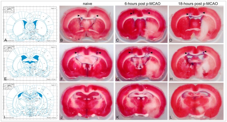
Methods: Male Wistar rats were subjected to right p-MCAO and clinically evaluated 6 and 18 hours post-occlusion, using the modified Neurological Stroke Scale (mNSS) and modified Bederson's Scale (mBS). Infarction volume, hemispheric edema, middle line dislocation and stereotaxia of the lateral ventricles were studied at the same time-points.
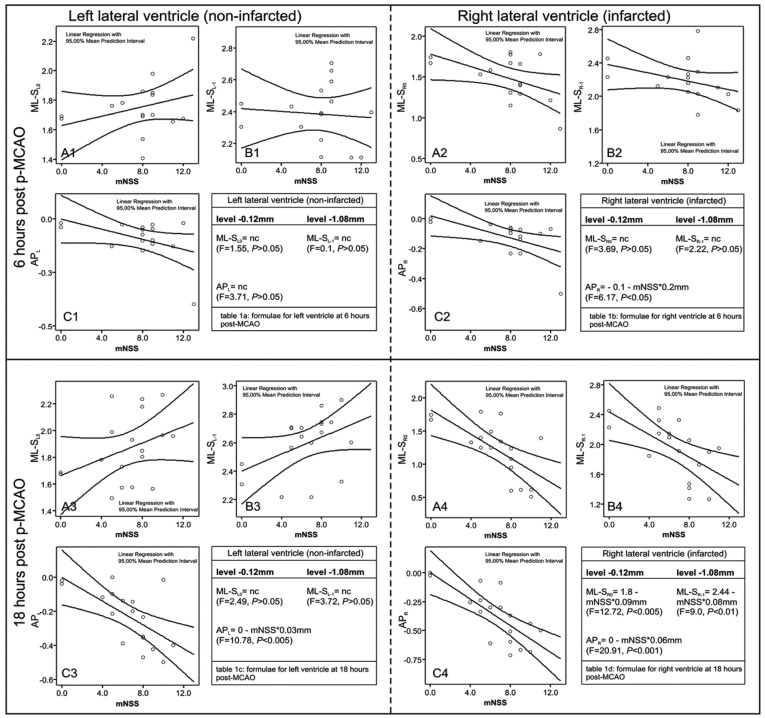
Results: P-MCAO induced mild but significant changes in the stereotaxia of the infarcted (ipsilateral) lateral ventricle on 18- (P<0.05), though not 6-hours (P>0.05) post-occlusion. These changes correlated with the mNSS and mBS scores (P<0.01) and allowed the expression of linear mathematical equations (stereotaxic coordinate = b0 + b1*mNSS; calculated by regression analysis) predicting the new ventricular position in each individual animal. The contralateral ventricular system was structurally unaffected on both time-points. Verification experiments indicated that the new coordinates were necessary on 18-hours post-occlusion for successful ICV invasion in all p-MCAO rats (Number Needed to Treat 2.28), compared to 56.25% success when using the classical coordinates for normal rats.
Conclusion: P-MCAO causes relatively late but predictable stereotaxia shifts for ICV invasion, which are different compared to t-MCAO.
Keywords: edema; intraventricular stereotaxic coordinates; lateral ventricles; permanent middle cerebral artery occlusion; stroke; transplantation.
Figures
References
-
- Belayev L, Alonso OF, Busto R, Zhao W, Ginsberg MD. Middle cerebral artery occlusion in the rat by intraluminal suture. Neurological and pathological evaluation of an improved model. Stroke. 1996;27:1616–1622. - PubMed
-
- Koizumi J, Yoshida Y, Nakazawa T, Ooneda G. Experimental studies of ischemic brain edema. 1. A new experimental model of cerebral embolism in rats in which recirculation can be introduced in ischemic area. Jpn J Stroke. 1986;8:1–8.
-
- Longa EZ, Weinstein PR, Carlson S, Cummins R. Reversible middle cerebral artery occlusion without craniectomy in rats. Stroke. 1989;20:84–91. - PubMed
-
- Lourbopoulos A, Karacostas D, Artemis N, Milonas I, Grigoriadis N. Effectiveness of a new modified intraluminal suture for temporary middle cerebral artery occlusion in rats of various weight. J Neurosci Methods. 2008;173:225–234. - PubMed
-
- Durukan A, Tatlisumak T. Acute ischemic stroke: overview of major experimental rodent models, pathophysiology, and therapy of focal cerebral ischemia. Pharmacol Biochem Behav. 2007;87:179–197. - PubMed
LinkOut - more resources
Full Text Sources