Review
. 2013 Dec;51(4):213-9.
doi: 10.1016/j.bcmd.2013.07.006. Epub 2013 Aug 7.

Erythroid development in the mammalian embryo

Affiliations
Review

Erythroid development in the mammalian embryo

Margaret H Baron et al. Blood Cells Mol Dis. 2013 Dec.

Abstract

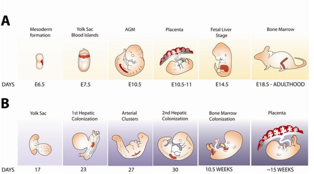
Erythropoiesis is the process by which progenitors for red blood cells are produced and terminally differentiate. In all vertebrates, two morphologically distinct erythroid lineages (primitive, embryonic, and definitive, fetal/adult) form successively within the yolk sac, fetal liver, and marrow and are essential for normal development. Red blood cells have evolved highly specialized functions in oxygen transport, defense against oxidation, and vascular remodeling. Here we review key features of the ontogeny of red blood cell development in mammals, highlight similarities and differences revealed by genetic and gene expression profiling studies, and discuss methods for identifying erythroid cells at different stages of development and differentiation.

Keywords: AGM; BFU-E; CFU-E; E#; E-Tmod; EMP; ES; ESRE; EryD; EryP; Erythroid differentiation; Fetal liver; GFP; HPP-CFC; MEP; Mammalian embryo; Primitive erythropoiesis; Transgenic mice; Yolk sac; aorta–gonad–mesonephros; bipotential megakaryocyte/erythroid progenitor; burst-forming unit erythroid; colony-forming cells erythroid; definitive, enucleated erythrocytes; embryonic day post-fertilization; embryonic stem; erythroid–myeloid progenitor; erythroid–tropomodulin; extensively self-renewing erythroid; green fluorescent protein; high proliferating progenitors-colony forming cell; primitive (nucleated) erythrocytes.


Figures

Figure 1
Figure 1. Ontogeny of mouse and human hematopoiesis
(A) Hematopoietic development in the mouse. The panels represent, from left to right, formation of mesoderm during gastrulation (E6.5), development of blood islands within the yolk sac (~E7.5), emergence of HSCs in the AGM region (E10.5) and placenta (E10.5–11), active fetal liver hematopoiesis (E14.5), and hematopoiesis in the bone marrow (~E18.5 in the late gestation fetus and throughout postnatal life). Myeloid and definitive erythroid potentials are found in the allantois and chorion (not shown) prior to their fusion to form the placenta [106]. The formation of HSCs is completed by mid-gestation. Lymphopoiesis is not represented in this figure. Cardiac function begins as early as ~E8.25, with active circulation by ~E9.0[107]. For a detailed review of mammalian hematopoiesis, see ref. [19]. (B) Hematopoietic development in the human embryo. The panels represent, from left to right, hematopoiesis at the yolk sac stage (day 17), at the time of the first hepatic colonization by HSCs (day 23), arterial cluster formation (day 27), the second hepatic colonization (day 30), and bone marrow colonization (10.5 weeks). For a review, see ref. [21]. In contrast with the mouse embryo, where HSCs are found in the placenta at around the same time as in the AGM region [108; 109], well before colonization of the bone marrow (~10.5 weeks [110]), human HSC activity is not detected in the placenta until several weeks after bone marrow colonization, at ~15 weeks [111]. Active circulation begins by ~day 21 [4].
Figure 2
Figure 2. Photographs of ε-globin-H2B-GFP transgenic mouse embryos at different developmental stages
Overlay of bright field and GFP channel of an ~E7.5 (A) and of an ~E8.5 embryo (B). Scale bars, 200µm. (C) GFP expression in an E10.5 embryo. The yolk sac (left) has been opened. Scale bar, 1mm. (D) Detail of yolk sac from an E10.5 ε-globin-H2BGFP; Flk1-Cre; Rosa26-tdTom transgenic embryo. Red fluorescence (tdTomato) is seen in endothelial cells of the yolk sac vasculature and results from the excision of a STOP cassette from the Rosa26-tdTom transgene [112] by Flk1 promoter-driven Cre. Scale bar, 100 µm.

References

    1. Haar J, Ackerman GA. A phase and electron microscopic study of vasculogenesis and erythropoiesis in the yolk sac of the mouse. Anat. Rec. 1971;170:199–224. - PubMed
    1. Palis J, Robertson S, Kennedy M, Wall C, Keller G. Development of erythroid and myeloid progenitors in the yolk sac and embryo proper of the mouse. Development. 1999;126:5073–5084. - PubMed
    1. Palis J, Yoder M. Yolk-sac hematopoiesis: The first blood cells of mouse and man. Exper. Hematol. 2001;29:927–936. - PubMed
    1. Tavian M, Peault B. Embryonic development of the human hematopoietic system. Int. J. Dev. Biol. 2005;49:243–250. - PubMed
    1. Wong PM, Chung SW, Chui DH, Eaves CJ. Properties of the earliest clonogenic hemopoietic precursors to appear in the developing murine yolk sac. Proc. Natl. Acad. Sci. U.S.A. 1986;83:3851–3854. - PMC - PubMed

Publication types

LinkOut - more resources