. 2013 Aug 19;23(16):1527-35.
doi: 10.1016/j.cub.2013.06.058. Epub 2013 Aug 8.

Dauer-specific dendrite arborization in C. elegans is regulated by KPC-1/Furin

Affiliations

Dauer-specific dendrite arborization in C. elegans is regulated by KPC-1/Furin

Nathan E Schroeder et al. Curr Biol. .

Abstract

Background: Dendrites often display remarkably complex and diverse morphologies that are influenced by developmental and environmental cues. Neuroplasticity in response to adverse environmental conditions entails both hypertrophy and resorption of dendrites. How dendrites rapidly alter morphology in response to unfavorable environmental conditions is unclear. The nematode Caenorhabditis elegans enters into a stress-resistant dauer larval stage in response to an adverse environment.

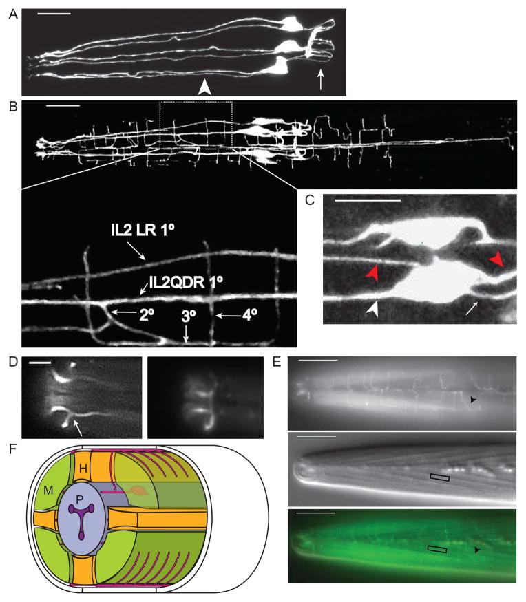
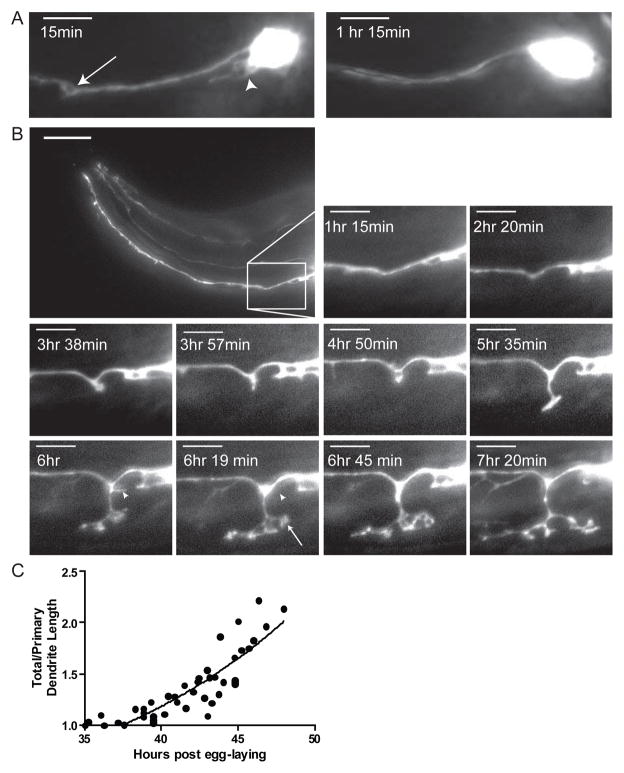
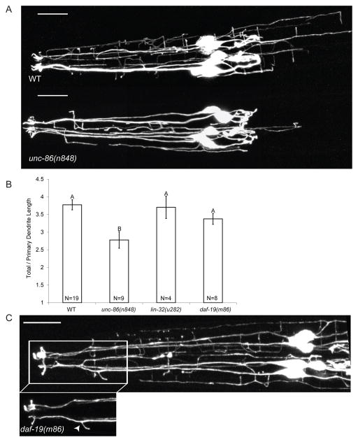
Results: Here we show that the IL2 bipolar sensory neurons undergo dendrite arborization and axon remodeling during dauer development. When dauer larvae are returned to favorable environmental conditions, animals resume reproductive development and IL2 dendritic branches retract, leaving behind remnant branches in postdauer L4 and adult animals. The C. elegans furin homolog KPC-1 is required for dauer IL2 dendritic arborization and dauer-specific nictation behavior. KPC-1 is also necessary for dendritic arborization of PVD and FLP sensory neurons. In mammals, furin is essential, ubiquitously expressed, and associated with numerous pathologies, including neurodegenerative diseases. While broadly expressed in C. elegans neurons and epithelia, KPC-1 acts cell autonomously in IL2 neurons to regulate dauer-specific dendritic arborization and nictation.

Conclusions: Neuroplasticity of the C. elegans IL2 sensory neurons provides a paradigm to study stress-induced and reversible dendritic branching, and the role of environmental and developmental cues in this process. The newly discovered role of KPC-1 in dendrite morphogenesis provides insight into the function of proprotein convertases in nervous system development.


Figures

Figure 1
Figure 1. The IL2 neurons show extensive remodeling during dauer
(A) Lateral Z-projection of a wild-type L3 expressing Pklp-6::GFP in IL2s. The six IL2s are arranged in a hexaradiate pattern with single 1° dendrites (arrowhead) anterior of the cell body. Axons (arrow) posterior from the cell body form a loop which innervate the nerve ring. Scale bar, 10 μm. (B) Dorsal Z-projection of wild-type dauer expressing Pklp-6::GFP in IL2s. The IL2Q (for quadrant) dendrites in dauer are highly branched. Zoomed inset: The IL2QDR 1° dendrite extends a 2° dendrite towards the dorsal midline which branches, forming a 3° dendrite that travels along the dorsal midline. This 3° dendrite branches and extends 4° dendrites into the body-wall muscle quadrants. Similar branching patterns are seen on the ventral body-wall. Scale bar, 10 μm. (C) Z-projection of IL2QDL dauer cell body expressing Pklp-6::GFP. The IL2Qs extend additional dauer-specific primary dendrites (1d°) (red arrowheads) in addition to the original non-dauer anteriorly directed 1° dendrite (white arrowhead), and posteriorly directed axon (white arrow). Scale bar, 5 μm. (D) Dorsal view of dauer nose expressing Plag-2::GFP showing (left) the single branch (arrow) extending from the lateral IL2LL 1° dendrite and (right) a body wall view of the same animal showing the formation of the crown from branches emerging from the IL2L dendrites. Scale bar, 5 μm. (E) Wild-type dauers extend fine dendritic processes along the body wall. Top: Dorsal/ventral view of body wall in wild-type dauer expressing Plag-2::GFP in IL2 neurons. Quaternary dendrites (arrowhead) extend from the dorsal/ventral midlines perpendicularly into the muscle quadrants. Middle: Same animal and focal plane with DIC Nomarski optics. Somatic muscle dense bodies (box), which serve as connections with the overlying hypodermis, are evident. Bottom: Merged image. Scale bars, 10 μm. (F) Oblique transverse schematic of dauer IL2 branching. M:muscle, P:pharynx, H:hypodermis.
Figure 2
Figure 2. Time-lapse imaging of IL2Qs during dauer formation reveals rapid dendrite arborization
(A) Z-projection time-lapse images of a single animal expressing Pklp-6::tdTomato following the onset of the L2d molt into dauer. 15 minutes following the L2d molt, 2° dendritic sprouts (arrow) and 1d° dendrites (arrowhead) appear on an IL2QDL neuron. 75 minutes after the onset of the L2d molt, these sprouts have retracted. Scale bar, 10 μm. (B) Lateral Z-projection time-lapse images of single animal expressing Pklp-6::tdTomato during the L2d molt into dauer. The formation of putative growth cones (arrow at 6 hr 19 min) as well as the retraction of branches (arrowheads at 6 hr – 6 hr 19 min) of IL2Qs is seen. Inset scale bar, 5 μm. (C)Quantification of relative IL2Q dendritic length during dauer formation following the onset of the L2d molt fits an exponential curve (Y= 0.849*e(0.066x), R2=0.7436). A ratio of total/primary dendritic length was used to adjust for changes in total body length. Each animal examined is represented by multiple time-points (N=9 animals, 5–25 time points per animal).
Figure 3
Figure 3. Following recovery from the dauer stage, IL2Q arbors undergo incomplete retraction
(A) Lateral Z-projection of post-dauer L4 expressing Pklp-6::GFP. Nematodes often will retain short remnant 2° (arrows) dendrites following recovery from dauer. Scale bar, 10 μm. (B) Quantification of relative IL2Q dendrite length following the return of dauers to favorable conditions (plentiful food, low population density) fits a sigmoidal response curve (Y=1.24+[2.518÷(1+10(0.183x-0.616))], R2=0.803). A ratio of total/primary dendritic length was used to adjust for changes in total body length. Each data point represents a separate animal (N=35 animals).
Figure 4
Figure 4. The transcription factors UNC-86 and DAF-19 regulate distinct components of dauer-specific IL2 remodeling
(A) Z-projections of WT (top) and unc-86(n848) (bottom)dauers expressing P F28A12.3::GFP. Scale bars, 10 μm. (B) unc-86(n848) dauers show significantly smaller IL2Q arbors than WT. Dendritic length was measured as a ratio of total/primary dendritic length to compensate for differences in body length between genotypes. See also Figure S3. Data are mean ± SEM. Genotypes with different letters above bars are statistically different (α=0.01) as determined by ANOVA followed by Tukey’s post-hoc test for comparison of multiple genotypes. (C) Dorsal Z-projection of daf-19(m86) dauer expressing PF28A12.3::GFP with inset of IL2LL dendrite showing supernumerary branching (arrowhead). Scale bar, 10 μm.
Figure 5
Figure 5. KPC-1 is required for multidendritic neuron arborization
(A) Z-projections of wild-type (top) and kpc-1(my24) (bottom)dauers expressing P klp-6::GFP. Disruption of kpc-1 results in disorganized IL2 dendritic arbors in 100% of animals examined (N>100). See also Figure S5. Scale bar, 10 μm. (B) Exon/Intron diagram of kpc-1. The kpc-1(my24) allele introduces a c→t missense mutation resulting in a P440L amino acid change at a highly conserved residue (highlighted yellow in alignment) four amino acids C-terminal from the catalytic serine (highlighted red in alignment). The kpc-1(gk8) deletion allele removes the majority of the catalytic domain. (C) Lateral Z-projections of PVD neurons in wild-type (left) and kpc-1(gk8) (right) adults expressing PF49H12.4::GFP. Similar to the IL2 neurons, disruption of kpc-1 leads to disorganized and truncated arbors in PVDs. Scale bar, 10 μm. See also Figure S5C.
Figure 6
Figure 6. kpc-1 is expressed broadly but acts cell autonomously to regulate IL2Q dauer-specific remodeling
(A) Ventral Z-projection of dauer expressing Pkpc-1::GFP with expression in numerous neuronal and non-neuronal cells throughout the head. Scale bar, 10 μm. (B) Ventral body-wall plane of same animal as in (A) showing GFP expression in IL2Q dauer-specific branches (arrow) as well as several additional neuronal commissures (arrowhead, amphid commisure). Scale bar, 10 μm. (C) Dauer expressing both the IL2-specific reporter Pklp-6::tdTomato (left) and Pkpc-1::GFP (middle). An overlay image (right) demonstrates that Pkpc-1::GFP is expressed in the IL2s. Scale bar, 5 μm. (D) kpc-1(gk8) defects in dauer-specific IL2Q arborization are rescued by constructs of either an IL2-specific promoter driving full length wild-type kpc-1 (Pklp-6::KPC-1) or full length kpc-1 tagged with dsRed and driven by the kpc-1 endogenous promoter (KPC-1::dsRed). Rescue was assessed by examining dauer-specific IL2Q arborization in kpc-1(gk8) dauers expressing Pklp-6::GFP in three independent transgenic lines (N=22–25 dauers per line). A mean ± SEM of the three lines is given. Statistical tests comparing the rescued lines with WT and kpc-1(gk8) cannot be performed due to a lack of variation in controls, however Pklp-6::KPC-1 and KPC-1::dsRed are not statistically different (Fisher’s Exact Test, p=0.9303). (E) Z-projection of kpc-1(gk8) dauer head expressing KPC-1::dsRed. In most neurons KPC-1::dsRed is localized exclusively within the cell bodies and is not observed in neuronal processes. However in the ventral nerve cord KPC-1::dsRed is found in both cell bodies and neuronal processes (See Figure S6B). Scale bar, 10 μm.
Figure 7
Figure 7. kpc-1 and unc-86 regulate nictation behavior
(A) Quantification of percent time spent nictating versus non-nictating [(Tnic/T)×100] in actively moving dauers as previously described [4]. kpc-1 and unc-86(n848) mutant dauers are defective in nictation ratio while IL2-specific rescue of kpc-1 restores nictation ratio to WT levels. (B) Quantification of nictation initiation index [N/(Tnic-T)×100] as previously described [4]. kpc-1 and unc-86(n848) mutants are defective while IL2-specific rescue of kpc-1 restores initiation index to WT levels. Data are means ± SEM. Genotypes with different letters above bars are statistically different (α=0.01) as determined by Kruskall-Wallis followed by Dunn’s multiple comparison. N=13–50 animals/genotype.

Comment in

References

    1. Brenner S. The genetics of Caenorhabditis elegans. Genetics. 1974;1:71–94. - PMC - PubMed
    1. Riddle DL, Albert PS. In: Genetic and environmental regulation of dauer larvae developmentC. elegans. Riddle DL II, Blumenthal T, Meyer BJ, Priess JR, editors. Plainview, NY: Cold Spring Harbor Laboratory Press; 1998. pp. 739–768. - PubMed
    1. Cassada RC, Russell RL. The dauerlarva, a post-embryonic developmental variant of the nematode Caenorhabditis elegans. Dev Biol. 1975;2:326–342. - PubMed
    1. Lee H, Choi MK, Lee D, Kim HS, Hwang H, Kim H, Park S, Paik YK, Lee J. Nictation, a dispersal behavior of the nematode Caenorhabditis elegans, is regulated by IL2 neurons. Nat Neurosci. 2011;1:107–112. - PubMed
    1. Golden JW, Riddle DL. The Caenorhabditis elegans dauer larva: developmental effects of pheromone, food, and temperature. Dev Biol. 1984;2:368–378. - PubMed

Publication types

MeSH terms