. 2013 Nov;34(33):8241-57.
doi: 10.1016/j.biomaterials.2013.07.052. Epub 2013 Aug 6.

Tuning mechanical performance of poly(ethylene glycol) and agarose interpenetrating network hydrogels for cartilage tissue engineering

Affiliations

Tuning mechanical performance of poly(ethylene glycol) and agarose interpenetrating network hydrogels for cartilage tissue engineering

Deena A Rennerfeldt et al. Biomaterials. 2013 Nov.

Abstract

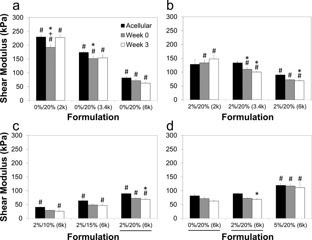
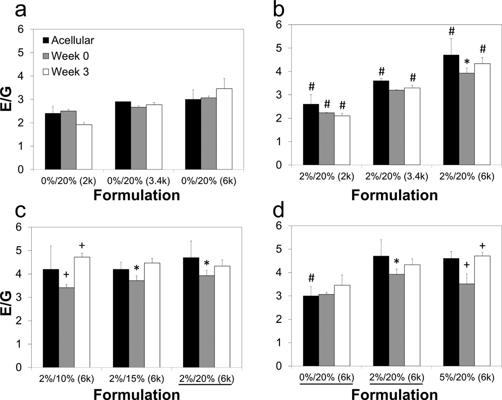
Hydrogels are attractive for tissue engineering applications due to their incredible versatility, but they can be limited in cartilage tissue engineering applications due to inadequate mechanical performance. In an effort to address this limitation, our team previously reported the drastic improvement in the mechanical performance of interpenetrating networks (IPNs) of poly(ethylene glycol) diacrylate (PEG-DA) and agarose relative to pure PEG-DA and agarose networks. The goal of the current study was specifically to determine the relative importance of PEG-DA concentration, agarose concentration, and PEG-DA molecular weight in controlling mechanical performance, swelling characteristics, and network parameters. IPNs consistently had compressive and shear moduli greater than the additive sum of either single network when compared to pure PEG-DA gels with a similar PEG-DA content. IPNs withstood a maximum stress of up to 4.0 MPa in unconfined compression, with increased PEG-DA molecular weight being the greatest contributing factor to improved failure properties. However, aside from failure properties, PEG-DA concentration was the most influential factor for the large majority of properties. Increasing the agarose and PEG-DA concentrations as well as the PEG-DA molecular weight of agarose/PEG-DA IPNs and pure PEG-DA gels improved moduli and maximum stresses by as much as an order of magnitude or greater compared to pure PEG-DA gels in our previous studies. Although the viability of encapsulated chondrocytes was not significantly affected by IPN formulation, glycosaminoglycan (GAG) content was significantly influenced, with a 12-fold increase over a three-week period in gels with a lower PEG-DA concentration. These results suggest that mechanical performance of IPNs may be tuned with partial but not complete independence from biological performance of encapsulated cells.

Keywords: Agarose; Cartilage; Hydrogels; Interpenetrating networks; Mechanical performance; PEG-DA.


Figures

Figure 1
Figure 1
(a) Maximum stress and (b) maximum strain percent of agarose and PEG-DA IPNs with PEG-DA molecular weights of 2000 (2k), 3400 (3.4k), and 6000 (6k) Da and agarose concentrations of 0% (pure PEG-DA gels), 2%, and 5%. Unlike most mechanical and swelling characteristics, PEG-DA molecular weight was the most significantly-contributing factor for these two failure properties. Values represent mean ± standard deviation with n=4. Values significantly different (p<0.05) from groups varying only in: #PEG-DA concentration, +agarose concentration, *PEG-DA molecular weight.
Figure 2
Figure 2
Images of an IPN sample (2% agarose, 20% 6k PEG-DA) (a) before unconfined compression, (b) at 50% strain, (c) at 95% strain, and (d) immediately after compression. This particular sample had a compressive modulus of 313 kPa and withstood a stress of 4.2 MPa without fracturing. After soaking in a phosphate buffered saline solution for 1 hour, the sample recovered to approximately 97% of its original height.
Figure 3
Figure 3
(a) Shear modulus and (b) ratio of compressive modulus to shear modulus of agarose (Ag) and PEG-DA IPNs. Nearly every formulation had a significantly higher shear modulus (p<0.05) with an increase in PEG-DA concentration, while E/G generally increased with increases in agarose concentration and PEG-DA molecular weight. Values represent mean ± standard deviation with n=4. Values significantly different (p<0.05) among groups varying only in: #PEG-DA concentration, +agarose concentration, *PEG-DA molecular weight.
Figure 4
Figure 4
Representative neo-Hookean elasticity model plots of stress versus strain function, demonstrating trends toward non-ideal elastic behavior for (a) increases in agarose concentration and (b) increases in PEG-DA molecular weight (in IPNs). The shear modulus, G, was taken as the slope of these plots up to a strain function value of 10 or until fracture. An E/G ratio of 3 is considered ideal elastic behavior, as is a linear stress-strain function profile. PEG-DA = polyethylene glycol) diacrylate, λ=L/L0, IPN = interpenetrating network, data points after fracture omitted in panel b.
Figure 5
Figure 5
(a) Equilibrium mass swelling ratio and (b) PEG-DA solids content. The mass swelling ratios generally showed an inverse relationship with shear modulus (Figure 2a), and the decreased PEG-DA content in IPNs versus pure PEG-DA gels (0% Ag) helps to explain corresponding decreases in shear modulus. Values represent mean ± standard deviation with n=4. Values significantly different (p<0.05) from groups varying only in: #PEG-DA concentration, +agarose concentration, *PEG-DA molecular weight; &value significantly higher (p<0.05) than all 26 other formulations.
Figure 6
Figure 6
(a) Mesh size and (b) crosslink density of PEG-DA in IPNs. Both properties were most significantly influenced by PEG-DA, though molecular weight was also an important factor. Values represent mean ± standard deviation with n=4. Values significantly different (p<0.05) from groups varying only in: #PEG-DA concentration, +agarose concentration, *PEG-DA molecular weight; &value significantly higher (p<0.05) than all 26 other formulations.
Figure 7
Figure 7
(a) total GAG accumulated per gel, (b) DNA content per gel, and (c) GAG normalized to DNA content per gel encapsulated with chondrocytes at 0 and 3 weeks, with formulations listed as agarose concentration/PEG-DA concentration (PEG-DA molecular weight). Halving the PEG-DA concentration in the 6k MW gels resulted in approximately double the normalized GAG accumulation for the two IPNs, while the pure PEG-DA gel did not provide a productive environment for chondrocytes in the absence of agarose. Values represent mean ± standard deviation with n=4. *Values significantly different from week 0 time point (p<0.005), #significant differences (p<0.05) among groups at the same time point. GAG, glycosaminoglycan.
Figure 8
Figure 8
Representative live/dead images for (a) agarose and (b) an IPN with 2% agarose and 20% PEG-DA (6000 Da molecular weight) 24 hours after encapsulation, and (c) an IPN with the same formulation 3 weeks after encapsulation. High cell viability 24 hours after encapsulation, even in the IPN formulation with the longest PEG-DA diffusion time and highest PEG-DA concentration, demonstrates the potential for this encapsulation process to work well with a wide variety of PEG-DA concentrations and molecular weights. Live cells are stained green with Calcein AM (green), while dead cells are stained red with ethidium homodimer-1. Scale bars = 50 µm.
Figure 9
Figure 9
Maximum strain of agarose and PEG-DA IPNs encapsulated with chondrocytes at 0 and 3 weeks. A total of 9 groups are compared three at a time, grouped as (a) pure 20% PEG-DA gels with varied molecular weights of 2000 (2k), 3400 (3.4k) and 6000 (6k) Da, (b) IPNs varying molecular weights, (c) IPNs with varying concentrations of PEG-DA, and (d) IPNs with varying agarose concentrations. As with maximum stress (Figure 8), cell-laden gels appeared to have a limit to the amount of strain they could withstand, regardless of the performance of their acellular counterparts. Formulations listed as agarose concentration/PEG-DA concentration (PEG-DA molecular weight), and underlines note the same formulation was shown in a previous panel. Values from acellular gels (taken from Fig. 1b) included for comparison. *Values significantly different from acellular gels with the same formulation (p<0.05), #significant differences among groups at the same time point within the same panel (p<0.05).
Figure 10
Figure 10
Maximum stress of agarose and PEG-DA IPNs encapsulated with chondrocytes at 0 and 3 weeks. A total of 9 groups are compared three at a time, grouped as (a) pure 20% PEG-DA gels with varied molecular weights of 2000 (2k), 3400 (3.4k) and 6000 (6k) Da, (b) IPNs varying molecular weights, (c) IPNs with varying concentrations of PEG-DA, and (d) IPNs with varying agarose concentrations. Unlike the shear and compressive moduli, values of the cell-encapsulated gels did not follow the same trends as their acellular counterparts due to an inability to withstand a stress of more than approximately 1500 kPa. Formulations listed as agarose concentration/PEG-DA concentration (PEG-DA molecular weight), and underlines note the same formulation was shown in a previous panel. Values from acellular gels (taken from Fig. 1a) included for comparison. *Values significantly different from acellular gels with the same formulation (p<0.05), #significant differences among groups at the same time point within the same panel (p<0.05).
Figure 11
Figure 11
Shear moduli of agarose and PEG-DA IPNs encapsulated with chondrocytes at 0 and 3 weeks. A total of 9 groups are compared three at a time, grouped as (a) pure 20% PEG-DA gels with varied molecular weights of 2000 (2k), 3400 (3.4k) and 6000 (6k) Da, (b) IPNs varying molecular weights, (c) IPNs with varying concentrations of PEG-DA, and (d) IPNs with varying agarose concentrations. Cell-laden gels typically followed the same trends as acellular gels, with little difference between time points. Formulations listed as agarose concentration/PEG-DA concentration (PEG-DA molecular weight), and underlines note the same formulation was shown in a previous panel. Values from acellular gels (taken from Fig. 2a) included for comparison. *Values significantly different from acellular gels with the same formulation (p<0.05), #significant differences among groups at the same time point within the same panel (p<0.05), +significant differences between week 0 and week 3 for a single group (p<0.05).
Figure 12
Figure 12
Ratio of compressive modulus to shear modulus (E/G) of agarose and PEG-DA IPNs encapsulated with chondrocytes at 0 and 3 weeks. A total of 9 groups are compared three at a time, grouped as (a) pure 20% PEG-DA gels with varied molecular weights of 2000 (2k), 3400 (3.4k) and 6000 (6k) Da, (b) IPNs varying molecular weights, (c) IPNs with varying concentrations of PEG-DA, and (d) IPNs with varying agarose concentrations. Very few significant differences were found in ideal elastic behavior between cell-laden and acellular gels. Formulations listed as agarose concentration/PEG-DA concentration (PEG-DA molecular weight), and underlines note the same formulation was shown in a previous panel. Values from acellular gels (taken from Fig. 2b) included for comparison. *Values significantly different from acellular gels with the same formulation (p<0.05), #significant differences among groups at the same time point within the same panel (p<0.05), +significant differences between week 0 and week 3 for a single group (p<0.05).

Similar articles

Cited by

References

    1. Gong JP, Katsuyama Y, Kurokawa T, Osada Y. Double-network hydrogels with extremely high mechanical strength. Adv Mater. 2003;15:1155–8.
    1. Na Y-H, Kurokawa T, Katsuyama Y, Tsukeshiba H, Gong JP, Osada Y, et al. Structural characteristics of double network gels with extremely high mechanical strength. Macromolecules. 2004;37:5370–4.
    1. Myung D, Waters D, Wiseman M, Duhamel P-E, Noolandi J, Ta CN, et al. Progress in the development of interpenetrating polymer network hydrogels. Polym Adv Technol. 2008;19:647–57. - PMC - PubMed
    1. Gong JP. Why are double network hydrogels so tough? Soft Matter. 2010;6:2583–90.
    1. Azuma C, Yasuda K, Tanabe Y, Taniguro H, Kanaya F, Nakayama A, et al. Biodegradation of high-toughness double network hydrogels as potential materials for artificial cartilage. J Biomed Mater Res A. 2007;81A:373–80. - PubMed

Publication types