. 2013 Sep 3;21(9):1690-7.
doi: 10.1016/j.str.2013.06.020. Epub 2013 Aug 8.

Conservation of protein structure over four billion years

Affiliations

Conservation of protein structure over four billion years

Alvaro Ingles-Prieto et al. Structure. .

Abstract

Little is known about the evolution of protein structures and the degree of protein structure conservation over planetary time scales. Here, we report the X-ray crystal structures of seven laboratory resurrections of Precambrian thioredoxins dating up to approximately four billion years ago. Despite considerable sequence differences compared with extant enzymes, the ancestral proteins display the canonical thioredoxin fold, whereas only small structural changes have occurred over four billion years. This remarkable degree of structure conservation since a time near the last common ancestor of life supports a punctuated-equilibrium model of structure evolution in which the generation of new folds occurs over comparatively short periods and is followed by long periods of structural stasis.


Figures

Figure 1
Figure 1. Overall structural features of extant thioredoxins and laboratory resurrections of Precambian thioredoxins
(A) Schematic phylogenetic tree showing the geological time (Perez-Jimenez et al., 2011) and the phylogenetic nodes targeted in this work. (B) Spatial course of the polypeptide chain for the human and E. coli thioredoxins, as well as for the several laboratory resurrections of Precambrian thioredoxins studied in this work. The color code is that given in panel A. (C) Sequences (Perez-Jimenez et al., 2011) and secondary structure assignments for the extant thioredoxins and the laboratory resurrections of Precambrian thioredoxins studied in this work. See Table S1 for RMSD and sequence identity values for all thioredoxin structure pairs.
Figure 2
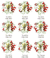
Figure 2. Ribbon representations of the thioredoxin structures studied in this work
General overview of the seven laboratory resurrections of Precambrian thioredoxins and the extant E. coli and human thioredoxins showing the canonical fold. See Figure S1 and Table S2 for energies of charge-charge interactions, accessible surface areas numbers of hydrogen bonds and salt bridges for all the thioredoxin structures studied in this work.
Figure 3
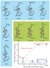
Figure 3. Changes in the size of helix α1 in thioredoxins over ~4 billion years as inferred from laboratory resurrections of Precambian proteins
Canonical α-helix hydrogen bonds are shown in red to highlight the changes in helix length. Different color backgrounds are used for short helices (blue) and long helices (green). A plot of helix length versus geological time is also included. See Table S3 for calculations supporting the robustness of the differences found in alpha helix 1 length.
Figure 4
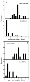
Figure 4. Statistical distribution of length of helix α1 for extant thioredoxin structures taken from the Protein Data Bank
(A) Query details are as follows: text search for “thioredoxin and X-ray as experimental method” was used obtaining a total of 494 structure hits. From these, all thioredoxin-related structures were discarded (i.e. thioredoxin reductases, glutaredoxins, etc); thioredoxins from chloroplast and mitochondria as well as thioredoxins from archaea were not considered either. A total of 39 thioredoxin structures from eukaryota and 32 from bacteria were used in our analysis. Note that in some cases the same protein structure might be overrepresented; this is the case, for instance, when different structures corresponding to mutants of the same protein are deposited in the PDB. (B) Here the search in the PDB was filtered in order to avoid overrepresentation indicated above. In particular, a single thioredoxin structure for each microorganism was selected (i.e., wild type protein) resulting in a total of 14 thioredoxin structures from eukaryota and 15 for bacteria.

References

    1. Adams PD, Afonine PV, Bunkoczi G, Chen VB, Davis IW, Echols N, Headd JJ, Hung LW, Kapral GJ, Grosse-Kunstleve RW, et al. PHENIX: a comprehensive Python-based system for macromolecular structure solution. Acta Crystallographica Section D. 2010;66:213–221. - PMC - PubMed
    1. Benner SA, Sassi SO, Gaucher EA. Molecular paleoscience: systems biology from the past. Advances in enzymology and related areas of molecular biology. 2007;75:1–132. xi. - PubMed
    1. Caetano-Anolles G, Wang M, Caetano-Anolles D, Mittenthal JE. The origin, evolution and structure of the protein world. Biochem J. 2009;417:621–637. - PubMed
    1. Chen VB, Arendall WB, 3rd, Headd JJ, Keedy DA, Immormino RM, Kapral GJ, Murray LW, Richardson JS, Richardson DC. MolProbity: all-atom structure validation for macromolecular crystallography. Acta crystallographica Section D, Biological crystallography. 2010;66:12–21. - PMC - PubMed
    1. Collaborative Computational Project N. The CCP4 suite: programs for protein crystallography. Acta crystallographica Section D, Biological crystallography. 1994;50:760–763. - PubMed

Publication types