. 2013 Nov 7;13(5):590-601.
doi: 10.1016/j.stem.2013.07.016. Epub 2013 Aug 8.

Myf5-positive satellite cells contribute to Pax7-dependent long-term maintenance of adult muscle stem cells

Affiliations

Myf5-positive satellite cells contribute to Pax7-dependent long-term maintenance of adult muscle stem cells

Stefan Günther et al. Cell Stem Cell. .

Erratum in

  • Cell Stem Cell. 2013 Dec 5;13(6):769

Abstract

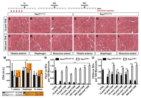
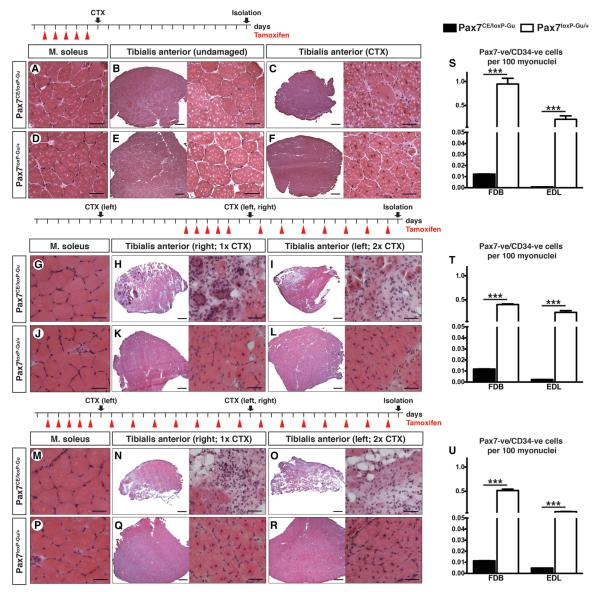
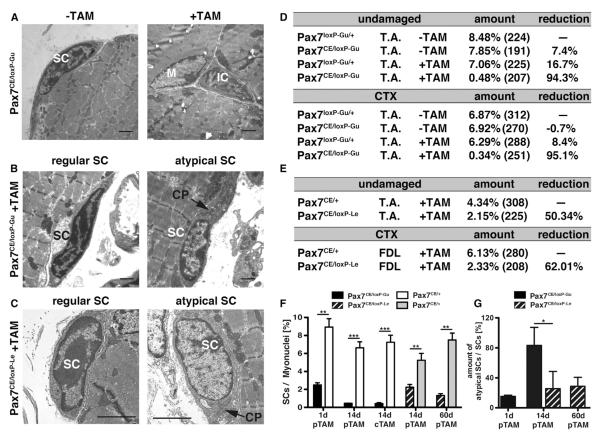
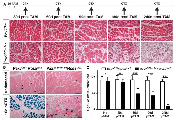
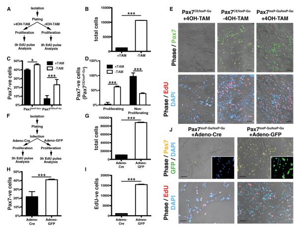
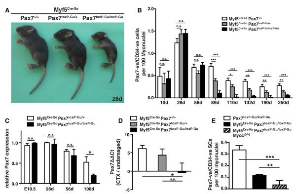
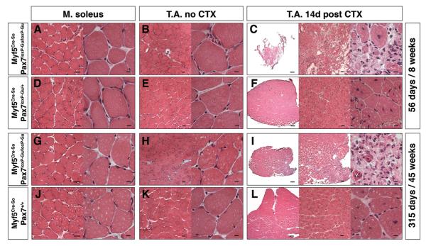
Skeletal muscle contains Pax7-expressing muscle stem or satellite cells, enabling muscle regeneration throughout most of adult life. Here, we demonstrate that induced inactivation of Pax7 in Pax7-expressing cells of adult mice leads to loss of muscle stem cells and reduced heterochromatin condensation in rare surviving satellite cells. Inactivation of Pax7 in Myf5-expressing cells revealed that the majority of adult muscle stem cells originate from myogenic lineages, which express the myogenic regulators Myf5 or MyoD. Likewise, the majority of muscle stem cells are replenished from Myf5-expressing myogenic cells during adult life, and inactivation of Pax7 in Myf5-expressing cells after muscle damage leads to a complete arrest of muscle regeneration. Finally, we demonstrate that a relatively small number of muscle stem cells are sufficient for efficient repair of skeletal muscles. We conclude that Pax7 acts at different levels in a nonhierarchical regulatory network controlling muscle-satellite-cell-mediated muscle regeneration.


Figures

Figure 1
Figure 1. Loss of Muscle Satellite Cells after Postnatal Inactivation of Pax7
Pax7CE/loxP-Gu and Pax7loxP-Gu/+ littermates (n = 3, in each group) were treated with TAM for 5 days. Muscle tissues were analyzed at different time points as indicated. (A–L) Hematoxylin/Eosin (HE) staining of muscle tissue sections from TAM-treated Pax7CE/loxP-Gu and Pax7loxP-Gu/+ mice at 1 day (A–C and G–I) and 2 months (D–F and J–L). (M) Statistical evaluation of cross-sectional areas (CSA) of muscle fibers of TAM-treated Pax7CE/loxP-Gu and Pax7loxP-Gu/+ mice at different time points. (N and O) Analysis of remaining SC numbers. SCs situated on isolated myotubes (n > 150) were identified by being stained with Pax7 (N) and CD34 (O) antibodies. SC numbers were calculated relative to the number of myonuclei (DAPI). Error bars represent standard deviation of the mean (t test: n.s. = not significant; ***p < 0.001; *p < 0.05). Scale bar: (A)–(L) = 20 mm. See also Figures S1 and S2.
Figure 2
Figure 2. Impaired Muscle Regeneration in Adult Mice after Enhanced Inactivation of Pax7
(A–F, G–L, and M–R) Morphological analysis of T.A. muscles of TAM-treated Pax7CE/loxP-Gu and Pax7loxP-Gu/+ mice injected with CTX to induce muscle regeneration. TAM treatment regimens and time points of analysis are indicated at the top of each panel. (A–F) HE staining of T.A. muscles of 12-week-old mice (n = 3) 14 days after CTX injection. Cre-recombinase-mediated inactivation of Pax7 was induced by five consecutive TAM injections. (G–R) HE staining of T.A. muscles of 20-week-old mice (n = 3), which received additional administration of TAM after one (H and K) or two (I and L) rounds of muscle regeneration. TAM was administered before and during the first round of muscle regeneration (G and L) or before and during two rounds of muscle regeneration (M–R). (S–U) Number of Pax7-positive and CD34-positive SC populations on isolated myofibers relative to the number of myonuclei (DAPI). Error bars represent standard deviation of the mean (t test: ***p < 0.001). Scale bar: overview = 200 μm; magnification = 25 μm. See also Figure S3.
Figure 3
Figure 3. Loss of SCs in Adult Mice Differs between Pax7 Conditional Alleles
(A–C) EM microphotographs of ultrathin muscle sections used for quantification of SCs in mice in Pax7CE/loxP-Gu allele (A) and Pax7CE/loxP-Le mice (C). (B and C) The majority of remaining SCs in Pax7CE/loxP-Le mutant mice are localized under the basal lamina as regular SCs but show a strong reduction of heterochromatin (C). Similar atypical stem cells were also present in Pax7CE/loxP-Gu mice but only shortly after Pax7 inactivation (B). (D) Number of SCs relative to myonuclei in undamaged and regenerated T.A. muscles of Pax7CE/loxP-Gu and Pax7loxP-Gu/+ mice 14 days after CTX injection. TAM was administered for 5 days before regeneration in the +TAM group. Numbers in brackets indicate the number of counted myonuclei, e.g., only a single SC was detected in TAM-treated Pax7CE/loxP-Gu mutants. (E) Comparison of the number of SCs 14 days after Pax7 deletion reveals differences in remaining SCs between the Pax7CE/loxP-Gu and the Pax7CE/loxP-Le strains. (F) Number of SCs identified by EM analysis in Pax7CE/loxP-Gu and Pax7CE/loxP-Le strains at different time points. Numbers include atypical SCs. (G) Number of atypical SCs identified by reduced heterochromatin content and abnormal amounts of cytoplasm and organelles. Abbreviations: M, myocyte; SC, satellite cell; IC, interstitial cell; CP, cytoplasm; pTAM, post TAM injection; cTAM, continuous TAM administration. Error bars represent standard deviation of the mean (t test: **p < 0.01; ***p < 0.001). Scale bar: (A)–(C) = 2 μm. See also Figure S4.
Figure 4
Figure 4. Loss of SCs and Impaired Skeletal Muscle Regeneration in Pax7CE/loxP-Le Mice Occurs Late after Cre Recombinase Activation
(A) HE staining of T.A. muscles at different time points after TAM treatment. CTX injections were performed as indicated following 5 days of TAM administration. (B) Representative images of HE and X-gal-stained muscles of mice 240 days after Cre recombinase activation and 10 days after CTX injection. The number of remaining lineage-traced SCs (arrowheads in upper panel) and their contribution to muscle regeneration (lower panel) is massively reduced after Pax7 inactivation. (C) Loss of SCs in Pax7CE/loxP-Le mice becomes apparent only late (60d) after Pax7 inactivation and reached a maximum at 240d. Error bars represent standard deviation of the mean (t test: n.s. = not significant; **p < 0.01; ***p < 0.001). Scale bar: 50 μm.
Figure 5
Figure 5. Impaired Proliferation of Adult SCs after Inactivation of Pax7 in Culture
(A–E) FACS-purified SCs from Pax7CE/loxP-Gu mice were treated in culture with or without 4OH-TAM under high serum concentrations (A). Total number of SCs after TAM-induced deletion of Pax7 (B). Number of Pax7-positive cells (C) and EdU-incorporating Pax7-positive cells (D and E) after addition of TAM. Cells were analyzed after 6d of cultivation. (F–J) Viral-mediated knockout of Pax7 in cultured SCs. FACS-sorted SCs from Pax7loxP-Gu/loxP-Gu mice were infected with Adeno-Cre and Adeno-eGFP viruses 24 hr after isolation to inactivate the Pax7 gene (F). Decreases of the total number of SC-derived cells (G), of Pax7-positive cells (H), and of EdU-incorporating Pax7-positive cells (I and J) 6d after Adeno-Cre mediated deletion of Pax7 are shown. Error bars represent standard deviation of the mean (t test: *p < 0.05; ***p < 0.001). Scale bar: 20 mm. See also Figures S5 and S6.
Figure 6
Figure 6. Loss of SCs and Impairment of Muscle Regeneration after Deletion of Pax7 in Myf5-Expressing Cells
(A) Growth retardation of 28d Myf5Cre-So/Pax7loxP-Gu/loxP-Gu mice compared to Myf5Cre-So/Pax7loxP-Gu/+ and Myf5Cre-So/Pax7+/+ littermates. The growth retardation persisted during postnatal life. (B) Numbers of Pax7-positive/CD34-positive SCs in flexor digitorum brevis (FDB) muscles of Myf5Cre-So/Pax7loxP-Gu/loxP-Gu (n = 3), Myf5Cre-So/Pax7loxP-Gu/+ (n = 3), and Myf5Cre-So/Pax7+/+ (n = 3) at different postnatal stages relative to the total number of myonuclei. A reduction of SCs is evident at 89d and thereafter. (B) Analysis of Pax7 mRNA expression in Myf5Cre-So/Pax7loxP-Gu/loxP-Gu mice during postnatal life in the T.A. (E). (C and D) Analysis of Pax7 mRNA expression in Myf5Cre-So/Pax7loxP-Gu/loxP-Gu mice during postnatal life (D) and after CTX-induced regeneration in the T.A. (E). (E) Quantitative analysis of SC numbers in Myf5Cre-So/Pax7loxP-Gu/loxP-Gu/MyoD(−/−) mice (striped) at an age of 100 days. Loss of MyoD results in a further loss of Pax7-positive cells. Error bars represent standard deviation of the mean (t test: n.s. = not significant; *p < 0.05; **p < 0.01; ***p < 0.001).
Figure 7
Figure 7. Age-Independent Impairment of Muscle Regeneration after Deletion of Pax7 in Myf5-Expressing Cells
Morphological analysis of T.A. muscles of Myf5Cre-So/Pax7loxP-Gu/loxP-Gu and Myf5Cre-So/Pax7loxP-Gu/+ mice of different ages with and without CTX injections to induce muscle regeneration. (A–F) HE staining of T.A. muscles of 8-week-old mice (n = 3) 14 days after CTX injection. (G–L) HE staining of T.A. muscles of 45-week-old mice (n = 3) 14 days after CTX injection. Myf5Cre-So-mediated deletion of Pax7 resulted in massive impairment of myofiber formation and increased fibrosis in regenerating muscles while no morphological abnormalities are apparent in nondamaged muscles. Cross-sections through nondamaged soleus muscles are shown as control (A, D, G, and J). Scale bar: overview = 20 μm; magnification = 5 μm. See also Figure S7.

References

    1. Bonaguidi MA, Wheeler MA, Shapiro JS, Stadel RP, Sun GJ, Ming GL, Song H. In vivo clonal analysis reveals self-renewing and multipotent adult neural stem cell characteristics. Cell. 2011;145:1142–1155. - PMC - PubMed
    1. Braun T, Gautel M. Transcriptional mechanisms regulating skeletal muscle differentiation, growth and homeostasis. Nat. Rev. Mol. Cell Biol. 2011;12:349–361. - PubMed
    1. Bulut-Karslioglu A, Perrera V, Scaranaro M, de la Rosa-Velazquez IA, van de Nobelen S, Shukeir N, Popow J, Gerle B, Opravil S, Pagani M, et al. A transcription factor-based mechanism for mouse heterochromatin formation. Nat. Struct. Mol. Biol. 2012;19:1023–1030. - PubMed
    1. Cerletti M, Jurga S, Witczak CA, Hirshman MF, Shadrach JL, Goodyear LJ, Wagers AJ. Highly efficient, functional engraftment of skeletal muscle stem cells in dystrophic muscles. Cell. 2008;134:37–47. - PMC - PubMed
    1. Cooper RN, Tajbakhsh S, Mouly V, Cossu G, Buckingham M, Butler-Browne GS. In vivo satellite cell activation via Myf5 and MyoD in regenerating mouse skeletal muscle. J. Cell Sci. 1999;112:2895–2901. - PubMed

Publication types

MeSH terms

Substances