Review
. 2013 Nov:37:68-89.
doi: 10.1016/j.preteyeres.2013.07.003. Epub 2013 Aug 9.

Aging is not a disease: distinguishing age-related macular degeneration from aging

Affiliations
Review

Aging is not a disease: distinguishing age-related macular degeneration from aging

Daniel Ardeljan et al. Prog Retin Eye Res. 2013 Nov.

Abstract

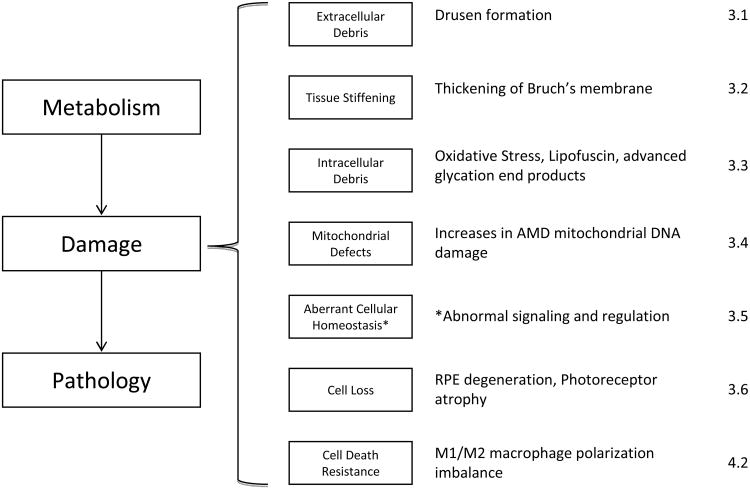
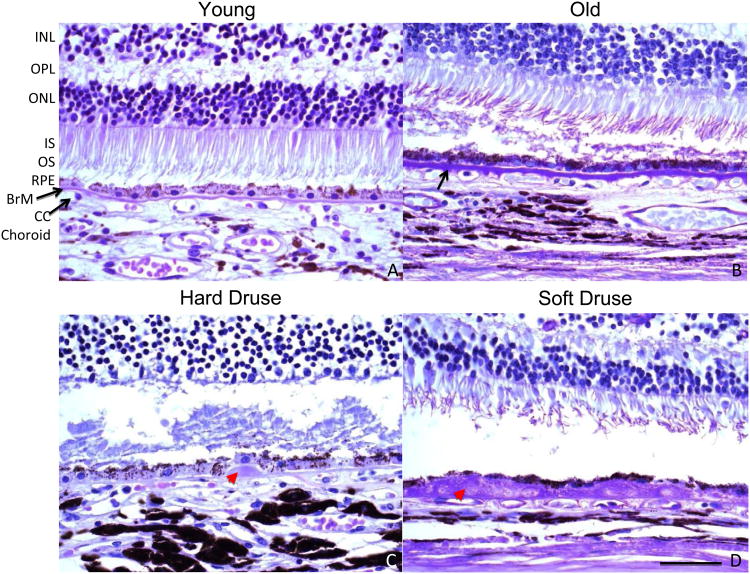
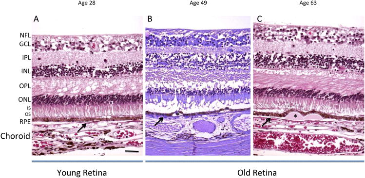
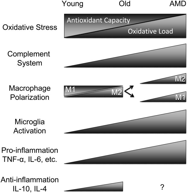
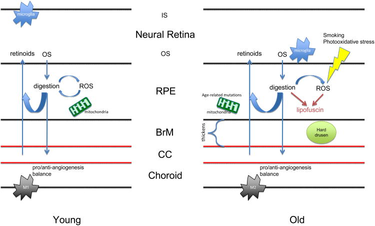
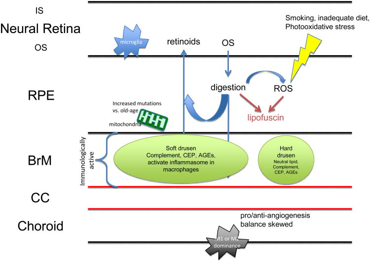
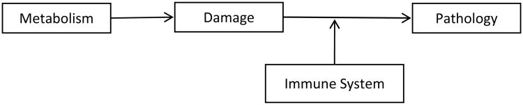
Age-related macular degeneration (AMD) is a disease of the outer retina, characterized most significantly by atrophy of photoreceptors and retinal pigment epithelium accompanied with or without choroidal neovascularization. Development of AMD has been recognized as contingent on environmental and genetic risk factors, the strongest being advanced age. In this review, we highlight pathogenic changes that destabilize ocular homeostasis and promote AMD development. With normal aging, photoreceptors are steadily lost, Bruch's membrane thickens, the choroid thins, and hard drusen may form in the periphery. In AMD, many of these changes are exacerbated in addition to the development of disease-specific factors such as soft macular drusen. Para-inflammation, which can be thought of as an intermediate between basal and robust levels of inflammation, develops within the retina in an attempt to maintain ocular homeostasis, reflected by increased expression of the anti-inflammatory cytokine IL-10 coupled with shifts in macrophage plasticity from the pro-inflammatory M1 to the anti-inflammatory M2 polarization. In AMD, imbalances in the M1 and M2 populations together with activation of retinal microglia are observed and potentially contribute to tissue degeneration. Nonetheless, the retina persists in a state of chronic inflammation and increased expression of certain cytokines and inflammasomes is observed. Since not everyone develops AMD, the vital question to ask is how the body establishes a balance between normal age-related changes and the pathological phenotypes in AMD.

Keywords: Age-related macular degeneration; Aging; Homeostasis; Oxidative stress; Para-inflammation; Retina.


Figures

Figure 1
Figure 1
The aging paradigm in AMD. The metabolism-damage-pathology rubric follows from the work of Zealley and de Grey (2012). The seven damaging events are indicated in the second column and the correlates observed in AMD in the third column. The final column indicates the sections that discuss these events in AMD and aging. The asterisk (*) is meant to distinguish that de Grey's model is slightly modified when applied to AMD since no cell proliferation is observed in AMD.
Figure 2
Figure 2
High magnifications of the outer retina, RPE, Batch's membrane, and choroid. The only visibly significant difference between young (A, 23 years old) and old (B, 65 years old) is the thickening of Batch's membrane with age. In retinas with hard drusen (C, 49 years old), deposits form underneath the RPE monolayer within the inner collagenous layer of Batch's membrane, and decreasing intact CC. In retinas with soft drusen (D, 73 years old), significant displacement of the flattened RPE from the CC is observed, likely contributing to loss of homeostasis in the eye. The appearance of the photoreceptors in C and D is a post-mortem artifact unrelated to the finding of drusen. INL: inner nuclear layer, OPL: outer plexiform layer, ONL: outer nuclear layer, IS/OS: inner/outer segments of photoreceptors, RPE: retinal pigment epithelium, BrM: Batch's membrane. The black arrow indicates Batch's membrane. The red arrowhead indicates drusen. Panels depict PAS staining. Scale bar, 50 μm. Paraffin sections cut at 4-6 μm.
Figure 3
Figure 3
Histological depiction of young and old. In the young image of a 28-year-old retina, the retinal layers are normal (A). The old retinas (49 and 63 years old) show thinning of the ONL and some RPE damage (B, C). In the drusen-containing retina of old, but healthy individuals, retinal layers appear histologically unaffected, except for the observed displacement of the RPE due to the physical presence of the drusen (asterisks). The arrows indicate Bruch's membrane. NFL: nerve fiber layer, GCL: ganglion cell layer, IPL: inner plexiform layer, INL: inner nuclear layer, OPL: outer plexiform layer, ONL: outer nuclear layer, IS/OS: inner/outer segments of photoreceptors, RPE: retinal pigment epithelium. Sections are paraffin at 4-6 μm. Left and Right panels: H&E; Middle panel: Periodic acid-Schiff (PAS). Scale bar, 50 μm. Paraffin sections cut at 4-6 μm.
Figure 4
Figure 4
Immune changes in aging vs. AMD. The aging eye sees shows increased oxidative load and diminished antioxidant capacity, increased immunoreactivity towards complement factors, shifts in macrophage polarization, general increased microglia activity, and upregulation of pro-and anti-inflammatory factors. In AMD, each of these balances is disturbed, evidenced by excessive increases in each of these categories that synergize to launch the system into a disease state. There is a lack of data regarding IL-10 and IL-4 levels in AMD patients with respect to age-matched controls (indicated by “?”).
Figure 5
Figure 5
Homeostasis of the healthy eye. Spatial organization of ocular homeostatic processes. IS: inner segments; OS: outer segments; RPE: retinal pigment epithelium; ROS: reactive oxygen species; BrM: Bruch's membrane; CC: choriocapillaris; Ml: Ml macrophages; VEGF: vascular endothelial growth factor. Hard drusen are primarily composed of neutral lipid and phospholipid (>40%).
Figure 6
Figure 6
Homeostasis of AMD eyes. Spatial organization of AMD-related processes. IS: inner segments; OS: outer segments; RPE: retinal pigment epithelium; ROS: reactive oxygen species; BrM: Bruch's membrane; CC: choriocapillaris; M1: M1 macrophages; M2: M2 macrophages; VEGF: vascular endothelial growth factor; CEP: carboxyethylpyrrole; AGEs: advanced glycation end products.
Figure 7
Figure 7
An AMD disease model. The Zealley and de Grey (2012) model of aging as applied to AMD, where a key aspect features activation of the immune system.

Similar articles

Cited by

References

    1. Ablonczy Z, Dahrouj M, Tang PH, Liu Y, Sambamurti K, Marmorstein AD, Crosson CE. Human retinal pigment epithelium cells as functional models for the RPE in vivo. Invest Ophthalmol Vis Sci. 2011;52:8614–8620. - PMC - PubMed
    1. Ahonen M, Poukkula M, Baker AH, Kashiwagi M, Nagase H, Eriksson JE, Kahari VM. Tissue inhibitor of metalloproteinases-3 induces apoptosis in melanoma cells by stabilization of death receptors. Oncogene. 2003;22:2121–2134. - PubMed
    1. Alsenz J, Schulz TF, Lambris JD, Sim RB, Dierich MP. Structural and functional analysis of the complement component factor H with the use of different enzymes and monoclonal antibodies to factor H. Biochem J. 1985;232:841–850. - PMC - PubMed
    1. Ambati J, Anand A, Fernandez S, Sakurai E, Lynn BC, Kuziel WA, Rollins BJ, Ambati BK. An animal model of age-related macular degeneration in senescent Ccl-2- or Ccr-2-deficient mice. Nat Med. 2003;9:1390–1397. - PubMed
    1. Anderson DH, Radeke MJ, Gallo NB, Chapin EA, Johnson PT, Curletti CR, Hancox LS, Hu J, Ebright JN, Malek G, Hauser MA, Rickman CB, Bok D, Hageman GS, Johnson LV. The pivotal role of the complement system in aging and age-related macular degeneration: hypothesis re-visited. Prog Retin Eye Res. 2010;29:95–112. - PMC - PubMed

Publication types

MeSH terms

Supplementary concepts