. 2013 Nov:249:1-7.
doi: 10.1016/j.expneurol.2013.07.019. Epub 2013 Aug 8.

Adult motor axons preferentially reinnervate predegenerated muscle nerve

Affiliations

Adult motor axons preferentially reinnervate predegenerated muscle nerve

M Abdullah et al. Exp Neurol. 2013 Nov.

Abstract

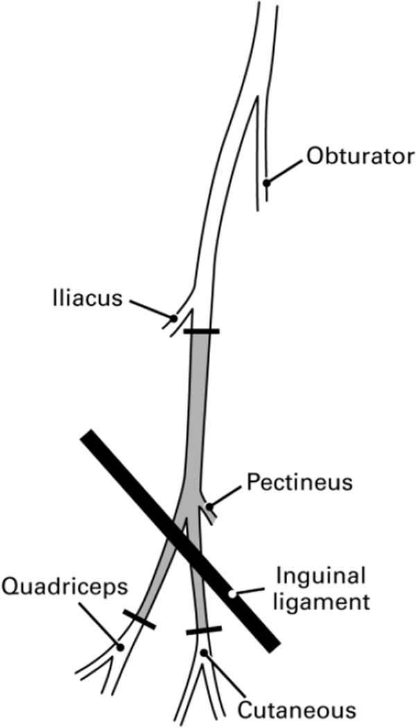
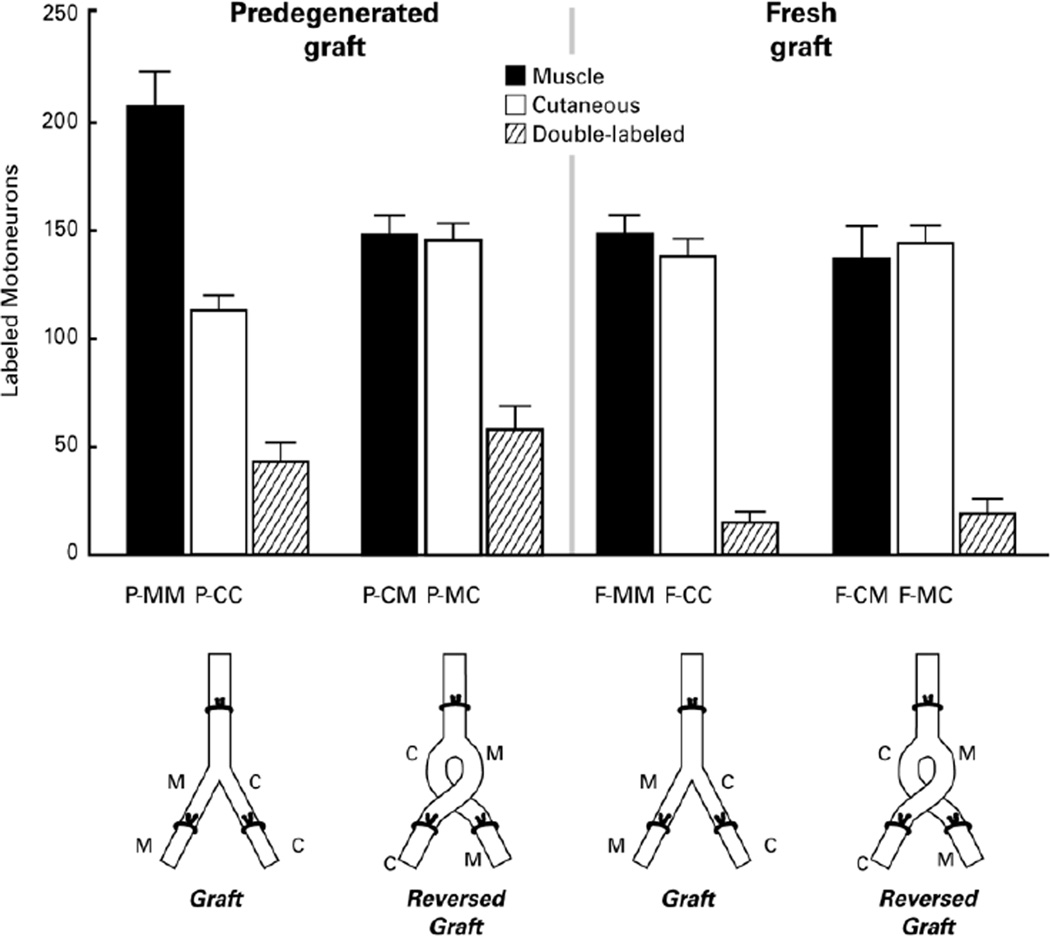
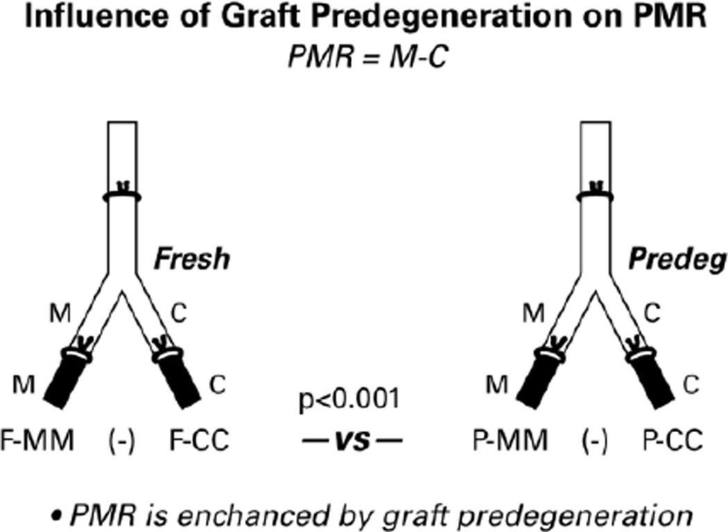
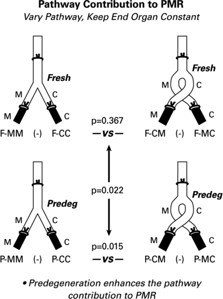
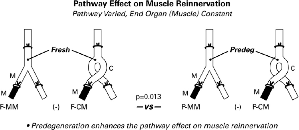
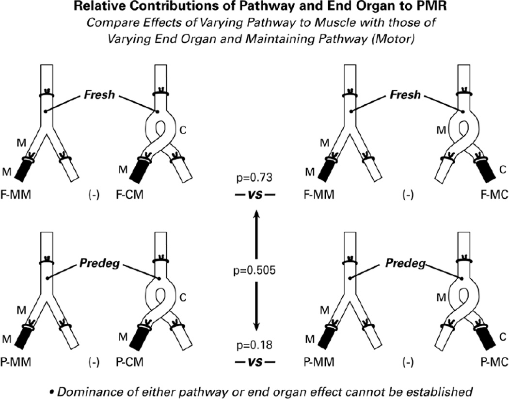
Preferential motor reinnervation (PMR) is the tendency for motor axons regenerating after repair of mixed nerve to reinnervate muscle nerve and/or muscle rather than cutaneous nerve or skin. PMR may occur in response to the peripheral nerve pathway alone in juvenile rats (Brushart, 1993; Redett et al., 2005), yet the ability to identify and respond to specific pathway markers is reportedly lost in adults (Uschold et al., 2007). The experiments reported here evaluate the relative roles of pathway and end organ in the genesis of PMR in adult rats. Fresh and 2-week predegenerated femoral nerve grafts were transferred in correct or reversed alignment to replace the femoral nerves of previously unoperated Lewis rats. After 8 weeks of regeneration the motoneurons projecting through the grafts to recipient femoral cutaneous and muscle branches and their adjacent end organs were identified by retrograde labeling. Motoneuron counts were subjected to Poisson regression analysis to determine the relative roles of pathway and end organ identity in generating PMR. Transfer of fresh grafts did not result in PMR, whereas substantial PMR was observed when predegenerated grafts were used. Similarly, the pathway through which motoneurons reached the muscle had a significant impact on PMR when grafts were predegenerated, but not when they were fresh. Comparison of the relative roles of pathway and end organ in generating PMR revealed that neither could be shown to be more important than the other. These experiments demonstrate unequivocally that adult muscle nerve and cutaneous nerve differ in qualities that can be detected by regenerating adult motoneurons and that can modify their subsequent behavior. They also reveal that two weeks of Wallerian degeneration modify the environment in the graft from one that provides no modality-specific cues for motor neurons to one that actively promotes PMR.

Keywords: Basal lamina; CC; CM; CSPG; F; Fluoro-Gold; MAG; MC; MM; Motoneuron; P; PMR; Peripheral nerve; Preferential motor reinnervation; Rat; Regeneration; Retrograde labeling; Specificity; WGA-ruby; chondroitin sulfate proteoglycans; fresh; graft cutaneous branch joined to recipient cutaneous nerve; graft cutaneous branch joined to recipient muscle branch; graft muscle branch joined to recipient cutaneous nerve; graft muscle nerve branch joined to recipient muscle nerve; myelin-associated glycoprotein; predegenerated; preferential motor reinnervation.


Figures

Figure 1
Figure 1
The right femoral nerve of the rat as seen through the ilio-inguinal approach. Femoral nerve grafts used in these experiments extended from just distal to the iliacus innervation to the distal-most unbranched portions of the femoral motor and sensory tributaries, the portion of the nerve shaded grey. The small pectineus branch was tied off at its origin. Sensory and motor axons intermingle within the nerve at the site of proximal transection. Distally, motor axons segregate into the quadriceps muscle branch, and are not normally found in the cutaneous branch.
Figure 2
Figure 2
The four types of experimental preparation and the regeneration specificity resulting from each. Grafts were either Fresh (F) or Predegenerated for 2 weeks (P), and were either aligned correctly (MM- graft Muscle branch joined to recipient Muscle nerve; CC- graft Cutaneous branch joined to recipient Cutaneous nerve) or reversed (CM- graft Cutaneous nerve to recipient Muscle nerve; MC- graft Muscle nerve to recipient Cutaneous nerve). Vertical bars represent the mean number of motoneurons in each group that correctly reinnervated the muscle branch (black bar), incorrectly reinnervated the cutaneous branch (open bar), or projected axon collaterals to both (striped bar). After 8 weeks of regeneration, PMR was pronounced in the correctly-aligned, predegenerated grafts, but not in other groups.
Figure 3
Figure 3
Diagram of the preparations that were used to determine the influence of pathway predegeneration on specificity generation. Data were obtained by retrograde labeling the distal pathways shown in black. Resulting motoneuron counts were subjected to longitudinal Poisson regression analysis. PMR is the difference between the number of motoneurons correctly reinnervating distal muscle nerve and/or muscle and the number incorrectly reinnervating distal cutaneous nerve and/or skin. PMR was significantly greater (p<0.001) when the pathway had been predegenerated.
Figure 4
Figure 4
To determine the contribution of the graft pathway to PMR, we analyzed the effects of reversing the alignment of the cutaneous and muscle branches of the graft while maintaining the constant alignment of the recipient nerve/end organ components. The pathway did not influence PMR when it was fresh (p=0.367), but influenced PMR significantly when it had been predegenerated for 2 weeks (p=0.15). When comparing these two effects, the pathway contribution to PMR was enhanced significantly by predegeneration (p=0.022).
Figure 5
Figure 5
Evaluation of the pathway effect on muscle reinnervation alone, without reference to motoneurons that project elsewhere. As in other diagrams, the pathways that provide data for the comparisons are solid black. Significantly more motoneurons reinnervated muscle through graft muscle nerve than through graft cutaneous nerve when the graft had been predegenerated (p=0.003), but not when it was fresh (p=0.442). Overall, predegeneration significantly enhanced the pathway effect on muscle reinnervation (p=0.013).
Figure 6
Figure 6
The relative contributions of graft pathway and recipient muscle to PMR were evaluated by comparing the effects of varying the pathway to muscle (on the left) with those of maintaining the muscle nerve pathway and varying the end organ (on the right). A significant difference between pathway and end organ contributions to PMR could not be demonstrated with the current data set.
Figure 7
Figure 7
The evolution of the distal nerve stump. As Wallerian degeneration proceeds, factors that promote regeneration, laminin and growth factors, are upregulated. MAG, an inhibitor of regeneration, is cleared within the first week, and the inhibitory CSPGs are upregulated transiently. The net effect (dotted line) is an initially inhibitory environment that becomes progressively more supportive of regeneration. The relative changes within each factor are derived from experimental data (see DISCUSSION); the relative impact of each factor in relation to the others is hypothetical .

References

    1. Al-Majed AA, Neumann CM, Brushart TM, Gordon T. Brief electrical stimulation promotes the speed and accuracy of motor axonal regeneration. J.Neurosci. 2000;20:2602–2608. - PMC - PubMed
    1. Bertelli JA, Taleb M, Mira JC, Ghizoni MF. Functional recovery improvement is related to aberrant reinnervation trimming. A comparative study using fresh or predegenerated nerve grafts. Acta Neuropathol.(Berl.) 2006;111:601–609. - PubMed
    1. Brushart TM. Preferential reinnervation of motor nerves by regenerating motor axons. J.Neurosci. 1988;8:1026–1031. - PMC - PubMed
    1. Brushart TM. Motor axons preferentially reinnervate motor pathways. J.Neurosci. 1993;13:2730–2738. - PMC - PubMed
    1. Brushart TM, Gerber J, Kessens P, Chen Y-G, Royall R. Contributions of pathway and neuron to preferential motor reinnervation. J. Neurosci. 1998;18:8674–8681. - PMC - PubMed

Publication types