. 2014 Jan 1;23(1):1-11.
doi: 10.1093/hmg/ddt387. Epub 2013 Aug 9.

Depletion of extracellular signal-regulated kinase 1 in mice with cardiomyopathy caused by lamin A/C gene mutation partially prevents pathology before isoenzyme activation

Affiliations

Depletion of extracellular signal-regulated kinase 1 in mice with cardiomyopathy caused by lamin A/C gene mutation partially prevents pathology before isoenzyme activation

Wei Wu et al. Hum Mol Genet. .

Abstract

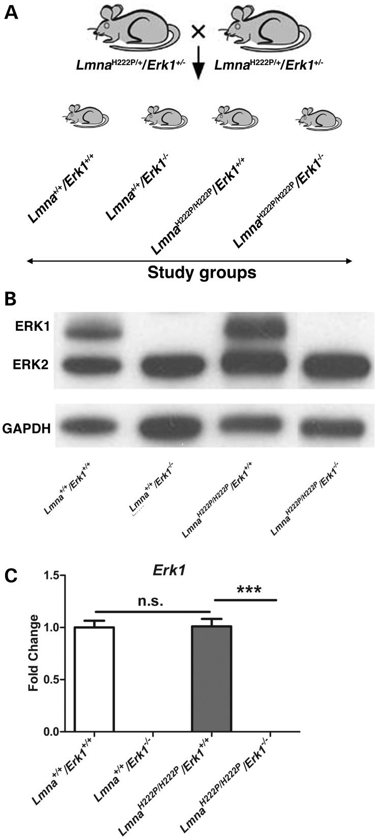
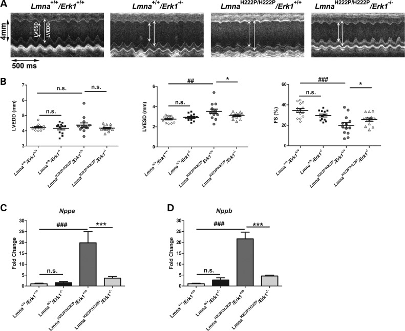
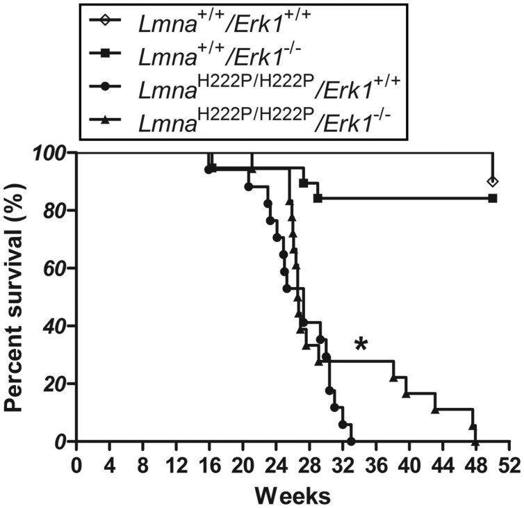
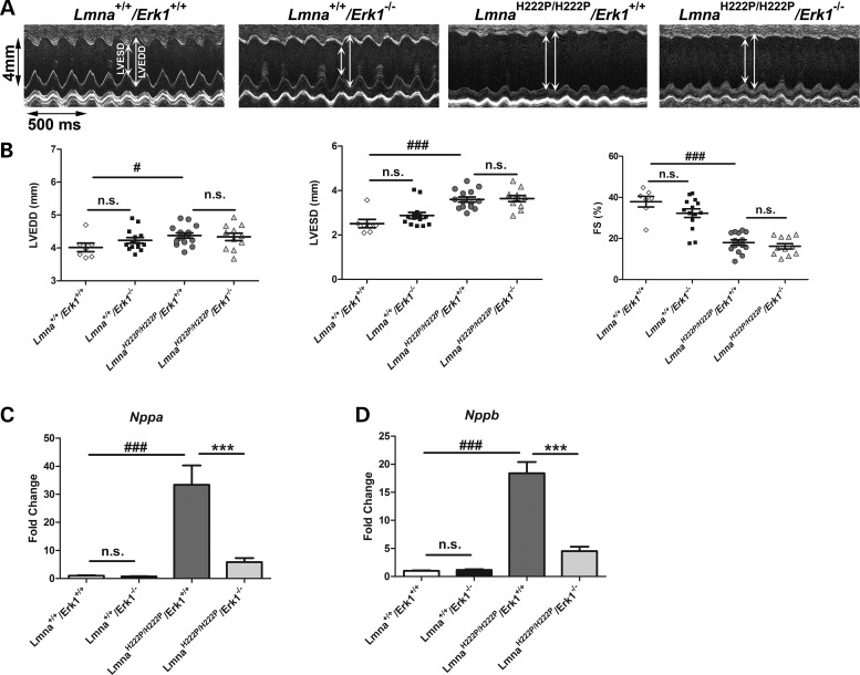
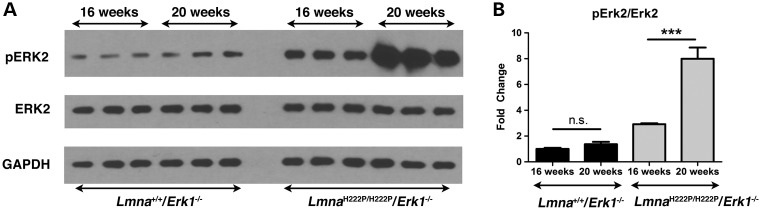
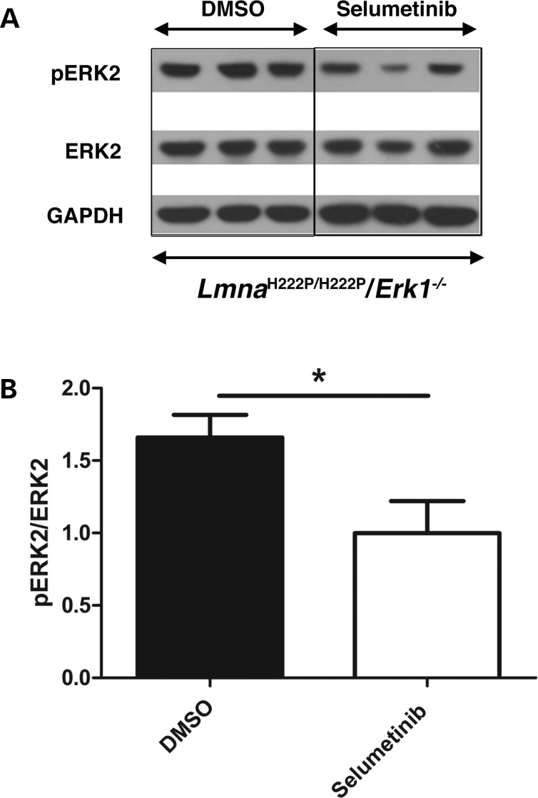
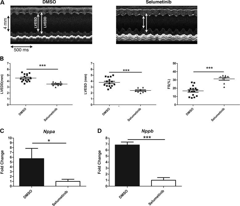
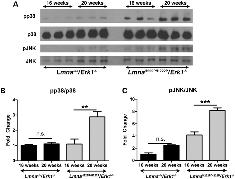
Mutations in the lamin A/C gene (LMNA) encoding A-type nuclear lamins cause dilated cardiomyopathy with variable muscular dystrophy. These mutations enhance mitogen-activated protein kinase signaling in the heart and pharmacological inhibition of extracellular signal-regulated kinase (ERK) 1 and 2 improves cardiac function in Lmna(H222P/H222P) mice. In the current study, we crossed mice lacking ERK1 to Lmna(H222P/H222P) mice and examined cardiac performance and survival. Male Lmna(H222P/H222P)/Erk1(-/-) mice lacking ERK1 had smaller left ventricular end systolic diameters and increased fractional shortening (FS) at 16 weeks of age than Lmna(H222P/H222P/)Erk1(+/+) mice. Their mean survival was also significantly longer. However, the improved cardiac function was abrogated at 20 weeks of age concurrent with an increased activity of ERK2. Lmna(H222P/H222P)/Erk1(-/-) mice treated with an inhibitor of ERK1/2 activation had smaller left ventricular diameters and increased FS at 20 weeks of age. These results provide genetic evidence that ERK1 and ERK2 contribute to the development of cardiomyopathy caused by LMNA mutations and reveal interplay between these isoenzymes in maintaining a combined pathological activity in heart.


Figures

Figure 1.
Figure 1.
Generation of mice lacking Erk1. (A) Schematic diagrams showing sibling mating of LmnaH222P/+/Erk1+/− mice to generate the study groups. (B) Representative immunoblots using antibodies against ERK1 and ERK2 and against GAPDH to probe proteins extracted from hearts of 16-week-old Lmna+/+/Erk1+/+, Lmna+/+/Erk1−/−, LmnaH222P/H222P/Erk1+/+ and LmnaH222P/H222P/Erk1−/− mice. (C) Expression of Erk1 mRNA in hearts from Lmna+/+/Erk1+/+, Lmna+/+/Erk1−/−, LmnaH222P/H222P/Erk1+/+ and LmnaH222P/H222P/Erk1−/− mice. Values represent fold change compared with Erk1 mRNA in hearts from Lmna+/+/Erk1+/+ mice and are given as means ± standard errors in Lmna+/+/Erk1+/+ (n = 6), Lmna+/+/Erk1−/− (n = 6), LmnaH222P/H222P/Erk1+/+ (n = 5) and LmnaH222P/H222P/Erk1−/− (n = 6) mice. ***P < 0.0005; n.s., not significant.
Figure 2.
Figure 2.
Genetic deletion of Erk1 improves left ventricular function in LmnaH222P/H222P mice at 16 weeks of age. (A) Representative M-mode transthoracic echocardiographic tracings from 16-week-old male Lmna+/+/Erk1+/+ (diamonds), Lmna+/+/Erk1−/− (squares), LmnaH222P/H222P/Erk1+/+ (circles) and LmnaH222P/H222P/Erk1−/− (triangles) mice. (B) Graphs showing mean LVEDD, LVESD and FS in 16-week-old male Lmna+/+/Erk1+/+, Lmna+/+/Erk1−/−, LmnaH222P/H222P/Erk1+/+ and LmnaH222P/H222P/Erk1−/− mice. Values for each individual mouse as well as means (long horizontal bars) and standard errors are shown. ##P < 0.005; ###P < 0.0005; *P < 0.05; n.s., not significant. (C) Expression of Nppa mRNA in hearts from 16-week-old Lmna+/+/Erk1+/+, Lmna+/+/Erk1−/−, LmnaH222P/H222P/Erk1+/+ and LmnaH222P/H222P/Erk1−/− mice. Values represent fold change compared with Nppa mRNA in hearts of Lmna+/+/Erk1+/+ mice and are given as means ± standard errors in Lmna+/+/Erk1+/+ (n = 6), Lmna+/+/Erk1−/− (n = 6), LmnaH222P/H222P/Erk1+/+ (n = 5) and LmnaH222P/H222P/Erk1−/− (n = 6) mice. ###P < 0.0005; ***P < 0.0005; n.s., not significant. (D) Expression of Nppb mRNA in hearts from 16-week-old Lmna+/+/Erk1+/+, Lmna+/+/Erk1−/−, LmnaH222P/H222P/Erk1+/+ and LmnaH222P/H222P/Erk1−/− mice. Values represent fold change of Nppb mRNA in hearts of Lmna+/+/Erk1+/+ mice and are given as means ± standard errors in Lmna+/+/Erk1+/+ (n = 6), Lmna+/+/Erk1−/− (n = 6), LmnaH222P/H222P/Erk1+/+ (n = 5) and LmnaH222P/H222P/Erk1−/− (n = 6) mice. ###P < 0.0005; ***P < 0.0005; n.s., not significant.
Figure 3.
Figure 3.
Mouse survival analysis. Kaplan–Meier survival curves for Lmna+/+/Erk1+/+ (n = 10), Lmna+/+/Erk1−/− (n = 19), LmnaH222P/H222P/Erk1+/+ (n = 18) and LmnaH222P/H222P/Erk1−/− (n = 17). *P < 0.05 for difference in mean survival between LmnaH222P/H222P/Erk1+/+ and LmnaH222P/H222P/Erk1−/− mice.
Figure 4.
Figure 4.
Abrogation of improved left ventricular function at 20 weeks of age in LmnaH222P/H222P mice with deletion of Erk1. (A) Representative M-mode transthoracic echocardiographic tracings from 20-week-old male Lmna+/+/Erk1+/+, Lmna+/+/Erk1−/−, LmnaH222P/H222P/Erk1+/+ and LmnaH222P/H222P/Erk1−/− mice. (B) Graphs showing mean LVEDD, LVESD and FS in 20-week-old male Lmna+/+/Erk1+/+ (diamonds), Lmna+/+/Erk1−/− (squares), LmnaH222P/H222P/Erk1+/+ (circles) and LmnaH222P/H222P/Erk1−/− (triangles) mice. Values for each individual mouse as well as means (long horizontal bars) and standard errors are shown. #P < 0.05; ###P < 0.0005; n.s., not significant. (C) Expression of Nppa mRNA in hearts from 20-week-old Lmna+/+/Erk1+/+, Lmna+/+/Erk1−/−, LmnaH222P/H222P/Erk1+/+ and LmnaH222P/H222P/Erk1−/− mice. Values represent fold change compared with Nppa mRNA in hearts of LmnaH222P/H222P/Erk1+/+ mice and are given as means ± standard errors in Lmna+/+/Erk1+/+ (n = 4), Lmna+/+/Erk1−/− (n = 4), LmnaH222P/H222P/Erk1+/+ (n = 3) and LmnaH222P/H222P/Erk1−/− (n = 5) mice. ###P < 0.0005; ***P < 0.0005; n.s., not significant. (D) Expression of Nppb mRNA in hearts from 20-week-old Lmna+/+/Erk1+/+, Lmna+/+/Erk1−/−, LmnaH222P/H222P/Erk1+/+ and LmnaH222P/H222P/Erk1−/− mice. Values represent fold change compared with Nppb mRNA in hearts of LmnaH222P/H222P/Erk1+/+ mice and are given as means ± standard errors in Lmna+/+/Erk1+/+ (n = 4), Lmna+/+/Erk1−/− (n = 4), LmnaH222P/H222P/Erk1+/+ (n = 3) and LmnaH222P/H222P/Erk1−/− (n = 5) mice. ###P < 0.0005; ***P < 0.0005; n.s., not significant.
Figure 5.
Figure 5.
Increased activation of ERK2 in hearts of 20-week-old LmnaH222P/H222P mice lacking Erk1. (A) Representative immunoblots using antibodies against phosphorylated ERK2, total ERK2 and GAPDH to probe proteins extracted from hearts of 16-week-old and 20-week-old Lmna+/+/Erk1−/− and LmnaH222P/H222P/Erk1−/− mice. (B) Bar graphs showing quantification of pERK2/ERK2 in hearts of 16-week-old and 20-week-old Lmna+/+/Erk1−/− and LmnaH222P/H222P/Erk1−/− mice. Values are means ± standard errors (n = 3). ***P < 0.0005; n.s., not significant.
Figure 6.
Figure 6.
Treatment with selumetinib blocks ERK2 phosphorylation in hearts of LmnaH222P/H222P mice lacking Erk1. (A) Representative immunoblots using antibodies against phosphorylated ERK2, ERK2 and GAPDH to probe proteins extracted from hearts of 20-week-old LmnaH222P/H222P/Erk1−/− mice treated with DMSO placebo or selumetinib. (B) Bar graphs showing quantification of pERK2/ERK2 in hearts 20-week-old LmnaH222P/H222P/Erk1−/− mice treated with DMSO placebo or selumetinib. Values are means ± standard errors (n = 3). *P < 0.05.
Figure 7.
Figure 7.
Treatment with selumetinib improves cardiac function in LmnaH222P/H222P mice lacking Erk1. (A) Representative M-mode transthoracic echocardiographic tracings from 20-week-old male LmnaH222P/H222P/Erk1−/− mice treated with DMSO placebo or selumetinib. (B) Graphs showing mean LVEDD, LVESD and FS in 20-week-old male LmnaH222P/H222P/Erk1−/− mice treated with DMSO placebo or selumetinib. Values for each individual mouse (squares and triangles) as well as means (long horizontal bars) and standard errors are shown. ***P < 0.0005. (C) Expression of Nppa mRNA in hearts from 20-week-old LmnaH222P/H222P/Erk1−/− mice treated with DMSO placebo or selumetinib. Values represent fold change compared with Nppa mRNA in hearts of selumetinib-treated LmnaH222P/H222P/Erk−/− mice and are given as means ± standard errors (n = 4). ***P < 0.0005. (D) Expression of Nppb mRNA in hearts from 20-week-old LmnaH222P/H222P/Erk1−/− mice treated with DMSO placebo or selumetinib. Values represent fold change compared with Nppb mRNA in hearts of selumetinib-treated LmnaH222P/H222P/Erk−/− mice and are given as means ± standard errors (n = 4). ***P < 0.0005.
Figure 8.
Figure 8.
Increased activation p38α and JNK in hearts of 20-week-old LmnaH222P/H222P mice lacking Erk1. (A) Representative immunoblots using antibodies against phosphorylated p38α (pp38), total p38α (p38), phosphorylated JNK (pJNK) and total JNK (JNK) to probe proteins extracted from hearts of 16-week-old and 20-week-old Lmna+/+/Erk1−/− and LmnaH222P/H222P/Erk1−/− mice. (B) Bar graphs showing quantification of the ratio of phosphorylated p38α to total p38α (pp38/p38) in hearts of 16-week-old and 20-week-old Lmna+/+/Erk1−/− and LmnaH222P/H222P/Erk1−/− mice. Values are means ± standard errors (n = 3). **P < 0.0005; n.s., not significant. (C) Bar graphs showing quantification of the ratio of phosphorylated JNK to total JNK (pJNK/JNK) in hearts of 16-week-old and 20-week-old Lmna+/+/Erk1−/− and LmnaH222P/H222P/Erk1−/− mice. Values are means ± standard errors (n = 3). ***P < 0.0005; n.s., not significant.

References

    1. Dauer W.T., Worman H.J. The nuclear envelope as a signaling node in development and disease. Dev. Cell. 2009;17:626–638. - PubMed
    1. Worman H.J., Fong L.G., Muchir A., Young S.G. Laminopathies and the long strange trip from basic cell biology to therapy. J. Clin. Invest. 2009;119:1825–1836. - PMC - PubMed
    1. Lu J., Muchir A., Nagy P.L., Worman H.J. LMNA cardiomyopathy: cell biology and genetics meet clinical medicine. Dis. Model. Mech. 2011;4:562–568. - PMC - PubMed
    1. Bonne G., Di Barletta M.R., Varnous S., Bécane H.M., Hammouda E.H., Merlini L., Muntoni F., Greenberg C.R., Gary F., Urtizberea J.A., et al. Mutations in the gene encoding lamin A/C cause autosomal dominant Emery–Dreifuss muscular dystrophy. Nat. Genet. 1999;21:285–288. - PubMed
    1. Fatkin D., MacRae C., Sasaki T., Wolff M.R., Porcu M., Frenneaux M., Atherton J., Vidaillet H.J., Spudich S., De Girolami U., et al. Missense mutations in the rod domain of the lamin A/C gene as causes of dilated cardiomyopathy and conduction-system disease. N. Engl. J. Med. 1999;341:1715–1724. - PubMed

Publication types

MeSH terms