. 2013 Dec 20;22(25):5229-36.
doi: 10.1093/hmg/ddt380. Epub 2013 Aug 9.

Congenital myopathy is caused by mutation of HACD1

Affiliations

Congenital myopathy is caused by mutation of HACD1

Emad Muhammad et al. Hum Mol Genet. .

Abstract

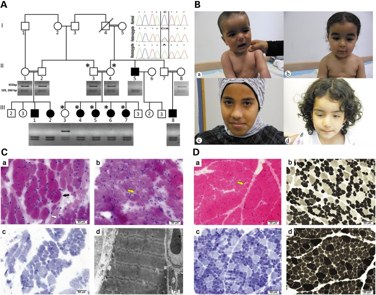
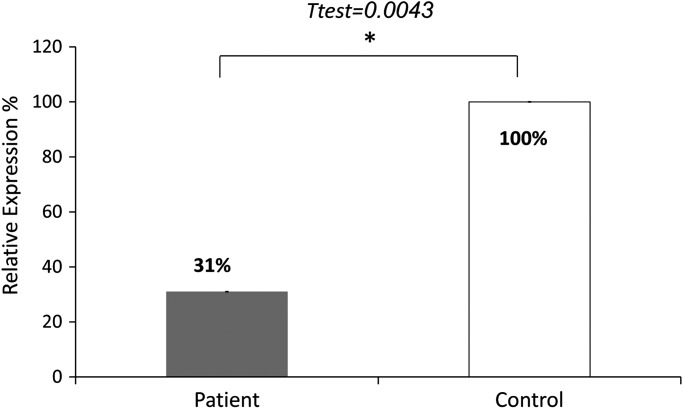
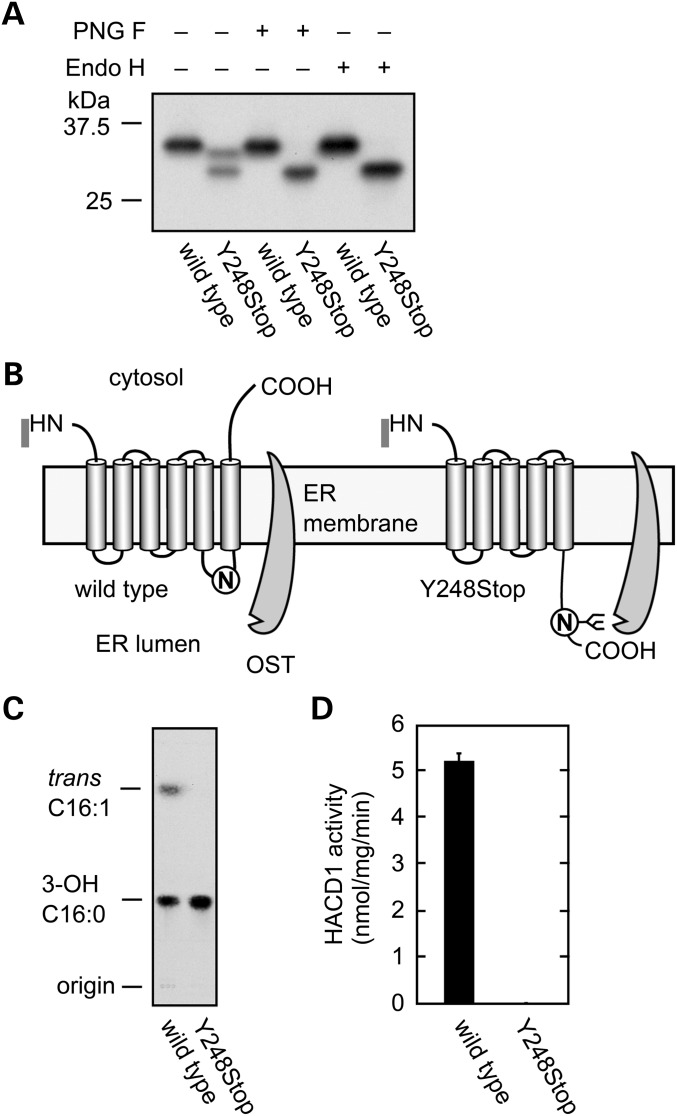
Congenital myopathies are heterogeneous inherited diseases of muscle characterized by a range of distinctive histologic abnormalities. We have studied a consanguineous family with congenital myopathy. Genome-wide linkage analysis and whole-exome sequencing identified a homozygous non-sense mutation in 3-hydroxyacyl-CoA dehydratase 1 (HACD1) in affected individuals. The mutation results in non-sense mediated decay of the HACD1 mRNA to 31% of control levels in patient muscle and completely abrogates the enzymatic activity of dehydration of 3-hydroxyacyl-CoA, the third step in the elongation of very long-chain fatty acids (VLCFAs). We describe clinical findings correlated with a deleterious mutation in a gene not previously known to be associated with congenital myopathy in humans. We suggest that the mutation in the HACD1 gene causes a reduction in the synthesis of VLCFAs, which are components of membrane lipids and participants in physiological processes, leading to congenital myopathy. These data indicate that HACD1 is necessary for muscle function.


Figures

Figure 1.
Figure 1.
The myopathy family: genetics, clinical histolopathological and EM findings. (A). Segregation of the HACD1 mutation in the pedigree. Digestion of the 455 bp amplicon of exon 6 with SspI containing the sequence variation NM_014241:c.744C > A results in cleavage into 195 and 260 bp fragments. Inset: sequence of the corresponding c.744C > A mutation resulting in p.Tyr248Stop. Patients were homozygous for the mutation; parents and the healthy sibling were heterozygous, and a healthy control is homozygous for the normal sequence. The genotyped individuals are marked by ‘*’. (B) Photographs of patients: (a) Patient III-1 at the age of 8 months sitting with support. Note facial weakness and dropping shoulders. (b) Patient III-2 at age 1 and 8 months. Note facial weakness, drooping shoulders and pectus excavatus. (c) and (d) correlate to Patients III-5 and III-6, ages 14 and 3 years, respectively. Note facial weakness and ptosis of the right eye in the latter. Permissions from guardians were granted for all shown photographs. (C) Histology and EM of core needle biopsy: (a)Frozen sections from Patient III-8 at the age of 2 years stained with H&E display focal variation in myofiber diameter (black arrow). Large, hypertrophic myofibers (most of them 35–40 μm in diameter) are scattered among smaller myofibers (normal diameter for age (∼20 μm) and small for age (∼13–16 μm in diameter)), occasionaly in small groups (white arrow). (b) Only isolated internally (centrally) displaced nuclei are seen (yellow arrow). (c) On NADH histochemical stain most hypertrophic myofibers are type 2, while most small fibers are type 1. There are no significant changes in the cytoarchitecture. (d) Electron microscopy is unremarkable. (D) Histology of open biopsy: (a) paraffin embedded and frozen sections from Patient III-5 at age 1 year stained with H&E display marked variation in myofiber diameter. In many areas, hypertrophic myofibers (most of them 20–30 μm in diameter) are scattered among smaller myofibers (normal diameter for age ∼18 μm in diameter and small for age ∼10–15 μm in diameter), occasionally in small groups. Only isolated internally (centrally) displaced nuclei are seen (yellow arrow). (b–d) On enzyme-histochemical staining (b, NADH; c, ATPase 4.3; d, ATPase 9.4), most scattered hypertrophic myofibers are type 2, while most (>90%) small fibers are type 1. There is also a relative increase of type 1 myofibers (∼80%).
Figure 2.
Figure 2.
Quantitative PCR of HACD1 cDNA in a frozen sample biopsy of a patient compared to controls. The results represent three experiments each done in duplicates. The qPCR data was analyzed with the ABI 7500 Software V2.0.3 (ΔCt method; normalization against GAPDH). Values are expressed as relative expression to the control muscle, means ± s.e.m. (not visible). Difference between groups was determined by the two-tailed Student's t-test (P = 0.0043). The HACD1 primers yielded a linear standard curve with an R2 = 0.99.
Figure 3.
Figure 3.
HACD1 Y248Stop exhibits no activity. (A). Production of 3xFLAG-tagged wild-type and mutant (Y248Stop) HACD1 proteins in HEK 293 T following purification using anti-FLAG M2 agarose. Proteins were incubated with buffer, peptide:N-glycosidase F (PNG F), or endoglycosidase H (Endo H) and detected by immunobloting with anti-FLAG antibodies. (B). Model for glycosylation of HACD1 (Y248Stop). OST: oligosaccharyltransferase. (C). Protein samples were incubated for 10 min at 37°C with [14C]3-hydroxypalmitoyl-CoA. After termination of the reactions, lipids were saponified, acidified, extracted and separated by TLC. 3-OH 16:0, 3-hydroxypalmitic acid; trans C16:1, 2,3-trans-hexadecenoic acid. (D). The radioactivity associated with the reaction product 2,3-trans-hexadecenoic acid quantified using bioimaging analyzer BAS-2500 (Fuji Photo Film, Tokyo, Japan) and represent the mean (± SD) from three independent experiments.

References

    1. Nance J.R., Dowling J.J., Gibbs E.M., Bönnemann C.G. Congenital myopathies: an update. Curr. Neurol. Neurosci. Rep. 2011;12:165–174. doi:10.1007/s11910-012-0255-x. - DOI - PMC - PubMed
    1. Kaplan J.C. The 2012 version of the gene table of monogenic neuromuscular disorders. Neuromuscul. Disord. 2011;21:833–861. doi:10.1016/j.nmd.2011.10.008. - DOI - PubMed
    1. Iannaccone S.T., Bove K.E., Vogler C., Azzarelli B., Muller J. Muscle maturation delay in infantile myotonic dystrophy. Arch. Pathol. Lab. Med. 1986;110:405–411. - PubMed
    1. Pelé M., Tiret L., Kessler J.L., Blot S., Panthier J.J. SINE Exonic insertion in the PTPLA gene leads to multiple splicing defects and segregates with the autosomal recessive centronuclear myopathy in dogs. Hum. Mol. Genet. 2005;14:1417–1427. doi:10.1093/hmg/ddi151. - DOI - PubMed
    1. Maquat L.E. Nonsense-mediated mRNA decay: splicing, translation and mRNP dynamics. Nat. Rev. Mol. Cell Biol. 2004;5:89–99. doi:10.1038/nrm1310. - DOI - PubMed

Publication types

MeSH terms