. 2013 Sep;16(9):1299-305.
doi: 10.1038/nn.3486. Epub 2013 Aug 11.

Suppression of eIF2α kinases alleviates Alzheimer's disease-related plasticity and memory deficits

Affiliations

Suppression of eIF2α kinases alleviates Alzheimer's disease-related plasticity and memory deficits

Tao Ma et al. Nat Neurosci. 2013 Sep.

Abstract

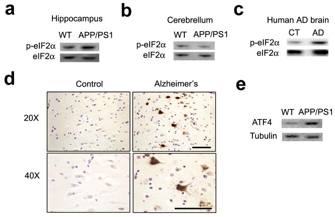
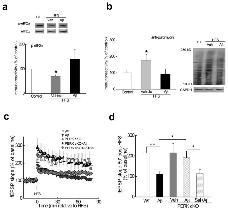
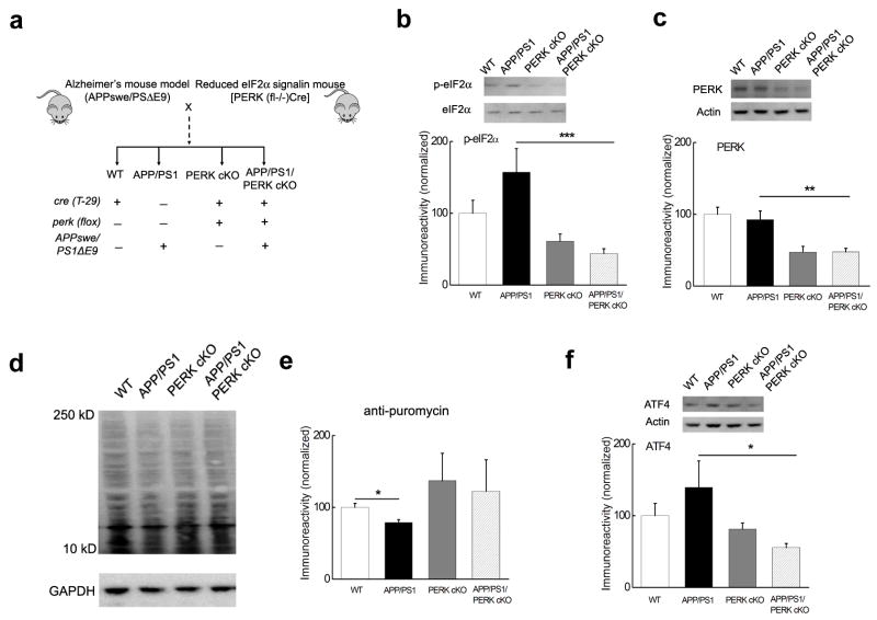
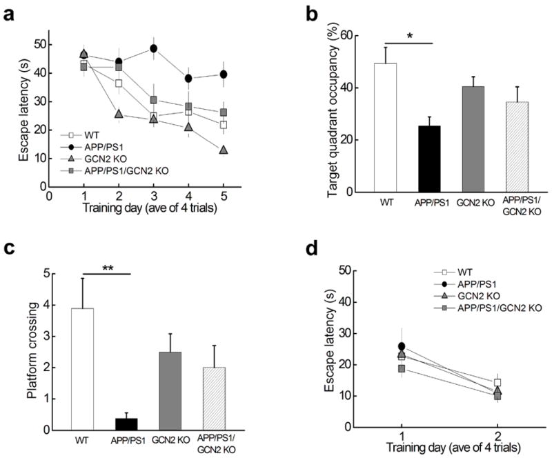
Expression of long-lasting synaptic plasticity and long-term memory requires protein synthesis, which can be repressed by phosphorylation of eukaryotic initiation factor 2 α-subunit (eIF2α). Elevated phosphorylation of eIF2α has been observed in the brains of Alzheimer's disease patients and Alzheimer's disease model mice. Therefore, we tested whether suppressing eIF2α kinases could alleviate synaptic plasticity and memory deficits in Alzheimer's disease model mice. Genetic deletion of eIF2α kinase PERK prevented enhanced phosphorylation of eIF2α and deficits in protein synthesis, synaptic plasticity and spatial memory in mice that express familial Alzheimer's disease-related mutations in APP and PSEN1. Similarly, deletion of another eIF2α kinase, GCN2, prevented impairments of synaptic plasticity and defects in spatial memory exhibited by the Alzheimer's disease model mice. Our findings implicate aberrant eIF2α phosphorylation as a previously unidentified molecular mechanism underlying Alzheimer's disease-related synaptic pathophysioloy and memory dysfunction and suggest that PERK and GCN2 are potential therapeutic targets for treatment of individuals with Alzheimer's disease.


Figures

Figure 1
Figure 1. Increased eIF2α phosphorylation in Alzheimer’s disease
(a) eIF2α phosphorylation was increased in the hippocampus of APP/PS1 AD model mice. n=7 for both groups. p=0.041.(b) eIF2α phosphorylation was not altered in cerebellum of APP/PS1 AD model mice. n=11 for WT and n=10 for APP/PS1. p=0.080. (c) eIF2α phosphorylation was elevated in postmortem human AD brain samples. n=4 for AD and age-matched control groups. p=0.012. (d) DAB staining revealed increased eIF2α phosphorylation in the hippocampus of postmortem human AD brains, representative of three independent experiments. Scale bar, 150 μm. (e) ATF4 levels were increased in the hippocampus of APP/PS1 mice compared to WT littermates. n=6 for each group. p=0.044 Full-length blots/gels are presented in Supplementary Figure 7.
Figure 2
Figure 2. Aβ-induced impairment in LTP is alleviated by deleting the eIF2α kinase PERK
(a) LTP-inducing stimulation decreased the phosphorylation of eIF2α (middle lane), which was reversed by Aβ(1–42) (right lane). Slices were harvested 30 minutes post-HFS and area CA1 was microdissected for Western blot analysis. n=8. *p<0.05. (b) De novo protein synthesis (assayed by SUnSET). LTP-inducing stimulation increased de novo protein synthesis, which was blunted by Aβ(1–42). Slices were harvested 30 minutes post-HFS and area CA1 was microdissected. n=4. *p<0.05. (c) Treatment of hippocampal slices from WT mice with 500 nM Aβ(1–42) resulted in impaired LTP (grey triangles, n=8) compared with LTP in vehicle-treated WT slices (open squares, n=7). In contrast, LTP was induced in slices from PERK cKO mice in the presence of Aβ(1–42) (half-filled triangles, n=6), which was blunted by application of 10 μM Sal003 (Sal, grey diamonds, n=7). In addition, HFS induced LTP in PERK cKO mice that was comparable to that in WT littermates (grey circles, n=7). (d) Cumulative data showing the mean fEPSP slope 80 min post-HFS from the LTP experiments in panel c. *p<0.05; **p<0.01. Full-length blots/gels are presented in Supplementary Figure 7.
Figure 3
Figure 3. Generation of AD model mice with reduced PERK-eIF2α signaling
(a) Diagram depicting the creation of mice with AD-associated transgenes and reduced PERK/eIF2α signaling. (b) eIF2α phosphorylation was reduced in hippocampal area CA1 of APP/PS1/PERK cKO mice compared to the increased levels of eIF2α phosphosphorylation in APP/PS1 mice, which was correlated with the expression of PERK (c). n=10 for APP/PS1/PERK cKO, n=6 the other three groups. (d) Representative Western blot showing that de novo protein synthesis (assayed by SUnSET) was reduced in APP/PS1 mice compared to WT littermates. In addition, de novo protein synthesis in PERK cKO and APP/PERK cKO mice was not different from WT mice. (e) Cumulative data showing densitometric analysis of experiments in panel d. n=4. *p<0.05. (f) Elevated levels of ATF4 in APP/PS1 mice were reduced to WT levels in APP/PS1/PERK cKO mice. Western blots were performed on tissue from area CA1 of the hippocampus. n=10. All data for the densitometric analysis of the Western blots were presented as mean ± SEM. *p< 0.05, **p< 0.01, ***p<0.001. Full-length blots/gels are presented in Supplementary Figure 7.
Figure 4
Figure 4. Spatial memory deficits in APP/PS1 AD model mice are alleviated by suppressing PERK/eIF2α signaling
(a) Escape latency in the Morris water maze plotted against the training days. WT n=14; APP/PS1, n=14; PERK cKO, n=16; APP/PS1/PERK cKO n=11. Repeated measures ANOVA followed by a post hoc Bonferroni multiple comparison test, p= 0.0028, F(3, 54)=8.440. APP/PS1 vs WT: p<0.01, t=4.815; APP/PS1 vs APP/PS1/PERK cKO: p<0.05, t=3.671. APP/PS1 vs PERK cKO: p>0.05, t=2.806. No difference was detected among WT, PERK cKO, and APP/PS1/PERK cKO groups. Repeated measures ANOVA. P=0.09, F(2,40)=3.302. (b) Percentage of time spent in the target quadrant during a 60 second probe trial of MWM test. One-way ANOVA, p = 0.0498, F=2.869. *p< 0.05. (c) Frequency of platform crossing during a 60 second probe trial of MWM test. One-way ANOVA, p =0.0109, F=4.171.*p< 0.05. (d) Visible platform test. Repeated measures ANOVA, p= 0.0943. F=5.653. (e) Object location task. Percentage of time interacting with the object at a new location (out of total time spent with objects) was calculated as discrimination ratio. WT, n=14; APP/PS1, n=10; PERK cKO, n=8; APP/PS1/PERK cKO, n=7. Independent t-test. *p< 0.05 (f) Y water maze task. Spatial memory was measured by percentage of correct arm choice. WT, n=18; APP/PS1, n=18; PERK cKO, n=15; APP/PS1/PERK cKO, Independent t-test. n=9. *p<0.05.
Figure 5
Figure 5. LTP impairments in APP/PS1 mice are rescued by decreasing PERK/eIF2α signaling
(a) LTP was inhibited in APP/PS1 mice (n=6) compared to LTP in WT mice (n=9). LTP was sustained in APP/PS1/PERK cKO mice (n=4) and PERK cKO mice (n=8). (b) Cumulative data showing mean fEPSP slopes 80 min post-HFS from the LTP experiments in panel a. Data were presented as mean +SEM. (c) LTP in APP/PS1/PERK cKO mice (n=7) was inhibited by anisomycin (40 μM). n=8 for WT, n=5 for anisomycin/WT, and n=7 for anisomycin/APP/PS1. Anisomycin was applied into recording chamber 30 min before HFS and present throughout the experiments. (d) Brain Aβ levels were decreased in APP/PS1/PERK cKO mice compared to APP/PS1 mice. n=6 for each group. Levels of full-length APP were not changed. n=9 for APP/PS1/PERK cKO group and n=6 for each of other three groups. (e) β-CTF, but not α-CTF was reduced in the hippocampus of APP/PS1/PERK cKO mice, compared with APP/PS1 mice. n=6. (f) Neprilysin expression was reduced in APP/PS1 mice and was corrected in APP/PS1/PERK cKO mice. n=7 for each group. (g, h) Representative blots (g) and cumulative data of densitometric analysis (h) showing that levels of activity-regulated cytoskeleton-associated protein (Arc, n=5), protein kinase M zeta (PKMζ, n=11), and synaptophysin (n=5) were reduced in hippocampal area CA1 of APP/PS1 mice and was corrected in APP/PS1/PERK cKO mice. Levels of calcium/calmodulin-dependent protein kinase II (CaMKII, n=5) and AMPA receptor subunit GluA1 (n=4) were not significantly altered. *p<0.05; **p<0.01. Full-length blots/gels are presented in Supplementary Figure 7.
Figure 6
Figure 6. Removal of GCN2 reverses AD-associated LTP failure
(a) Normal LTP was induced in slices from GCN2 KO mice in the presence of Aβ(1–42) (half-filled triangles, n=7), which was blunted by application of 10 μM Sal003 (Sal, grey diamonds, n=7). In contrast, slices of WT littermates treated with Aβ(1–42) (grey circles, n=5) exhibited impaired LTP. In addition, HFS induced hippocampal LTP in GCN2 KO mice that was enhanced (grey triangles, n=5) compared to WT littermates (open squares, n=5). (b) Cumulative data showing mean fEPSP slopes 80 min post-HFS from the LTP experiments in panel a. (c) HFS-induced hippocampal LTP was inhibited in APP/PS1 mice (light grey triangles, n=6) compared to LTP in WT mice (open squares, n=8). In contrast, hippocampal LTP was sustained in APP/PS1/GCN2 KO mice (half-filled triangles, n=5) and GCN2 KO mice (grey circles, n=6). (c) Representative fEPSP traces for data shown in panel b. (d) Cumulative data showing mean fEPSP slopes 80 min post-HFS from the LTP experiments in panel b. Data were presented as mean ± SEM. *p<0.05.
Figure 7
Figure 7. Spatial memory deficits in APP/PS1 AD model mice are alleviated by deleting eIF2α kinase GCN2
(a) Escape latency in the Morris water maze plotted against the training days. APP/PS1 mice (black circles, n=8) were slower to learn than WT mice (open squares, n=9), APP/PS1/GCN2 KO (grey squares, n=9) or GCN2 KO mice (grey triangles, n=12). Repeated measures ANOVA followed by a post hoc Bonferroni multiple comparison test, p= 0.0014, F(3, 37)=9.960. APP/PS1 vs WT: p<0.01, t=4.532; APP/PS1 vs APP/PS1/GCN2 KO: p<0.05, t=3.360; APP/PS1/GCN2 KO vs WT: p>0.05, t=1.172. (b) Percentage of time spent in the target quadrant during a 60 second probe trial of MWM test. One-way ANOVA, p = 0.0261, F=3.584. (c) Frequency of platform crossing during a 60 second probe trial of MWM test. One-way ANOVA, p = 0.0144, F=4.057. Number of platform crossing of APP/PS1/GCN2 KO mice is not significantly different from that of WT mice; APP/PS1 vs APP/PS1/GCN2 KO: p=0.0526. **p< 0.01. (d) In the visible platform test no difference was observed for escape latency among the four genotypes of mice. Repeated measures ANOVA, p=0.3735. F=1.500.

Comment in

References

    1. Holtzman DM, Morris JC, Goate AM. Alzheimer’s disease: the challenge of the second century. Sci Transl Med. 2011;3:77sr71. - PMC - PubMed
    1. Querfurth HW, LaFerla FM. Alzheimer’s disease. N Engl J Med. 2010;362:329–344. - PubMed
    1. Selkoe DJ. Resolving controversies on the path to Alzheimer’s therapeutics. Nat Med. 2011;17:1060–1065. - PubMed
    1. Haass C, Selkoe DJ. Soluble protein oligomers in neurodegeneration: lessons from the Alzheimer’s amyloid beta-peptide. Nat Rev Mol Cell Biol. 2007;8:101–112. - PubMed
    1. Costa-Mattioli M, Sossin WS, Klann E, Sonenberg N. Translational control of long-lasting synaptic plasticity and memory. Neuron. 2009;61:10–26. - PMC - PubMed

Publication types

MeSH terms

LinkOut - more resources