. 2013 Sep;16(9):1257-65.
doi: 10.1038/nn.3489. Epub 2013 Aug 11.

The Parkinson's disease-linked proteins Fbxo7 and Parkin interact to mediate mitophagy

Affiliations

The Parkinson's disease-linked proteins Fbxo7 and Parkin interact to mediate mitophagy

Victoria S Burchell et al. Nat Neurosci. 2013 Sep.

Abstract

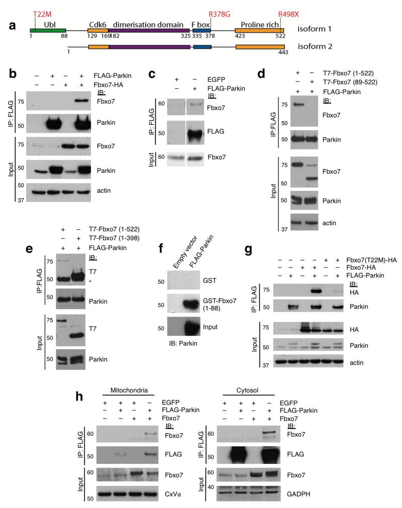
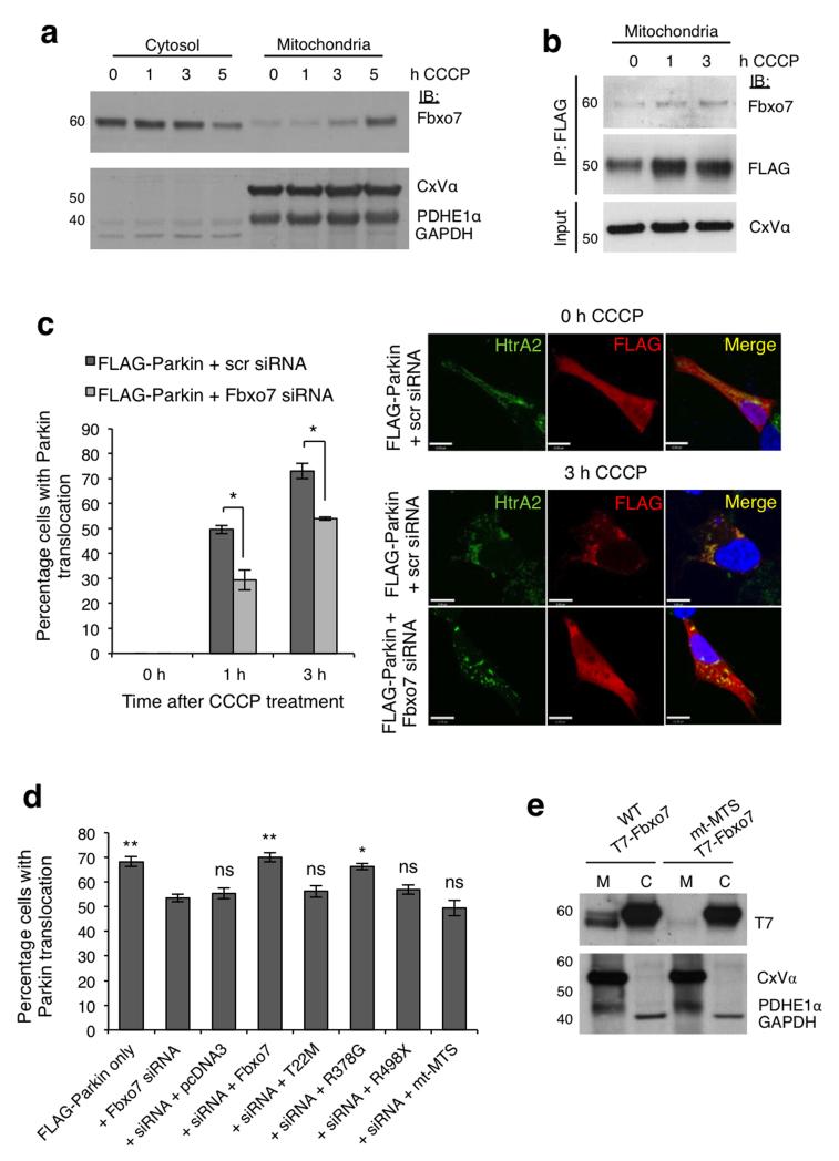
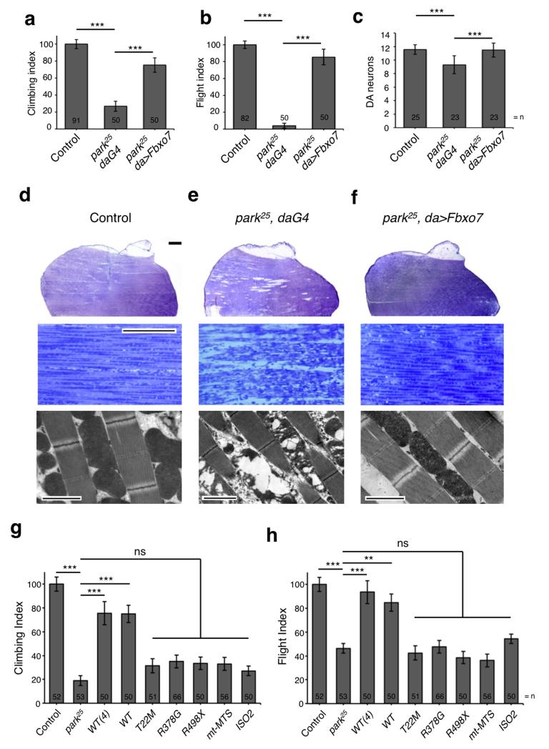
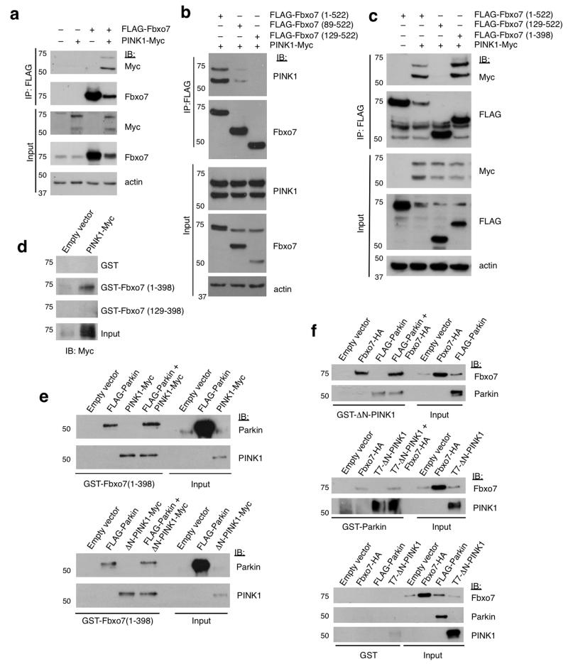
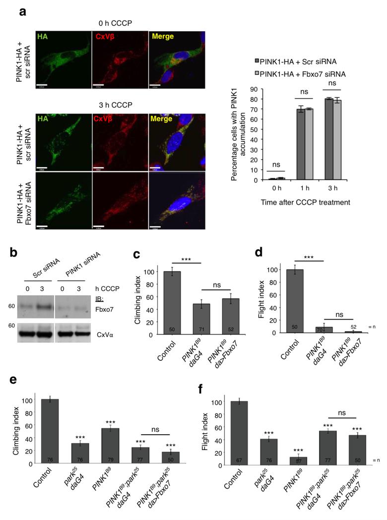
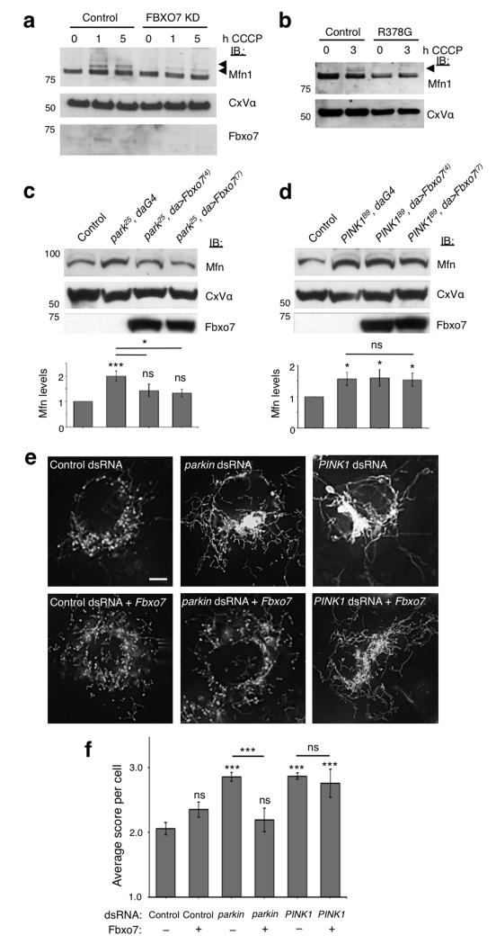
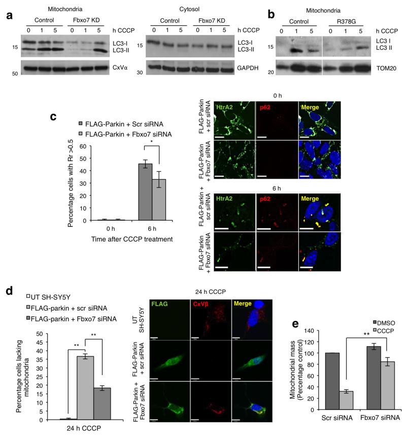
Compelling evidence indicates that two autosomal recessive Parkinson's disease genes, PINK1 (PARK6) and Parkin (PARK2), cooperate to mediate the autophagic clearance of damaged mitochondria (mitophagy). Mutations in the F-box domain-containing protein Fbxo7 (encoded by PARK15) also cause early-onset autosomal recessive Parkinson's disease, by an unknown mechanism. Here we show that Fbxo7 participates in mitochondrial maintenance through direct interaction with PINK1 and Parkin and acts in Parkin-mediated mitophagy. Cells with reduced Fbxo7 expression showed deficiencies in translocation of Parkin to mitochondria, ubiquitination of mitofusin 1 and mitophagy. In Drosophila, ectopic overexpression of Fbxo7 rescued loss of Parkin, supporting a functional relationship between the two proteins. Parkinson's disease-causing mutations in Fbxo7 interfered with this process, emphasizing the importance of mitochondrial dysfunction in Parkinson's disease pathogenesis.


Figures

Figure 1
Figure 1. The amino-terminal Ubl domain of Fbxo7 interacts directly with Parkin
a, Schematic diagram of Fbxo7 isoforms 1 and 2, showing the location of functional domains and the Parkinson’s disease-associated mutations. b, Co-immunoprecipitation of Fbxo7-HA and FLAG-Parkin in whole cell lysates from U2OS cells overexpressing both proteins. c, Co-immunoprecipitation of endogenous Fbxo7 with FLAG-Parkin in HEK293T cells transfected with FLAG-Parkin or a control protein (EGFP). d, FLAG-immunoprecipitation was repeated in U2OS cells transfected with FLAG-Parkin and either full length (1-522) T7-Fbxo7 or an N-terminal truncation lacking the Ubl domain (89-522). e, as with (d) using lysates from U2OS cells expressing FLAG-Parkin and T7-Fbxo7, either full length (1-522) or a C-terminal deletion of the proline rich region (1-398). * indicates IgG heavy chain. f, In vitro translated (IVT) FLAG-Parkin was incubated with bacterially expressed glutathione-S-transferase (GST) or GST fused to the Fbxo7 Ubl domain (1-88) immobilised on glutathione beads. Bead-bound proteins and inputs were analysed by immunoblotting with anti-Parkin antibodies. g, The disease-causing mutation T22M interferes with Fbxo7’s interaction with Parkin. Co-immunoprecipitation was performed as with (b) using lysates from U2OS cells expressing FLAG-Parkin and wild-type or T22M Fbxo7-HA. h, Co-immunoprecipitation of FLAG-Parkin and Fbxo7 in the mitochondrial and cytosolic fractions of HEK293T cells overexpressing both proteins. All representative western blots were performed at least three times. Full-length blots are presented in Supplementary Figure S9.
Figure 2
Figure 2. Fbxo7 participates in CCCP-induced accumulation of Parkin at the mitochondria
a, Fbxo7 relocates from the cytosolic to the mitochondrial fractions of HEK293T cells treated with CCCP (10 μ M). b, Fbxo7 levels are increased in FLAG-Parkin complexes immunoprecipitated from the mitochondrial fraction of HEK293T cells transfected with FLAG-Parkin and untagged Fbxo7 following 1 or 3 h treatment with CCCP (10 μM). c, Parkin localisation at the mitochondria was assessed by immunocytochemistry in SH-SY5Y cells transfected with FLAG-Parkin plus scrambled (scr) or Fbxo7 siRNA, following 1 or 3 h treatment with CCCP (10 μM). Cells were scored visually for the co-localisation of FLAG-Parkin with HtrA2, a mitochondrial marker. Representative images are displayed for cells transfected as indicated, following 0 or 3 h CCCP treatment. For corresponding images at 0 h and 1 h treatment, see Supplementary Figure S2c. Scale bar, 10 μm. d, Loss of FLAG-Parkin translocation upon Fbxo7 silencing is rescued by WT and R378G Fbxo7, but not by T22M Fbxo7, R498X Fbxo7, or by Fbxo7 in which the mitochondrial targeting sequence is mutated (mt-MTS). For c, and d, histograms indicate the percentage of cells in which Parkin localised to the mitochondria. Data are presented as mean of three experiments ± S.E.M. * p < 0.05, ** p < 0.01 compared to cells transfected with FLAG-Parkin plus Fbxo7 siRNA. e, T7-tagged WT Fbxo7 localises to both cytosolic (C) and mitochondrial (M) fractions of transfected HEK293T cells, but MTS mutant (mt-MTS) Fbxo7 localises only to the cytosolic fraction. All representative western blots were performed at least three times. Full-length blots are presented in Supplementary Figure S9.
Figure 3
Figure 3. Expression of Fbxo7 rescues parkin mutant phenotypes
a-b, Overexpression of Fbxo7 suppresses (a) climbing and (b) flight defects of parkin mutants. c, Overexpression of Fbxo7 also suppresses dopaminergic neurodegeneration in the PPL1 cluster of parkin mutants. d-f, (top and middle panels) Toluidine blue stained sections of adult thorax and (bottom panels) TEM images of muscle show that Fbxo7 overexpression suppresses muscle degeneration and mitochondrial disruption in parkin mutants. Toluidine blue scale bars show 200μm (top) and 20μm (middle). TEM scale bars show 2μm. Images are representative of three animals per genotype. g-h, Overexpression of Fbxo7 pathogenic mutants, mt-MTS or isoform 2 by da-GAL4 fails to rescue (g) climbing and (h) flight deficits in parkin mutants. Control genotype is park/+; da-GAL4/+. Histograms indicate mean ± S.E.M. Significance was determined by one-way ANOVA with Bonferroni correction (*** p < 0.001, ** p < 0.01). For climbing and flight assays at least 50 flies were assessed.
Figure 4
Figure 4. PINK1 interacts directly with the amino-terminus of Fbxo7
a, Co-immunoprecipitation of PINK1-Myc and FLAG-Fbxo7 in whole cell lysates from U2OS cells overexpressing both proteins. b, Co-immunoprecipitation of PINK1-Myc with full length and two N-terminal deletions of FLAG-Fbxo7. PINK1-Myc is detected at low levels in complex with FLAG-Fbxo7(89-522) but not with FLAG-Fbxo7(129-522). c, as with (b) using lysates from U2OS cells expressing PINK1-Myc, and FLAG-Fbxo7 containing either N- or C-terminal truncations. d, In vitro pull down experiments were performed using in vitro translated (IVT) PINK1-Myc and either GST or GST fusions of Fbxo7 containing (1-398) or (129-398) immobilised on glutathione beads. e, Competitive binding assays using immobilised GST-Fbxo7(1-398) incubated with IVT FLAG-Parkin and/or full length (top panel) or N-terminally truncated (bottom panel) PINK1-Myc. Input and bead-bound proteins were analysed by immunoblotting as indicated. f, as with (e) immobilised GST-ΔN-PINK1 (top panel), GST-Parkin (middle panel) and immobilised GST alone (bottom panel) were incubated with combinations of IVT FLAG-Parkin, T7-ΔN-PINK1 and Fbxo7-HA as indicated. Input and bead-bound proteins were analysed by immunoblotting with the indicated antibodies. All western blots were performed a minimum of three times. Full-length blots are presented in Supplementary Figure S9.
Figure 5
Figure 5. Functional interaction of Fbxo7 with PINK1
a, PINK1 localisation at the mitochondria was assessed by immunocytochemistry in SH-SY5Y cells transfected with PINK1-HA plus scrambled (scr) or Fbxo7 siRNA following 1 or 3 h treatment with CCCP (10 μM). Cells were scored visually for the co-localisation of PINK1-HA with complex V β subunit (CxVβ), a mitochondrial marker. Histograms indicate the percentage of cells in which PINK1-HA accumulated at the mitochondria. Data are presented as mean ± S.E.M., * p < 0.05. Representative images are displayed for cells transfected as indicated, following 0 or 3 h CCCP treatment. For corresponding images at 0 h and 1 h treatment, see Supplementary Figure 5a. Scale bar, 10 μm. b, Fbxo7 accumulation in the mitochondrial fraction following treatment with CCCP (10 μM) is impaired in SH-SY5Y cells transfected with PINK1 siRNA compared to scrambled siRNA (scr). Full-length blots are presented in Supplementary Figure S9. c-f, Overexpression of Fbxo7 does not rescue (c, e) climbing or (d, f) flight defects in PINK1 male mutants (PINK1B9) or PINK1:parkin double mutants (PINK1B9;park, daG4). Control genotype is (c, d) PINK1B9/+;da-GAL4/+ and (e, f) da-GAL4/+. Histograms indicate mean ± S.E.M. Significance was determined by one-way ANOVA with Bonferroni correction (*** p < 0.001). For climbing and flight assays at least 50 flies were assessed.
Figure 6
Figure 6. Fbxo7 promotes Mfn1 ubiquitination and restores Mfn levels and mitochondrial morphology in Parkin but not PINK1 deficient cells
a-b, Ubiquitination of Mfn1 following treatment with CCCP (10 μM) is reduced in the mitochondrial fraction of both (a) SH-SY5Y cells stably expressing Fbxo7 shRNA (Fbxo7 KD) compared to an empty vector control line and (b) patient fibroblasts with homozygous R378G mutation compared to fibroblasts from healthy controls. Arrows indicate ubiquitinated Mfn1. c-d, Fbxo7 expression restores elevated Mfn steady state levels in (c) parkin but not (d) PINK1 mutant Drosophila. Histograms show mean ± S.E.M. of densitometry analysis of Mfn immunoblots above, normalised to Complex V α (CxV α). Control genotype is (c) park/+;da-GAL4/+ and (d) PINK1B9/+;da-GAL4/+. e, Mitochondria in control Drosophila S2R+ cells stained with MitoTracker Red show a heterogeneous morphology, with a mixture of tubules and fragmented mitochondria. RNAi knockdown of parkin or PINK1 causes excessive fusion and elongated mitochondria compared to control dsRNA (C. elegans gene ZK686.3). Expression of Fbxo7 restores parkin but not PINK1 knockdown phenotype to wild type appearance. Scale bar shows 5 μm. f, Quantification of mitochondrial morphology in dsRNA treated cells. Score system; 1=fragmented, 2=wild type, 3=tubular, 4=hyper-fused (clumped). Histograms indicate mean ± S.E.M. Significance was determined by two-tailed Student t-tests (*** p < 0.001, * p < 0.05). All western blots were performed a minimum of three times and images are representative of 100 cells scored per condition. Full-length blots are presented in Supplementary Figure S9.
Figure 7
Figure 7. Fbxo7 is important for mitophagy
a, Treatment with CCCP (10 μM) results in an increase in LC3-II in the mitochondrial but not the cytosolic fraction of cells stably expressing the empty shRNA vector (control), and this is delayed in stable Fbxo7 knockdown SH-SY5Y cells. b, As in (a), an accumulation of LC3-II was observed in the mitochondrial fraction of healthy control fibroblasts following 1 and 5 h CCCP treatment, but this was reduced in fibroblasts from a patient carrying the R378G mutation. Western blots were performed a minimum of three times. c, Mitochondrial accumulation of p62 following CCCP treatment is inhibited by Fbxo7 siRNA. FLAG-Parkin overexpressing SH-SY5Y cells were transfected with scrambled (scr) or Fbxo7 siRNA as indicated and treated with either DMSO or CCCP (10 μM) for 6 h. Colocalisation of p62 with HtrA2, a mitochondrial marker, was assessed by Pearson’s correlation co-efficient (Rr) on a cell by cell basis. Histogram shows the percentage of cells in which Rr was greater than 0.5. Data are represented as mean ± S.E.M., * p < 0.05. Scale bar, 10 μm. d, Mitophagy was analysed in untransfected (UT) SH-SY5Y cells or in stable FLAG-Parkin overexpressing SH-SY5Y cells transfected with either scrambled (scr) or Fbxo7 siRNA. Histogram indicates the percentage of cells with no remaining mitochondria following 24 h treatment with CCCP (10 μM) for each condition. Complex V β subunit (CxVβ) was used as a mitochondrial marker. Data are presented as mean ± S.E.M., ** p < 0.01. Scale bar, 10 μm. e, Mitochondrial mass was measured in FLAG-Parkin overexpressing SH-SY5Y cells transfected with scrambled (scr) or Fbxo7 siRNA and treated for 24 h with either DMSO or CCCP (10 μM). For representative images, see Supplementary Fig. S8d. Full-length blots are presented in Supplementary Figure S9.

References

    1. Narendra D, Tanaka A, Suen DF, Youle RJ. Parkin is recruited selectively to impaired mitochondria and promotes their autophagy. J Cell Biol. 2008;183:795–803. - PMC - PubMed
    1. Vives-Bauza C, et al. PINK1-dependent recruitment of Parkin to mitochondria in mitophagy. Proc Natl Acad Sci U S A. 2010;107:378–83. - PMC - PubMed
    1. Narendra DP, et al. PINK1 is selectively stabilized on impaired mitochondria to activate Parkin. PLoS Biol. 2010;8:e1000298. - PMC - PubMed
    1. Geisler S, et al. PINK1/Parkin-mediated mitophagy is dependent on VDAC1 and p62/SQSTM1. Nat Cell Biol. 2010;12:119–31. - PubMed
    1. Deas E, Wood NW, Plun-Favreau H. Mitophagy and Parkinson’s disease: the PINK1-parkin link. Biochim Biophys Acta. 2011;1813:623–33. - PMC - PubMed

Publication types

MeSH terms