. 2014 Feb;45(1):81-9.
doi: 10.1007/s10735-013-9530-7. Epub 2013 Aug 10.

Stimulation of primary osteoblasts with ATP induces transient vinculin clustering at sites of high intracellular traction force

Affiliations

Stimulation of primary osteoblasts with ATP induces transient vinculin clustering at sites of high intracellular traction force

Toh Weng Tan et al. J Mol Histol. 2014 Feb.

Abstract

Adenosine 5'-triphosphate (ATP), released in response to mechanical and inflammatory stimuli, induces the dynamic and asynchronous protrusion and subsequent retraction of local membrane structures in osteoblasts. The molecular mechanisms involved in the ligand-stimulated herniation of the plasma membrane are largely unknown, which prompted us to investigate whether the focal-adhesion protein vinculin is engaged in the cytoskeletal alterations that underlie the ATP-induced membrane blebbing. Using time-lapse fluorescence microscopy of primary bovine osteoblast-like cells expressing green fluorescent protein-tagged vinculin, we found that stimulation of cells with 100 μM ATP resulted in the transient and rapid clustering of recombinant vinculin in the cell periphery, starting approximately 100 s after addition of the nucleotide. The ephemeral nature of the vinculin clusters was made evident by the brevity of their mean assembly and disassembly times (66.7 ± 13.3 s and 99.0 ± 6.6 s, respectively). Traction force vector maps demonstrated that the vinculin-rich clusters were localized predominantly at sites of high traction force. Intracellular calcium measurements showed that the ligand-induced increase in [Ca(2+)]i clearly preceded the clustering of vinculin, since [Ca(2+)]i levels returned to normal within 30 s of exposure to ATP, indicating that intracellular calcium transients trigger a cascade of signalling events that ultimately result in the incorporation of vinculin into membrane-associated focal aggregates.


Figures

Fig. 1
Fig. 1
Detection of osteogenic marker proteins in cultured bovine osteoblast-like cells. Shown are immunocytochemical stainings of methanol/acetone-fixed cells for expression of osteocalcin (a), procollagen type I (b), osteopontin (c), and bone sialoprotein (d) using horseradish peroxidase-conjugated secondary antibodies. Positive immunoreactivity was visualized by incubating the enzyme with the substrate α-chloronaphthol, which produced a dark staining against a white background (bar 30 μm)
Fig. 2
Fig. 2
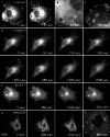
ATP and bradykinin stimulation of bovine primary osteoblasts results in the formation of vinculin-rich membrane clusters. a Fluorescence micrographs of a bovine osteoblast expressing recombinant vinculin fused to green-fluorescent protein (GFP) before (0 s) and 190 s after addition of 100 μM ATP. The intracellular distribution of GFP-vinculin before and after ATP treatment at low (left two images, bar 10 μm) and higher magnification (right two images, bar 2 μm) is shown. b Time-lapse fluorescence microscopy showing de novo formation of vinculin-containing aggregates upon stimulation of cells with ATP (exposure started at t = 0 s). Arrowheads mark typical vinculin clusters. c No membrane clustering was observed upon ATP treatment in cells expressing GFP-tagged STAT1, used as a control. d Transient cluster formation of GFP-vinculin in osteoblasts treated with 1 μM bradykinin (+BK) for 0, 600 and 1,400 s, respectively. The image on the right shows vinculin clusters in the perinuclear region at higher magnification
Fig. 3
Fig. 3
a Kinetics of the assembly and disassembly of single GFP-vinculin-containing membrane clusters. Changes in fluorescence intensity were averaged over several aggregate-containing ROIs in the cytoplasm and plotted over time. The peak of fluorescence intensity was set to t = 0 s. b Vinculin-rich aggregates co-localize at sites of high traction force magnitude. Intracellular traction forces were measured by substrate deformation using fluorescent latex micro-beads embedded in a collagen-coated flexible polyacrylamide sheet. The fluorescent micro-beads scattered throughout the substratum were clearly distinguishable from the larger and more weakly labelled vinculin clusters. The images depict a fluorescence micrograph of a GFP-vinculin-expressing osteoblast (left image) exhibiting dot-like aggregates at its cell periphery (marked with arrowheads, bar 10 µm) and the corresponding vector map of traction forces within the same cell (right image). The scale vector represents 500 N/m2 (total force of 4,500 μN/cell)
Fig. 4
Fig. 4
Kinetics of calcium influx in bovine osteoblasts stimulated with high concentrations of ATP. a Cultured cells were loaded with Fluo-3/AM and subsequently treated with 100 μM of the nucleotide. Exposure to ATP resulted in an increase in Fluo-3/AM fluorescence intensity. A phase contrast microscopical image (left, bar 8 μm) and the corresponding fluorescence staining before (middle, 0 sec) and after addition of ATP (right, 10 sec) are shown. b Fura-2AM fluorescence signal intensities emitted at 505 nm were measured before (0 s), and 6 and 100 s after ATP treatment using excitation wavelengths of 340 and 380 nm, respectively. c Changes in the ratio F340/F380 were calculated from a typical cell before and after stimulation with ATP (marked with an arrow)

References

    1. Baldini G, Ponti C, Bortul R, Narducci P, Grill V, Martelli AM. Sparc localizes to the blebs of Hobit cells and human primary osteoblasts. J Cell Biochem. 2008;104(6):2310–2323. doi: 10.1002/jcb.21789. - DOI - PubMed
    1. Bowler WB, Buckley KA, Gartland A, Hipskind RA, Bilbe G, Gallagher JA. Extracellular nucleotide signaling: a mechanism for integrating local and systemic responses in the activation of bone remodeling. Bone. 2001;28(5):507–512. doi: 10.1016/S8756-3282(01)00430-6. - DOI - PubMed
    1. Buckley KA, Hipskind RA, Gartland A, Bowler WB, Gallagher JA. Adenosine triphosphate stimulates human osteoclast activity via upregulation of osteoblast-expressed receptor activator of nuclear factor-κB ligand. Bone. 2002;31(5):582–590. doi: 10.1016/S8756-3282(02)00877-3. - DOI - PubMed
    1. Curtze S, Dembo M, Miron M, Jones DB. Dynamic changes in traction forces with DC electric field in osteoblast-like cells. J Cell Sci. 2004;117(Pt 13):2721–2729. doi: 10.1242/jcs.01119. - DOI - PubMed
    1. Dubyak GR. P2X7 receptor regulation of non-classical secretion from immune effector cells. Cell Microbiol. 2012;14(11):1697–1706. doi: 10.1111/cmi.12001. - DOI - PMC - PubMed

LinkOut - more resources