. 2013 Jul;123(7):2862-72.
doi: 10.1172/JCI66966. Epub 2013 Jun 24.

Reversal of gene dysregulation in cultured cytotrophoblasts reveals possible causes of preeclampsia

Affiliations

Reversal of gene dysregulation in cultured cytotrophoblasts reveals possible causes of preeclampsia

Yan Zhou et al. J Clin Invest. 2013 Jul.

Erratum in

  • J Clin Invest. 2013 Oct 1;123(10):4541

Abstract

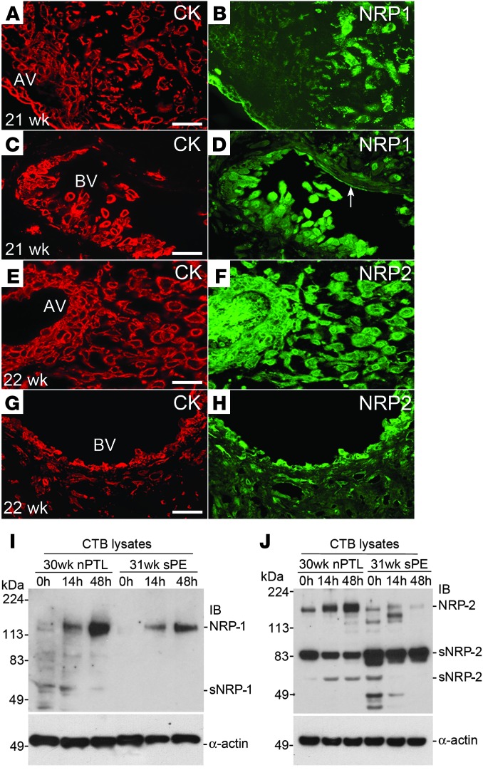
During human pregnancy, a subset of placental cytotrophoblasts (CTBs) differentiates into cells that aggressively invade the uterus and its vasculature, anchoring the progeny and rerouting maternal blood to the placenta. In preeclampsia (PE), CTB invasion is limited, reducing placental perfusion and/or creating intermittent flow. This syndrome, affecting 4%-8% of pregnancies, entails maternal vascular alterations (e.g., high blood pressure, proteinuria, and edema) and, in some patients, fetal growth restriction. The only cure is removal of the faulty placenta, i.e., delivery. Previously, we showed that defective CTB differentiation contributes to the placental component of PE, but the causes were unknown. Here, we cultured CTBs isolated from PE and control placentas for 48 hours, enabling differentiation and invasion. In various severe forms of PE, transcriptomics revealed common aberrations in CTB gene expression immediately after isolation, including upregulation of SEMA3B, which resolved in culture. The addition of SEMA3B to normal CTBs inhibited invasion and recreated aspects of the PE phenotype. Additionally, SEMA3B downregulated VEGF signaling through the PI3K/AKT and GSK3 pathways, effects that were observed in PE CTBs. We propose that, in severe PE, the in vivo environment dysregulates CTB gene expression; the autocrine actions of the upregulated molecules (including SEMA3B) impair CTB differentiation, invasion and signaling; and patient-specific factors determine the signs.


Figures

Figure 1
Figure 1. sPE-associated aberrations in CTB gene expression returned to control values after 48 hours of culture.
RNA was analyzed immediately after the cells were isolated (0 hour) and after 12, 24, and 48 hours in culture. The relative gene expression levels for CTBs isolated from placentas of patients who delivered due to nPTL (n = 5) or sPE (n = 5) are shown as a heat map, ranging from high (red) to low (blue). The sPE CTBs were from the following cases (tiled from left to right): (a) HELLP syndrome and IUGR; (b) sPE; (c) sPE and IUGR; (d) superimposed sPE; and (e) HELLP syndrome. One sample of nPTL CTBs collected at 48 hours was omitted for technical reasons. The fold changes for each time point (sPE vs. nPTL) are shown on the right. ns, no significant difference (LIMMA); t, no significant difference in expression (sPE vs. nPTL) by 48 hours (maSigPro).
Figure 2
Figure 2. SEMA3B expression was high in the placenta and upregulated in sPE.
(A) Binding of a 32P-SEMA3B probe to a multiple tissue expression array revealed high placental expression (coordinate B8). (B) Northern hybridization of polyA+ RNA extracted from chorionic villi and pooled from 3 placentas showed that SEMA3B expression increased over gestation and was highest in sPE (n = 3 replicates). (C) In situ hybridization (3 placentas per group) confirmed enhanced SEMA3B mRNA expression in the STB layer of the chorionic villi in sPE (25 weeks) as compared with normal pregnancy (23 weeks) and nPTL (34 weeks). (D) Immunoblotting of CTB lysates (15 μg per lane) showed that SEMA3B protein expression was low to undetectable in control cells from normal placentas (15–39 weeks). In all cases, expression was higher in sPE (26–33 weeks) as compared with nPTL (30, 33 weeks). A protein of the expected Mr was detected in COS-1 cells transfected with SEMA3B but not in those transfected with SEMA3A-Fc. Vertical lines denote noncontiguous lanes from the same gel. The relative intensity of the bands quantified by densitometry is also shown. The values for each sample type were averaged and expressed relative to the α-actin loading controls. The entire experiment was repeated twice. (E) Staining tissue sections with anti-SEMA3B showed a sPE-associated upregulation of immunoreactivity associated with the trophoblast components of chorionic villi and among extravillous CTBs within the basal plate (n = 5 per group). Trophoblasts were identified by staining adjacent tissue sections with anti–cytokeratin-8/18 (data not shown). Scale bars: 100 μm (C and E). NB, Northern blot; GA, gestational age; RP, recombinant protein.
Figure 3
Figure 3. NRP-1 and NRP-2 (protein) expression at the maternal-fetal interface in normal pregnancy and in sPE.
Tissue sections were double stained with anti–cytokeratin-8/18 (CK), which reacts with all trophoblast subpopulations, and anti–NRP-1 or NRP-2. (A and B) NRP-1 expression was detected in association with villous trophoblasts. Within the uterine wall, immunoreactivity associated with invasive CTBs was upregulated as the cells moved from the surface to the deeper regions. (C and D) Endovascular CTBs that lined a maternal blood vessel (BV) were also stained. (EH) Anti–NRP-2 reacted with trophoblast and nontrophoblast cells in anchoring villi (AV) as well as interstitial and endovascular CTBs. Essentially the same staining patterns, but with weaker intensity, were observed in sPE (data not shown). CTBs were isolated from the placentas of control nPTL cases and from the placentas of women who experienced sPE. (I) Over 48 hours in culture, NRP-1 expression was upregulated in both instances but to a lesser degree in sPE. (J) Control nPTL CTBs also upregulated NRP-2. Expression of this receptor was reduced in sPE and the soluble form was more abundant. (AJ) The data shown are representative of the analysis of a minimum of 3 samples from different placentas. Scale bars: 100 μm. AV, anchoring villi.
Figure 4
Figure 4. Exogenous SEMA3B mimicked the effects of sPE on CTBs and endothelial cells and inhibited angiogenesis.
(A) The addition of anti-VEGF or SEMA3B protein significantly inhibited CTB invasion as compared with the addition of a control protein, CD6-Fc. The removal of both ligands (anti–VEGF/NRP1-Fc and anti–VEGF/NRP-2–Fc) restored invasion to control levels. (B) The variables tested in A had the opposite effects on CTB apoptosis, suggesting that increased programmed cell death contributed to decreased invasion. (C) Exogenous VEGF stimulated the migration of UtMVECs, which was inhibited by SEMA3B. (D) The results in C were quantified relative to the addition of CD6-Fc. (E) In UtMVECs, VEGF promoted survival and SEMA3B increased apoptosis relative to control levels. (F) In the chick chorioallantoic membrane angiogenesis assay, VEGF promoted angiogenesis by approximately 3 fold and SEMA3B inhibited this process approximately 5 fold relative to the effects of CD6-Fc. Arrows mark the edge of the filter paper used to apply the protein. The area of the CAM beneath the filter paper is shown in the bottom row. Scale bar: 200 μm (top row); 100 μm (bottom row). n = 6 replicates (AD); n = 3 replicates (E and F). Mean ± SEM; 2-tailed Student’s t test. *P < 0.05, **P < 0.01, ***P < 0.001.
Figure 5
Figure 5. SEMA3B inhibited PI3K/AKT and GSK3β signaling in CTBs and the same effects were observed in sPE.
(A) SEMA3B and wortmannin (WM) inhibited PI3K activity, which was stimulated by VEGF. DMSO was used as a vehicle control. Mean ± SEM, 2-tailed Student’s t test. *P < 0.05, **P < 0.01. (B) The addition of SEMA3B to UtMVECs resulted in the dissociation of the p85 and the p110α subunits of PI3K, which was rescued by the addition of VEGF. (C) In COS-1 cells, SEMA3B inhibited AKT Ser473 phosphorylation (activation), which increased during CTB differentiation/invasion (0–12 hours). The addition of SEMA3B inhibited AKT phosphorylation, which was enhanced by exogenous VEGF. (D) In COS-1 cells, SEMA3B inhibited GSK3β Ser9 phosphorylation (inactivation), which increased during CTB differentiation/invasion (0–12 hours). Exogenous SEMA3B inhibited GSK3β phosphorylation, which was enhanced by VEGF. GSK3α Ser21 phosphorylation was variable. (E) In CTBs, sPE correlated with dissociation of the p85 and p110α (and γ) subunits of PI3K relative to control cells isolated from normal third trimester placentas. (F) In freshly isolated CTBs, sPE was associated with decreased phosphorylation of AKT Ser473 and GSK3β Ser9. α-Actin was used as a loading control. (G) In chorionic villi, sPE was associated with phosphorylation (inactivation) of β-catenin. (AD) The same results were obtained in 3 separate experiments that used different preparations of cells. (F and G) The results shown are representative of analyses of a total of 6 CTB isolates from different placentas of women diagnosed with sPE.
Figure 6
Figure 6. Model of SEMA3B effects on CTBs in sPE and normal pregnancy.

Comment in

References

    1. Maltepe E, Bakardjiev AI, Fisher SJ. The placenta: transcriptional, epigenetic, and physiological integration during development. J Clin Invest. 2010;120(4):1016–1025. doi: 10.1172/JCI41211. - DOI - PMC - PubMed
    1. Pijnenborg R, Vercruysse L, Hanssens M. The uterine spiral arteries in human pregnancy: facts and controversies. Placenta. 2006;27(9–10):939–958. doi: 10.1016/j.placenta.2005.12.006. - DOI - PubMed
    1. Zhou Y, et al. Human cytotrophoblasts adopt a vascular phenotype as they differentiate. A strategy for successful endovascular invasion? J Clin Invest. 1997;99(9):2139–2151. doi: 10.1172/JCI119387. - DOI - PMC - PubMed
    1. Red-Horse K, Kapidzic M, Zhou Y, Feng KT, Singh H, Fisher SJ. EPHB4 regulates chemokine-evoked trophoblast responses: a mechanism for incorporating the human placenta into the maternal circulation. Development. 2005;132(18):4097–4106. doi: 10.1242/dev.01971. - DOI - PubMed
    1. Hunkapiller NM, et al. A role for Notch signaling in trophoblast endovascular invasion and in the pathogenesis of pre-eclampsia. Development. 2011;138(14):2987–2998. doi: 10.1242/dev.066589. - DOI - PMC - PubMed

Publication types

MeSH terms