Review
. 2014 Feb;71(3):479-92.
doi: 10.1007/s00018-013-1439-5. Epub 2013 Aug 11.

Myosin II in mechanotransduction: master and commander of cell migration, morphogenesis, and cancer

Affiliations
Review

Myosin II in mechanotransduction: master and commander of cell migration, morphogenesis, and cancer

Rocío Aguilar-Cuenca et al. Cell Mol Life Sci. 2014 Feb.

Abstract

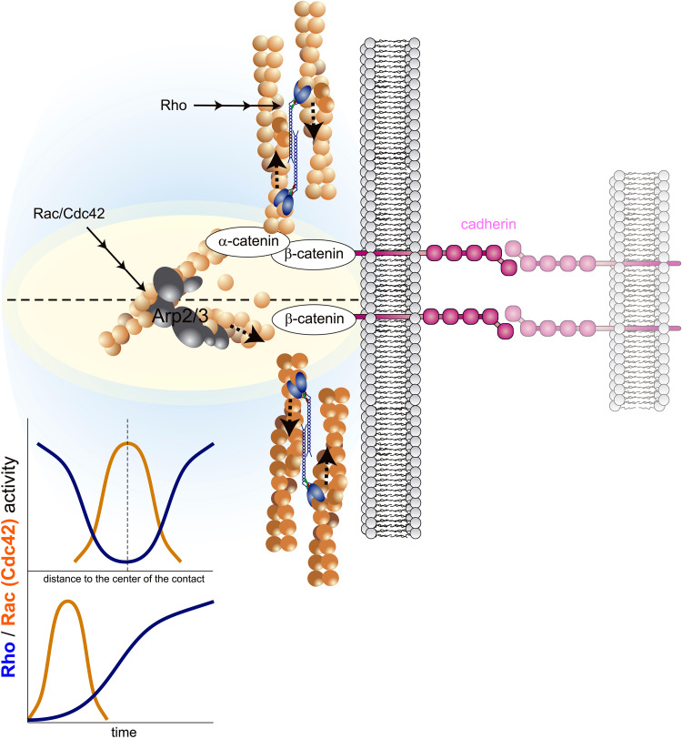
Mechanotransduction encompasses the role of mechanical forces in controlling cell behavior by activating signal transduction pathways. Most forces at a cellular level are caused by myosin II, which contracts and cross-links actin. Myosin II-dependent forces are transmitted through the actin cytoskeleton to molecular endpoints that promote specific cellular outcomes, e.g., cell proliferation, adhesion, or migration. For example, most adhesive and migratory phenomena are mechanically linked by a molecular clutch comprised of mechanosensitive scaffolds. Myosin II activation and mechanosensitive molecular mechanisms are finely tuned and spatiotemporally integrated to coordinate morphogenetic events during development. Mechanical events dependent on myosin II also participate in tumor cell proliferation, invasion, and metastatic dissemination. Specifically, tumor cells alter the mechanical properties of the microenvironment to create favorable conditions for proliferation and/or dissemination. These observations position myosin II-dependent force generation and mechanotransduction at the crossroads between normal development and cancer.


Figures

Fig. 1
Fig. 1
Mechanical regulation of the molecular clutch that controls cell adhesion and migration. Diagrams show the mechanical regulation of talin that controls its interaction with vinculin and force-dependent integrin activation. a α and β integrin chains are represented in an extended, intermediate affinity conformation that binds to RGD motifs in ECM proteins. Integrin is bound to non-stretched talin (represented as a coiled spring), through the tail of the β chain. Talin is bound to actin. Also shown is non-muscle myosin II (NMII). Arrows point to the prospective sliding direction of the actin filaments upon force generation induced by NMII. Numbers represent the critical points of the mechanotransduction point prior to force application (b). b Conformation change of the NMII head (1′) slides actin filaments in the indicated directions. Actin bound to the talin tail stretches the molecule (represented as an extended spring; a two-headed arrow signals the extension) and exposes a binding site (2′) for binding to vinculin. Vinculin binds to actin and strengthens the integrin-actin linkage. Also, mechanical force separates the cytoplasmic domains of the α and β integrin chain (3′) and evokes a conformational movement that extends the head domain of the integrin (4′), increasing its affinity. Force is transmitted to the extracellular matrix and stretches fibronectin, exposing a cryptic site (5′, shown in green) that cooperates to binding. c Same as (b), except vinculin is shown recruiting an extra actin bundle, increasing actin cross-linking at adhesions. d The integrin-actin linkage. Some of the mechanosensitive scaffolds are represented. Black dotted lines represent the talin-integrin force-dependent binding and activation pathway outlined in (a). Red dotted lines indicate the possible routes of mechanical activation of p130CAS. CAS has a cryptic, stretch-dependent Src-phosphorylable Tyr residue (Tyr165. NMII-dependent sliding of the actin filaments generates mechanical force that is transmitted to CAS through zyxin, exposing Tyr165 in CAS. Also depicted, focal adhesion kinase (FAK), which interacts with talin and activates Src, Crk-II, and paxillin, which link FAK-CAS to vinculin and could constitute an alternative pathway of force transmission from actomyosin to CAS
Fig. 2
Fig. 2
NMII participates in cell–cell contact formation and stabilization. Diagram shows actin polymerization and NMII-mediated contraction and bundling in forming cell–cell contacts. Cadherins recruit different proteins, most notably β and α-catenin, which links to actin and may act as a force transducer [115]. Upon cadherin engagement (right), an early, transient wave of Rac/Cdc42-mediated actin polymerization pushes the membrane to form the cell–cell contact. Later, Rho-dependent NMII activation in the periphery of the initial contact extends the contact area and solidifies the cell–cell contact. Bottom left, graphs are non-scale, schematic representations of the Rac/Cdc42 and Rho localization (top) and kinetics (bottom)
Fig. 3
Fig. 3
Role of NMII and actin polymerization in morphogenetic epithelial movements. a Apical constriction mediated by NMII. The initial signal is represented by a morphogenetic hormone or peptide, but other signals may accomplish the same effect. The hormone binds a G protein-coupled receptor that, acting through G proteins, activates RhoGEFs, which in turn activate Rho, which induces NMII phosphorylation and activation via ROCK [116]. NMII-mediated actin contraction mediates constriction of the apical pole. b Basolateral extension in the direction of migration is induced by actin polymerization. Constriction at this area is prevented by accumulation of RhoGAPs, which inactivate Rho and locally deactivate NMII. This is simultaneous to the clustering of Rac/Cdc42 activators (GEFs), which depends of the presence of the polarity proteins Par3/Par6 and may require atypical PKC activation [72]. Rac/Cdc42 promote actin polymerization through the Arp2/3 complex via WASP/WAVE proteins
Fig. 4
Fig. 4
Morphology relates to the migratory properties of the cell. The four most typical morphologies related to cell migration are depicted: non-migratory epithelial, mesenchymal, bleb-based, and amoeboid. Magenta represents Rac/Cdc42-dependent protrusive areas, blue RhoA-NMII-dependent retraction areas, black RhoA-NMII-dependent membrane blebs, green cell–matrix adhesions, and brown lines actin bundles. Text next to each of the morphologies describes receptors implicated in adhesion, protrusiveness, and predominant geometry of the actin, small Rho GTPase usage, and migration speed (in bold) and cell types that most commonly display the indicated morphology. Known transitions between the different migratory modes are indicated between morphologies; dashed lines with question marks indicate transitions that have been postulated but lack definitive experimental proof

References

    1. Scott JD, Pawson T. Cell signaling in space and time: where proteins come together and when they’re apart. Science. 2009;326(5957):1220–1224. - PMC - PubMed
    1. Engler AJ, Sen S, Sweeney HL, Discher DE. Matrix elasticity directs stem cell lineage specification. Cell. 2006;126(4):677–689. - PubMed
    1. Klein EA, Yin L, Kothapalli D, Castagnino P, Byfield FJ, Xu T, Levental I, Hawthorne E, Janmey PA, Assoian RK. Cell-cycle control by physiological matrix elasticity and in vivo tissue stiffening. Curr Biol. 2009;19(18):1511–1518. - PMC - PubMed
    1. Lo CM, Wang HB, Dembo M, Wang YL. Cell movement is guided by the rigidity of the substrate. Biophys J. 2000;79(1):144–152. - PMC - PubMed
    1. Raab M, Swift J, PC PD, Shah P, Shin JW, Discher DE (2012) Crawling from soft to stiff matrix polarizes the cytoskeleton and phosphoregulates myosin-II heavy chain. J Cell Biol. doi:10.1083/jcb.201205056 - PMC - PubMed

Publication types

LinkOut - more resources