. 2013 Oct;79(20):6312-24.
doi: 10.1128/AEM.01226-13. Epub 2013 Aug 9.

The periplasmic HrpB1 protein from Xanthomonas spp. binds to peptidoglycan and to components of the type III secretion system

Affiliations

The periplasmic HrpB1 protein from Xanthomonas spp. binds to peptidoglycan and to components of the type III secretion system

Jens Hausner et al. Appl Environ Microbiol. 2013 Oct.

Abstract

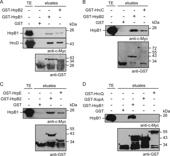
The plant-pathogenic bacterium Xanthomonas campestris pv. vesicatoria employs a type III secretion (T3S) system to translocate bacterial effector proteins into eukaryotic host cells. The membrane-spanning secretion apparatus consists of 11 core components and several associated proteins with yet unknown functions. In this study, we analyzed the role of HrpB1, which was previously shown to be essential for T3S and the formation of the extracellular T3S pilus. We provide experimental evidence that HrpB1 localizes to the bacterial periplasm and binds to peptidoglycan, which is in agreement with its predicted structural similarity to the putative peptidoglycan-binding domain of the lytic transglycosylase Slt70 from Escherichia coli. Interaction studies revealed that HrpB1 forms protein complexes and binds to T3S system components, including the inner membrane protein HrcD, the secretin HrcC, the pilus protein HrpE, and the putative inner rod protein HrpB2. The analysis of deletion and point mutant derivatives of HrpB1 led to the identification of amino acid residues that contribute to the interaction of HrpB1 with itself and HrcD and/or to protein function. The finding that HrpB1 and HrpB2 colocalize to the periplasm and both interact with HrcD suggests that they are part of a periplasmic substructure of the T3S system.


Figures

Fig 1
Fig 1
Complementation studies with HrpB1 derivatives. (A) Genomic hrpB1 expressed under the control of the native promoter restores the pathogenicity of hrpB1 deletion mutant strains. X. campestris pv. vesicatoria strains 85-10 (wild type [wt], hrpG wild type), 85-10ΔhrpB1hrpB1, hrpG wild type), 85* (wild type, hrpG*), and 85*ΔhrpB1hrpB1, hrpG*) with plasmid pBRM (empty vector [EV]) or encoding HrpB1 or HrpB1GTG downstream of the native (pnat) or the lac (plac) promoter in trans or in cis, as indicated, were inoculated at a density of 108 CFU ml−1 into leaves of susceptible ECW and resistant ECW-10R pepper plants. Disease symptoms were photographed at 8 dpi. For the better visualization of the HR, leaves were bleached in ethanol at 2 dpi. Dashed lines mark the infiltrated areas. For the immunodetection of HrpB1 and derivatives thereof, bacteria were grown in NYG medium and equal amounts of total cell extracts were analyzed by immunoblotting using a HrpB1-specific antibody. Note that cis-encoded HrpB1 is expressed only in the presence of hrpG*, which activates hrp gene expression. (B) Ectopic expression of hrpB1 exerts a negative effect on bacterial pathogenicity. X. campestris pv. vesicatoria strain 85-10 (wild type) without an expression construct (−), carrying plasmid pBRM (EV) or encoding HrpB1 derivatives, as indicated, was inoculated into the leaves of susceptible ECW and resistant ECW-10R pepper plants. Disease symptoms and the HR were documented as described for panel A. (C) In cis-encoded HrpB1 restores HrpF secretion in strain 85*ΔhrpB1. Strains 85* (wild type) and 85*ΔhrpB1hrpB1) with plasmid pBRM (EV) or expressing hrpB1 in trans or in cis (HrpB1in cis) under the control of the native (pnat) or the lac (plac) promoter, as indicated, were incubated in secretion medium. Total cell extracts (TE) and culture supernatants (SN) were analyzed by immunoblotting, using HrpF-specific antibodies. The upper band represents HrpF; the lower bands correspond to degradation products.
Fig 2
Fig 2
HrpB1 is a periplasmic protein and binds to peptidoglycan. (A) HrpB1 and HrpB2 colocalize to the bacterial periplasm. Strains 85* (wild type), 85*ΔhrpB2hrpB2), and 85*ΔhrpB1hrpB1) were incubated in secretion medium, and fractions enriched in the cytoplasm (C), IM, periplasm (P), and OM were separated by ultracentrifugation. Bacterial total cell extracts and different fractions were analyzed by immunoblotting using antibodies specific for HrpB1, HrpB2, HrcJ, and HrcC. (B) HrpB1 binds to peptidoglycan in vitro. Lysozyme, purified HrpB1-His6, derivatives thereof, and AvrBs3Δ2-His6, as indicated, were incubated in the absence (−) or presence (+) of peptidoglycan. Total protein extracts (TE) as well as proteins in the supernatant (SN) and the pellet (P) after centrifugation were analyzed by SDS-PAGE, Coomassie staining, or immunoblotting using His6 epitope-specific antibodies.
Fig 3
Fig 3
Interaction studies with HrpB1 and HrpB2. (A) GST pulldown assays with HrpB1, HrpB2, and HrcD. GST, GST-HrpB1, and GST-HrpB2 were immobilized on glutathione-Sepharose and incubated with bacterial lysates containing HrpB1–c-Myc or HrcD–c-Myc. Total cell extracts (TE) and eluted proteins (eluates) were analyzed by immunoblotting, using c-Myc epitope- and GST-specific antibodies, respectively. Asterisks mark GST and GST fusion proteins; additional signals presumably correspond to degradation products. (B) HrpB1 interacts with the OM secretin HrcC. GST, GST-HrpB2, and GST-HrcC were immobilized on glutathione-Sepharose and incubated with a bacterial lysate containing HrpB1–c-Myc. Total cell extracts and eluates were analyzed as described for panel A. (C) HrpB1 interacts with the pilus protein HrpE. GST, GST-HrpB2, and GST-HrpE were immobilized on glutathione-Sepharose and incubated with a bacterial lysate containing HrpB1–c-Myc. Total cell extracts and eluates were analyzed as described for panel A. (D) Interaction studies with HrpB1, XopA, and HrcQ. GST, GST-HrpB1, GST-XopA, and GST-HrcQ were immobilized on glutathione-Sepharose and incubated with a bacterial lysate containing HrpB1–c-Myc. Total cell extracts and eluates were analyzed as described for panel A.
Fig 4
Fig 4
HrpB1 forms protein complexes. (A) HrpB1 complex formation in E. coli. E. coli carrying plasmid pBRM (empty vector [EV]) or encoding HrpB1–c-Myc was grown in LB medium and incubated without (−) or with increasing concentrations (final concentrations, 0.05 to 5 mM) of the cross-linking reagent DSS. Protein extracts were analyzed by immunoblotting using HrpB1-specific antibodies. (B) Complex formation of purified HrpB1. Purified HrpB1-His6 was incubated without (−) or with DSS as described for panel A. Protein extracts were analyzed by immunoblotting using HrpB1-specific antibodies. (C) HrpB1 complex formation in X. campestris pv. vesicatoria. Strain 85-10ΔhrpB1 carrying plasmid pBRM (EV) or construct pBRMhrpB1Stop (HrpB1) was grown in NYG medium and incubated without (−) or with increasing concentrations of DSS, as indicated. Protein extracts were analyzed as described for panel A. (D) HrpB1 coimmunoprecipitates with HrpB1–c-Myc. X. campestris pv. vesicatoria strains 85* and 85*ΔhrpB1 containing HrpB1–c-Myc or plasmid pBRM (EV), as indicated, were grown in secretion medium. Bacterial lysates were incubated in the presence (+) or absence (−) of c-Myc epitope-specific antibodies coupled to protein G Sepharose. Bacterial total cell extracts (TE) and precipitated proteins (eluate) were analyzed by immunoblotting using HrpB1-specific antibodies.
Fig 5
Fig 5
Interaction studies with HrpB1 deletion derivatives. (A) Amino acids 11 to 20 of HrpB1 contribute to the interaction of HrpB1 with itself, HrpB2, HrpE, and HrcD. GST, GST-HrpB1, GST-HrpB2, GST-HrpE, and GST-HrcD were immobilized on glutathione-Sepharose and incubated with bacterial lysates containing HrpB1–c-Myc, HrpB111-159–c-Myc, HrpB121-159–c-Myc, HrpB11-139–c-Myc, and HrpB11-149–c-Myc, as indicated. Total cell extracts (TE) and eluted proteins (eluates) were analyzed by immunoblotting, using c-Myc epitope- and GST-specific antibodies. Asterisks mark GST and GST fusion proteins; additional signals presumably correspond to degradation products. (B) Protein-protein interaction studies with HrpB1 derivatives containing internal deletions. GST, GST-HrpB1, GST-HrpB2, GST-HrpE, and GST-HrcD were immobilized on glutathione-Sepharose and incubated with bacterial lysates containing HrpB1–c-Myc and deletion derivatives thereof, as indicated. Total cell extracts and eluates were analyzed as described for panel A.
Fig 6
Fig 6
Interaction studies with HrpB1 point mutant derivatives. (A) GST, GST-HrpB1, GST-HrpB2, GST-HrpE, GST-HrcD, and GST-HrcC were immobilized on glutathione-Sepharose and incubated with bacterial lysates containing HrpB1–c-Myc or point mutant derivatives thereof, as indicated. Total cell extracts (TE) and eluted proteins (eluates) were analyzed by immunoblotting, using c-Myc epitope- and GST-specific antibodies. Asterisks mark GST and GST fusion proteins; additional signals presumably correspond to degradation products. (B) L22A and L97A mutations in HrpB1 interfere with complex formation. X. campestris pv. vesicatoria strain 85*ΔhrpB1 containing plasmid pBRM (−) or expression constructs encoding HrpB1, HrpB1L22A, or HrpB1L97A under the control of the native promoter were incubated in secretion medium. Equal amounts of total cell extracts were analyzed by immunoblotting using HrpB1-specific antibodies. The lower band corresponds to HrpB1; the upper band that is detected in all lanes results from unspecific binding of the antibody.

Similar articles

Cited by

References

    1. Leyns F, De Cleene M, Swings J, De Ley J. 1984. The host range of the genus Xanthomonas. Bot. Rev. 50:305–355
    1. Chan JWYF, Goodwin PH. 1999. The molecular genetics of virulence of Xanthomonas campestris. Biotechnol. Adv. 17:489–508 - PubMed
    1. Ghosh P. 2004. Process of protein transport by the type III secretion system. Microbiol. Mol. Biol. Rev. 68:771–795 - PMC - PubMed
    1. Francis MR, Sosinsky GE, Thomas D, DeRosier DJ. 1994. Isolation, characterization and structure of bacterial flagellar motors containing the switch complex. J. Mol. Biol. 235:1261–1270 - PubMed
    1. DePamphilis ML, Adler J. 1971. Fine structure and isolation of the hook-basal body complex of flagella from Escherichia coli and Bacillus subtilis. J. Bacteriol. 105:384–395 - PMC - PubMed

Publication types

MeSH terms

LinkOut - more resources