. 2014 Feb 15;134(4):997-1007.
doi: 10.1002/ijc.28429. Epub 2013 Sep 3.

STAT3 inhibition sensitizes colorectal cancer to chemoradiotherapy in vitro and in vivo

Affiliations

STAT3 inhibition sensitizes colorectal cancer to chemoradiotherapy in vitro and in vivo

Melanie Spitzner et al. Int J Cancer. .

Abstract

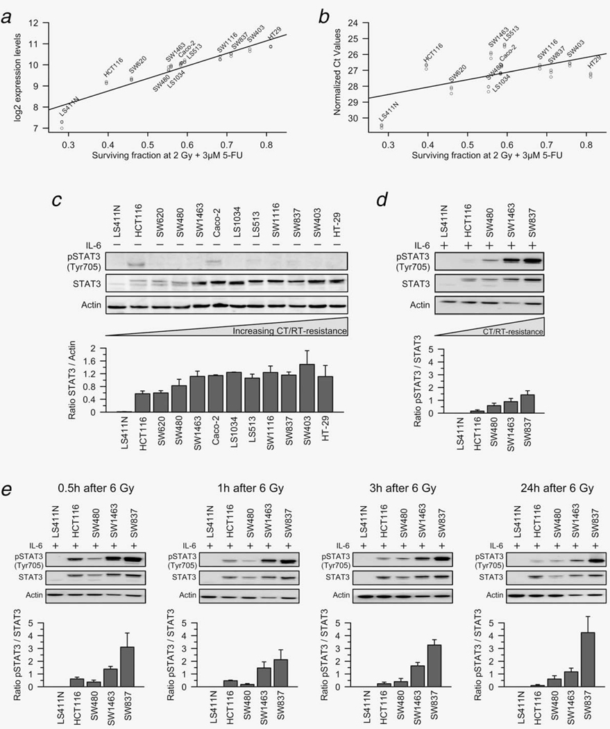
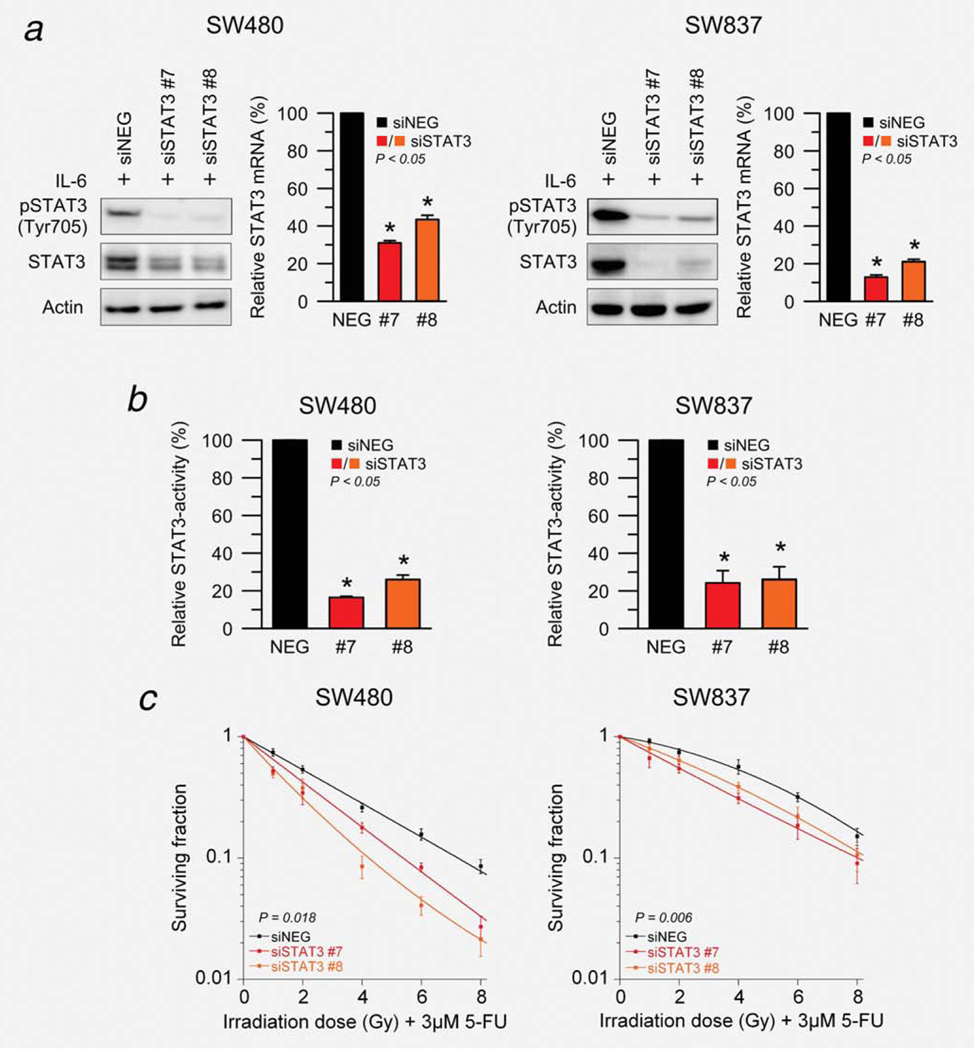
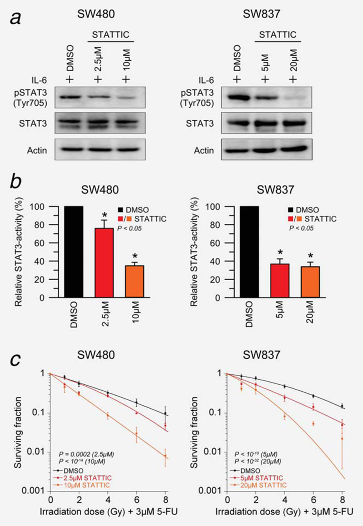
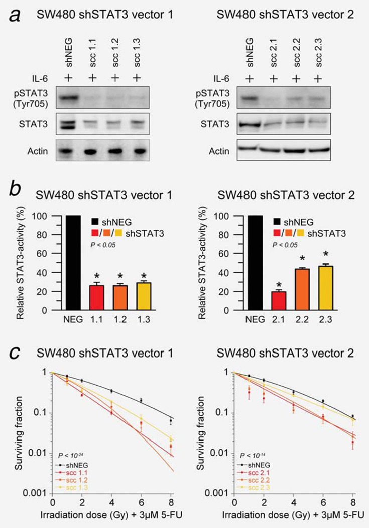
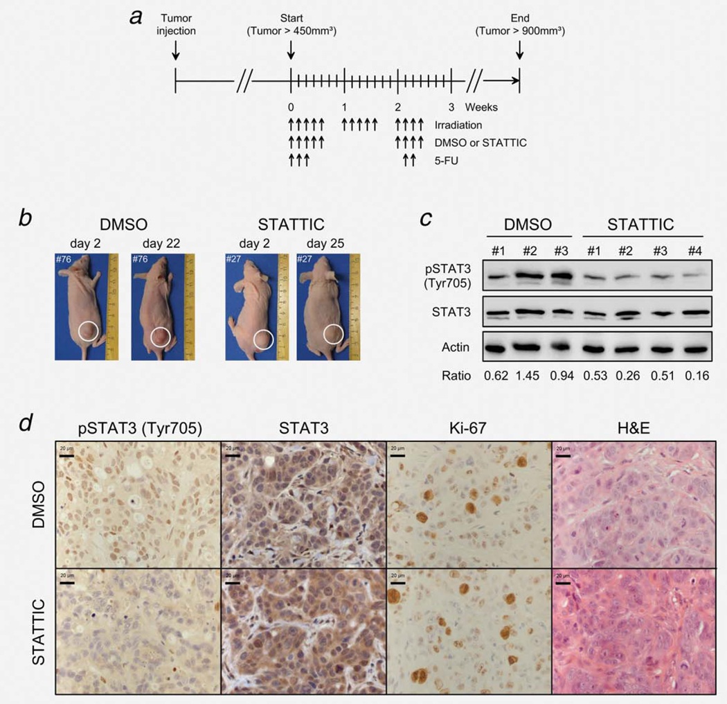
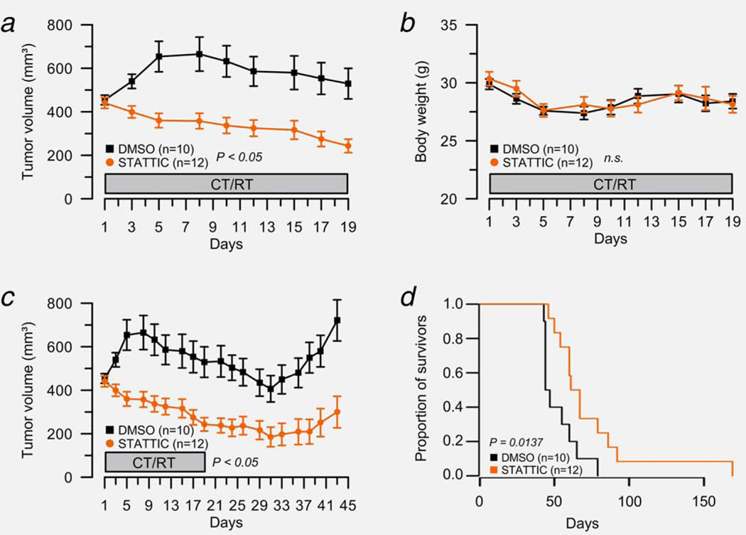
Increased activity of signal transducer and activator of transcription 3 (STAT3) is common in human malignancies, including colorectal cancers (CRCs). We have recently reported that STAT3 gene expression correlates with resistance of CRC cell lines to 5-fluorouracil (5-FU)-based chemoradiotherapy (CT/RT). This is of considerable clinical importance, because a large proportion of rectal cancers are resistant to preoperative multimodal treatment. To test whether STAT3 contributes to CT/RT-resistance, we first confirmed that STAT3 protein expression correlated positively with increasing resistance. While STAT3 was not constitutively active, stimulation with interleukin-6 (IL-6) resulted in remarkably higher expression levels of phosphorylated STAT3 in CT/RT-resistant cell lines. A similar result was observed when we determined IL-6-induced expression levels of phosphorylated STAT3 following irradiation. Next, STAT3 was inhibited in SW480 and SW837 using siRNA, shRNA and the small-molecule inhibitor STATTIC. Successful silencing and inhibition of phosphorylation was confirmed using Western blot analysis and a luciferase reporter assay. RNAi-mediated silencing as well as STATTIC treatment resulted in significantly decreased clonogenic survival following exposure to 3 µM of 5-FU and irradiation in a dose-dependent manner, with dose-modifying factors of 1.3-2.5 at a surviving fraction of 0.37. Finally, STAT3 inhibition led to a profound CT/RT-sensitization in a subcutaneous xenograft model, with a significantly delayed tumor regrowth in STATTIC-treated mice compared with control animals. These results highlight a potential role of STAT3 in mediating treatment resistance and provide first proof of concept that STAT3 represents a promising novel molecular target for sensitizing resistant rectal cancers to CT/RT.

Keywords: STAT3; chemoradiotherapy-resistance; chemoradiotherapy-sensitization; molecular target; rectal cancer.


Figures

Figure 1.
Figure 1.
STAT3 expression and IL-6-induced phosphorylation of STAT3 correlate with resistance to chemoradiotherapy. (a and b) STAT3 mRNA expression levels correlated positively with increasing resistance of 12 CRC cell lines to CT/RT, measured by microarrays (a, data modified from reference 9) and RT-PCR (b). (c) Western blot analysis confirmed this positive correlation for total STAT3 protein expression, but there was no pSTAT3Tyr705 expression in 10 of 12 cell lines. In HCT116 and Caco-2, very low expression levels of pSTAT3Tyr705 were detected. (d) After incubation with 100 ng/ml IL-6 for 30 min, pSTAT3Tyr705 levels were remarkably higher in chemoradiotherapy-resistant cell lines. (e) When cell lines were exposed to 6 Gy of X-rays, followed by stimulation with 100 ng/ml IL-6 0.5, 1, 3 and 24 hr after irradiation, we again detected remarkably higher levels of phosphorylated STAT3 in chemoradiotherapy-resistant cell lines. This difference remained stable over time. All experiments were done in triplicate and showed similar results (exemplified is one representative experiment with the respective densitometry data ± SEM (pSTAT3/STAT3 ratios) of all replicates).
Figure 2.
Figure 2.
siRNA-mediated silencing of STAT3 results in a significant sensitization to chemoradiotherapy. (a) Total STAT3 and pSTAT3Tyr705 protein levels decreased 96 hr after transfection (and stimulation with 100 ng/ml IL-6 for 30 min) with two different siRNAs targeting STAT3 compared to a nonspecific negative-control (siNEG) in SW480 (left) and SW837 (right). STAT3 mRNA silencing was established 72 hr after transfection using RT-PCR (P < 0.05). (b) STAT3 transcriptional activity was determined 96 hr after transfection (and stimulation with 100 ng/ml IL-6 for 30 min) using a dual luciferase reporter assay in SW480 (left) and SW837 (right). A negative reporter plasmid served as control. Silencing of STAT3 led to a significant reduction of STAT3 transcription factor activity (P < 0.05). (c) Seventy-two hours after transfection, SW480 (left) and SW837 cells (right) were preincubated with 3 μM of 5-FU for 16 hr, and subsequently irradiated at 1, 2, 4, 6 and 8 Gy of X-rays (without stimulation with IL-6). Silencing of STAT3 significantly increased the sensitivity of both SW480 (P = 0.018; ANOVA model) and SW837 (P = 0.006) to 5-FU-based chemoradiotherapy. Each experiment was repeated three times. Data are displayed as mean values, n = 3, error bars ± SEM.
Figure 3.
Figure 3.
Small molecule-mediated inhibition of pSTAT3Tyr705 sensitizes SW480 and SW837 cells to chemoradiotherapy in a dose-dependent manner. (a) SW480 (left) and SW837 cells (right) were treated with DMSO or different doses of STATTIC for 30 min (SW480: 2.5 and 10 μM; SW837: 5 and 20 μM, respectively), and successful inhibition of pSTAT3Tyr705 was confirmed after IL-6 stimulation (100 ng/ml) for 30 min using Western blot analysis. (b) STAT3 reporter plasmid or control reporter plasmid transfected cells were treated with DMSO or STATTIC, and a dual luciferase reporter assay demonstrated, after stimulation with 100 ng/ml IL-6 (SW480: 90 min; SW837: over night), decreased STAT3 activity following STAT3 inhibitor treatment in both cell lines (P < 0.05). (c) Cell lines were treated with DMSO or STATTIC, incubated with 3 μM of 5-FU over night and subsequently irradiated at 1, 2, 4, 6 and 8 Gy of X-rays (without stimulation with IL-6). STATTIC treatment resulted in a significant sensitization to chemoradiotherapy in a dose-dependent manner in both SW480 (2.5 μM, P = 0.0002; 10 μM, P < 10−14; ANOVA model) and SW837 (5 μM, P < 10−10; 20 μM, P < 10−32). Experiments were done in triplicates, error bars ± SEM.
Figure 4.
Figure 4.
shRNA-mediated silencing of STAT3 sensitizes SW480 cells to chemoradiotherapy. (a) Stable single-cell clone populations were established for each inducible shRNA vector targeting STAT3, and successful silencing was confirmed 120 hr after addition of 1 μg/ml doxycycline to induce shRNA expression and stimulation with 100 ng/ml IL-6 for 30 min using Western blotting. (b) Reduced STAT3 transcription factor activity was demonstrated using a dual luciferase reporter assay (P < 0.05). (c) Ninety-six hours after doxycycline induction, cell clones were incubated with 3 μM of 5-FU and irradiated at 1, 2, 4, 6 and 8 Gy of X-rays (without stimulation with IL-6). shRNA-mediated silencing of STAT3 led to a significantly increased treatment sensitivity (vector 1, P < 10−24; vector 2, P < 10−14; ANOVA model). All experiments were done as triplicates and showed similar results, error bars ± SEM.
Figure 5.
Figure 5.
Establishment of a subcutaneous xenograft model that recapitulates clinical conditions, i.e. fractionated doses of both chemotherapy and irradiation. (a) Two million SW837 cells (100 μl) were injected subcutaneously into the right flank of NMRI-Foxn1 nude mice. At a volume of ~450 mm3, mice were separated into two groups, DMSO (n = 10) and STATTIC (n = 12) and treated as indicated (black arrows pointing upward) at irradiation doses of 1.8 Gy (14×), intraperitoneal injections of 50 mg/kg 5-FU (5×) and intratumoral injections of 23% DMSO or 10 mg/kg STATTIC (9×). (b) To document tumor development, pictures of each mouse were taken once weekly. (c) To confirm inhibition of STAT3 phosphorylation in vivo, selected tumor-bearing mice were randomly sacrificed 2 hr after STATTIC or DMSO injection and subjected to Western blot analysis. Compared to DMSO-treated mice, we detected remarkably reduced pSTAT3Tyr705 levels in STATTIC-treated mice. Displayed is a representative blot from three DMSO- and four STATTIC-treated mice with the respective densitometry data (pSTAT3/STAT3 ratios). (d) Immunohistochemical analysis also confirmed remarkably reduced pSTAT3Tyr705 levels in STATTIC-treated mice compared to DMSO-treated mice (exemplified is one representative DMSO- and STATTIC-treated tumor, randomly selected). STATTIC- and DMSO-treated tumors showed similar rates of total STAT3 expression, proliferation (Ki-67 staining) and cellular morphologies (H&E staining).
Figure 6.
Figure 6.
STAT3 inhibition sensitizes SW837 cells to chemoradiotherapy in vivo. (a) Tumor size was measured thrice weekly to calculate the respective tumor volume during the administration of 5-FU and radiation therapy. While the tumor volume of DMSO-treated mice (n = 10) remained relatively stable, the tumor volume of STATTIC-treated mice (n = 12) decreased significantly over time (P = 0.046, linear mixed effects model). (b) With respect to the body weight, there was no significant difference between both groups. (c) All mice were monitored until the tumor volume exceeded 900 mm3 (start volume ×2). Tumor regrowth was significantly delayed in STATTIC-treated mice compared with control animals (P = 2.8 10−6, linear mixed effects model). (d) Kaplan–Meier curves were calculated to evaluate a potential survival benefit for mice under STATTIC treatment. The median survival for the DMSO group was 45.5 days, compared to 64.0 days for the STATTIC group, indicating a clear survival benefit for STATTIC-treated mice (P = 0.0137; hazard ratio = 0.33, 95% confidence interval: 0.13–0.83).

References

    1. Yu H, Jove R. The STATs of cancer—new molecular targets come of age. Nat Rev Cancer 2004;4: 97–105. - PubMed
    1. Haura EB, Turkson J, Jove R. Mechanisms of disease: insights into the emerging role of signal transducers and activators of transcription in cancer. Nat Clin Pract Oncol 2005;2: 315–24. - PubMed
    1. Bromberg JF, Wrzeszczynska MH, Devgan G, et al. Stat3 as an oncogene. Cell 1999;98:295–303. - PubMed
    1. Darnell JE Jr., STATs and gene regulation. Science 1997;277:1630–5. - PubMed
    1. Yu H, Kortylewski M, Pardoll D. Crosstalk between cancer and immune cells: role of STAT3 in the tumour microenvironment. Nat Rev Immunol 2007;7:41–51. - PubMed

Publication types

MeSH terms