. 2013 Oct;41(18):e174.
doi: 10.1093/nar/gkt690. Epub 2013 Aug 8.

In-gel probing of individual RNA conformers within a mixed population reveals a dimerization structural switch in the HIV-1 leader

Affiliations

In-gel probing of individual RNA conformers within a mixed population reveals a dimerization structural switch in the HIV-1 leader

Julia C Kenyon et al. Nucleic Acids Res. 2013 Oct.

Abstract

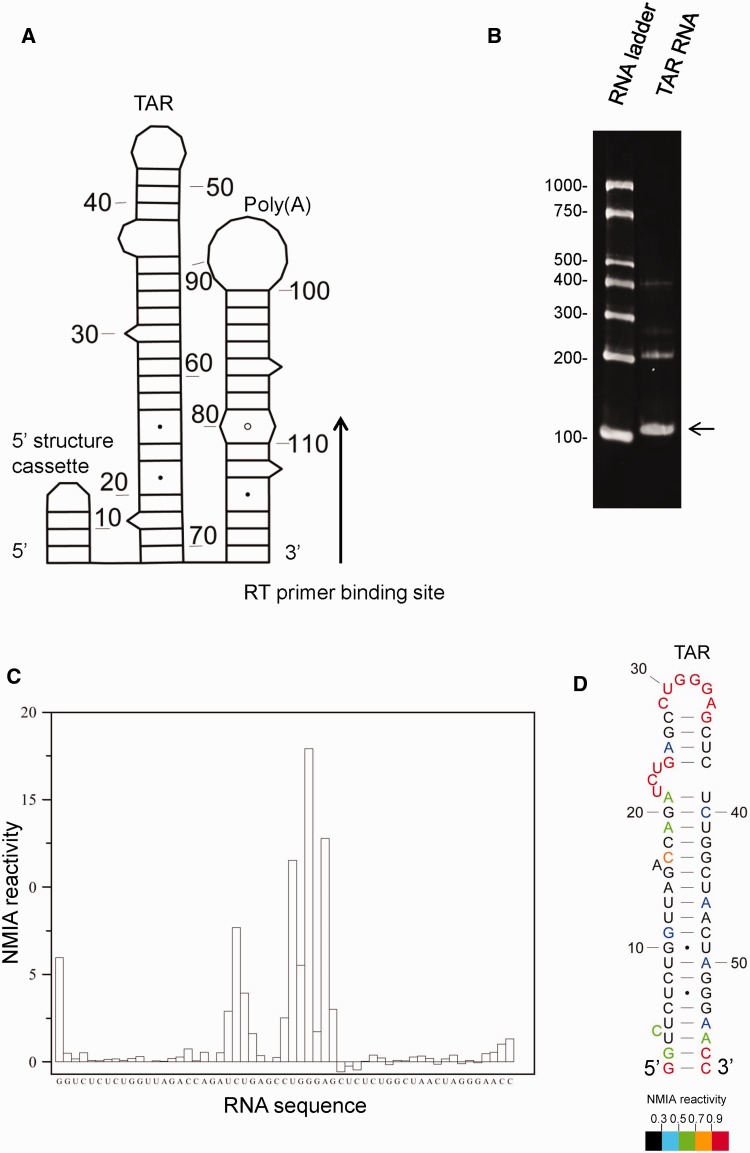
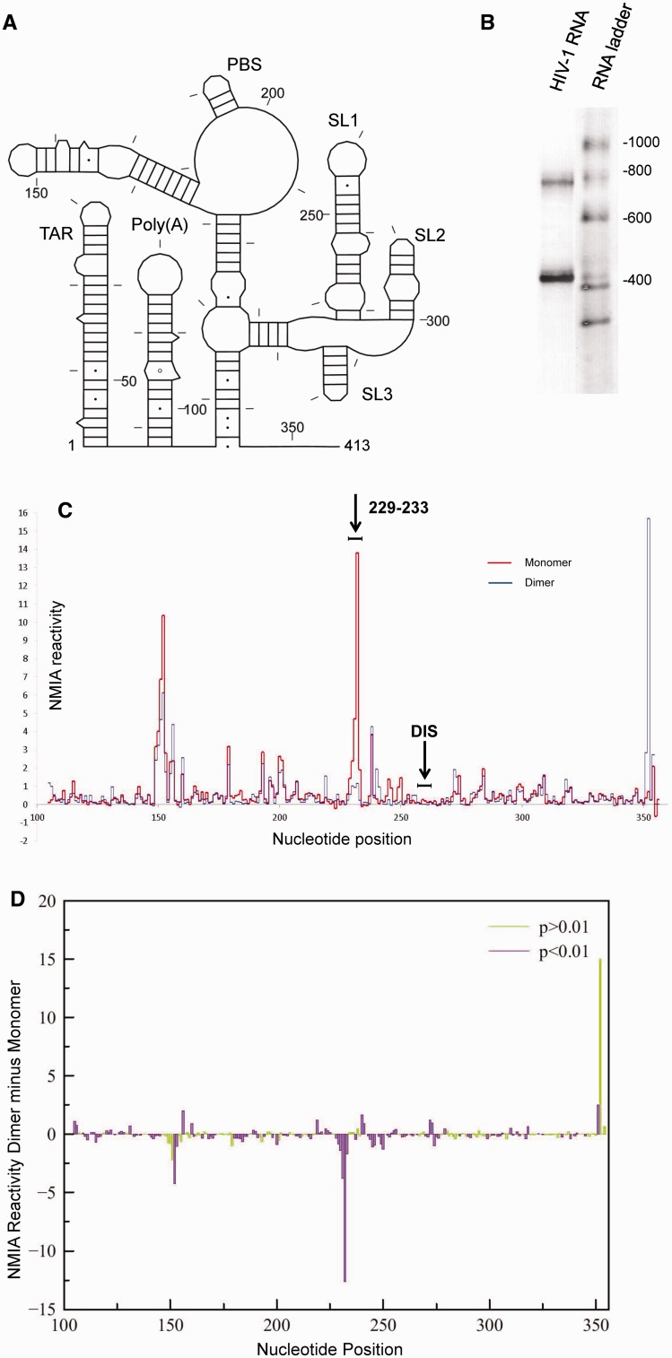
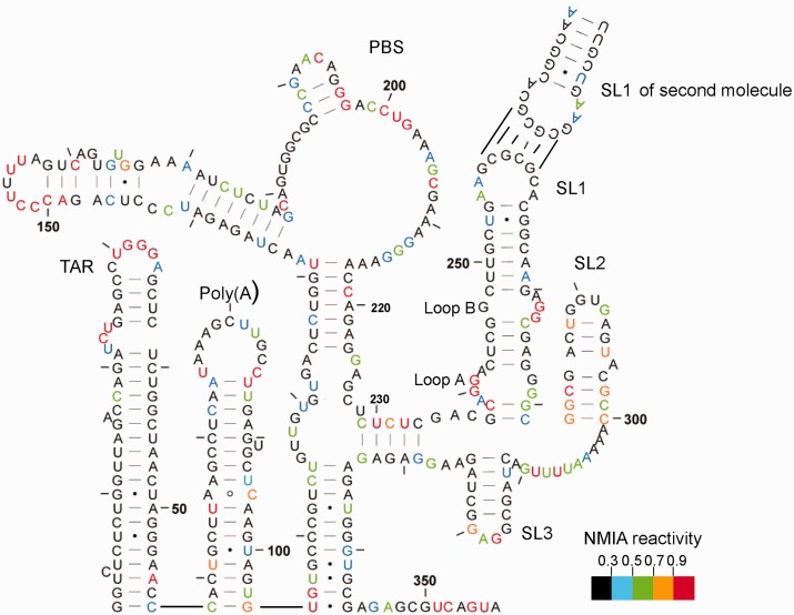
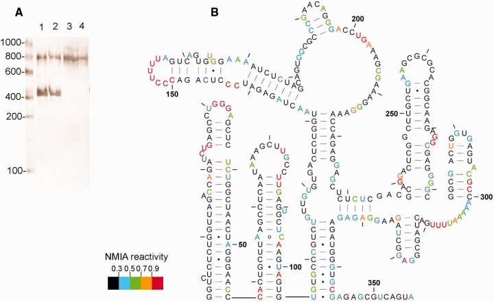
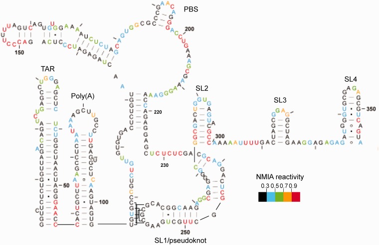
Definitive secondary structural mapping of RNAs in vitro can be complicated by the presence of more than one structural conformer or multimerization of some of the molecules. Until now, probing a single structure of conformationally flexible RNA molecules has typically relied on introducing stabilizing mutations or adjusting buffer conditions or RNA concentration. Here, we present an in-gel SHAPE (selective 2'OH acylation analysed by primer extension) approach, where a mixed structural population of RNA molecules is separated by non-denaturing gel electrophoresis and the conformers are individually probed within the gel matrix. Validation of the technique using a well-characterized RNA stem-loop structure, the HIV-1 trans-activation response element, showed that authentic structure was maintained and that the method was accurate and highly reproducible. To further demonstrate the utility of in-gel SHAPE, we separated and examined monomeric and dimeric species of the HIV-1 packaging signal RNA. Extensive differences in acylation sensitivity were seen between monomer and dimer. The results support a recently proposed structural switch model of RNA genomic dimerization and packaging, and demonstrate the discriminatory power of in-gel SHAPE.


Figures

Figure 1.
Figure 1.
In-gel SHAPE probing of a well-characterized stable RNA structure generates accurate reproducible results. (A) HIV-1 TAR RNA was appended with a 5′ structure cassette and poly(A) stem-loop. Nucleotides are numbered every 10 bases. (B) Ethidium bromide-stained native polyacrylamide gel. Arrow shows the monomeric TAR band that was excised for in-gel structural analysis. (C) NMIA reactivity trace of the in-gel probed monomeric TAR RNA. (D) RNAstructure prediction of TAR RNA, using in-gel SHAPE data as pseudo-free energy constraints. Individual NMIA reactivities are shown using different colours. Nucleotides are numbered every 10 bases.
Figure 2.
Figure 2.
In-gel SHAPE of monomeric and dimeric HIV-1 packaging signal RNA shows significant differences in reactivity between monomer and dimer. (A) Schematic diagram of the HIV-1 RNA examined. Nucleotides are numbered every 50 bases and marked every 10 bases. (B) Ethidium bromide-stained polyacrylamide gel slice showing HIV-1 RNA monomer and dimer excised for probing. (C) NMIA reactivity of monomer (red) and dimer (blue) for nucleotides 100–354. The position of the 6 nt DIS is marked above. Results are an average of 7–10 independent experiments. (D) Plot of average SHAPE reactivity of the monomer subtracted from the average NMIA reactivity of the dimer at each nt position. Colour shows statistical significance by t-test, where purple bars are statistically significant and green are not significant, to P < 0.01.
Figure 3.
Figure 3.
In-gel SHAPE data mapped onto the dimeric RNA structure proposed by Lu et al. (22). RNA was renatured as in Materials and Methods and probed with 10 mM NMIA. Nucleotides are numbered every 50 bases and marked every 10 bases. Reactivities represented by each colour are shown in the key. Data are an average of seven independent experiments.
Figure 4.
Figure 4.
In-gel SHAPE data from HIV-1 RNA renatured using snap-cooling refolding conditions. (A) Ethidium bromide-stained gel slice showing the relative amounts of monomer and dimer present when RNA is renatured by slow-cooling as described in Materials and Methods (lanes 1 and 2), or using Lu et al. conditions (22) (lanes 3 and 4). (B) NMIA reactivities mapped onto the proposed structure, shown by colour according to the key. Data are an average of four independent experiments.
Figure 5.
Figure 5.
In-gel SHAPE data from monomeric HIV-1 RNA mapped onto the proposed pseudo-knot structure. RNA was renatured using slow-cooling conditions, as in Materials and Methods, and probed using 10 mM NMIA. Reactivities are shown by colour according to the key.

References

    1. Lockard RE, Kumar A. Mapping tRNA structure in solution using double-strand-specific ribonuclease V1 from cobra venom. Nucleic Acids Res. 1981;9:5125–5140. - PMC - PubMed
    1. Tijerina P, Mohr S, Russell R. DMS footprinting of structured RNAs and RNA-protein complexes. Nat. Protoc. 2007;2:2608–2623. - PMC - PubMed
    1. Merino EJ, Wilkinson KA, Coughlan JL, Weeks KM. RNA structure analysis at single nucleotide resolution by selective 2'-hydroxyl acylation and primer extension (SHAPE) J. Am. Chem. Soc. 2005;127:4223–4231. - PubMed
    1. Gherghe CM, Shajani Z, Wilkinson KA, Varani G, Weeks KM. Strong correlation between SHAPE chemistry and the generalized NMR order parameter (S2) in RNA. J. Am. Chem. Soc. 2008;130:12244–12245. - PMC - PubMed
    1. Wilkinson KA, Gorelick RJ, Vasa SM, Guex N, Rein A, Mathews DH, Giddings MC, Weeks KM. High-throughput SHAPE analysis reveals structures in HIV-1 genomic RNA strongly conserved across distinct biological states. PLoS Biol. 2008;6:e96. - PMC - PubMed

Publication types