Clinical Trial
. 2013 Sep 20;288(38):27423-27433.
doi: 10.1074/jbc.M113.497248. Epub 2013 Aug 9.

The transcription factor Twist1 limits T helper 17 and T follicular helper cell development by repressing the gene encoding the interleukin-6 receptor α chain

Affiliations
Clinical Trial

The transcription factor Twist1 limits T helper 17 and T follicular helper cell development by repressing the gene encoding the interleukin-6 receptor α chain

Duy Pham et al. J Biol Chem. .

Abstract

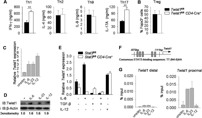
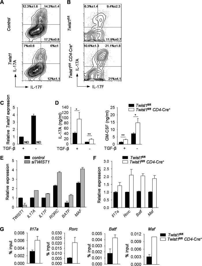
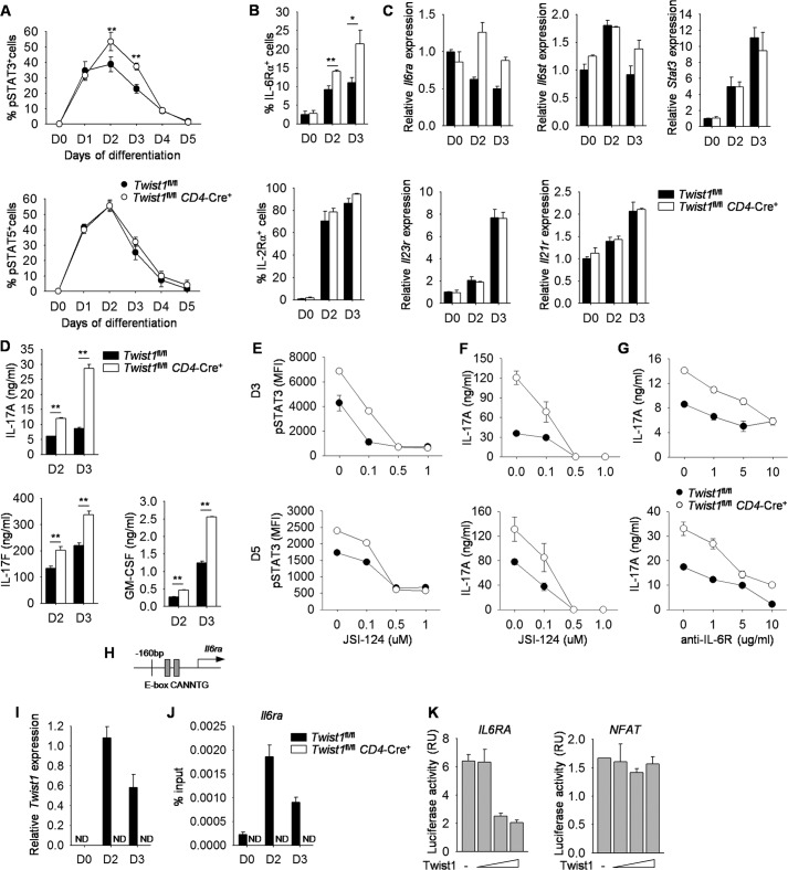
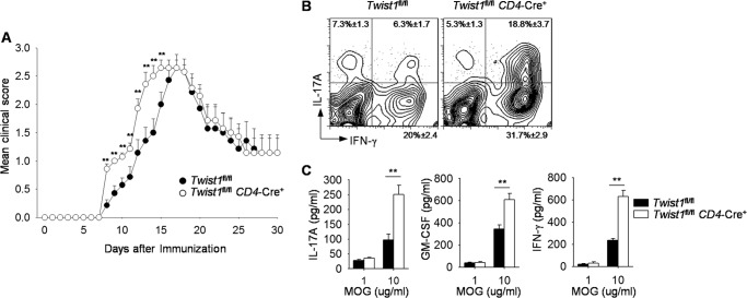
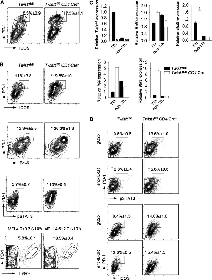
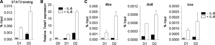
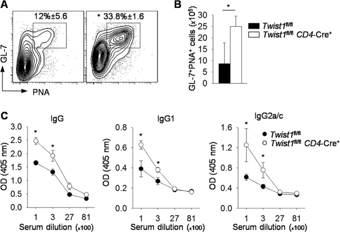
Cytokine responsiveness is a critical component of the ability of cells to respond to the extracellular milieu. Transcription factor-mediated regulation of cytokine receptor expression is a common mode of altering responses to the external environment. We identify the transcription factor Twist1 as a component of a STAT3-induced feedback loop that controls IL-6 signals by directly repressing Il6ra. Human and mouse T cells lacking Twist1 have an increased ability to differentiate into Th17 cells. Mice with a T cell-specific deletion of Twist1 demonstrate increased Th17 and T follicular helper cell development, early onset experimental autoimmune encephalomyelitis, and increased antigen-specific antibody responses. Thus, Twist1 has a critical role in limiting both cell-mediated and humoral immunity.

Keywords: Autoimmunity; Differentiation; Inflammation; Interleukin; STAT Transcription Factor; STAT3; T Cell Biology; T Helper Cell; Transcription Factors.


Figures

FIGURE 1.
FIGURE 1.
Twist1 is regulated by STAT3-activating cytokines in Th17 cells. A, naive wild type and Twist1-deficient CD4+ T cells were cultured under Th1, Th2, Th9, Th17, and Treg cell polarizing conditions. Th1, Th2, Th9, and Th17 cells were restimulated with anti-CD3 for 24 h to access cytokine production by ELISA. B, percentage of Foxp3 expression in Treg cells following in vitro differentiation. C and D, on day 5, differentiated wild type Th17 cells generated as described in A were rested or stimulated with IL-6, IL-23, or IL-12 for 2 h before gene expression analysis by qRT-PCR (C) and Twist1 expression by immunoblot (IB) with densitometry normalized against β-actin (D). E, naïve wild type and Stat3-deficient CD4+ T cells were activated with anti-CD3 and anti-CD28 in the presence or absence of IL-6, TGF-β, or IL-12 and gene expression was analyzed by qRT-PCR after 3 days. F, schematic of Twist1 promoter containing STAT3 binding sites. G, cells prepared as described in C were used for ChIP analysis using STAT3 antibody. Data are mean of four independent experiments ± S.E. (A and B), or are mean of replicate samples ± S.D. and representative of three independent experiments with similar results (C–G).**, p < 0.01. unstim, unstimulated.
FIGURE 2.
FIGURE 2.
Twist1 suppresses cytokine production in Th17 cells. A, naïve CD4+ T cells were isolated from wild type mice and differentiated under Th17 culture conditions. On day 2, cells were transduced with either control or Twist1-GFP (Twist1)-expressing retrovirus. On day 5, cells were stimulated with PMA and ionomycin for 6 h before intracellular staining (ICS) for cytokine production. Data are gated on GFP+ cells. B, differentiated wild type and Twist1-deficient Th17 cells were stimulated with PMA and ionomycin for 6 h before ICS analysis. C and D, naïve wild type and Twist1-deficient CD4+ T cells were cultured under Th17 polarizing conditions with or without TGF-β. On day 5, cells were left unstimulated for gene expression analysis by qRT-PCR (C) or reactivated with anti-CD3 for 24 h to assess cytokine production by ELISA (D). E, naïve CD4+ T cells were isolated from PBMCs and differentiated under Th17 culture conditions. On day 5, cells were transfected with control or siRNA targeting TWIST1, rested overnight, and stimulated with anti-CD3 to assess gene expression by qRT-PCR. F and G, differentiated wild type and Twist1-deficient Th17 cells were used for gene expression analysis by qRT-PCR before (Rorc, Batf, and Maf) or after (Il17a) 6 h anti-CD3 stimulation (F) and ChIP analysis using STAT3 antibody (G). Data are mean of four to five independent experiments ± S.D (A–D) or are mean of replicate samples ± S.D. and representative of three independent experiments with similar results (E–G). *, p < 0.05; **, p < 0.01. ND, not detectable.
FIGURE 3.
FIGURE 3.
Twist1 impairs IL-6-STAT3 signaling in Th17 cells. A–D, naïve CD4+ T cells were isolated from WT and Twist1fl/flCD4-Cre mice and differentiated under Th17 polarizing conditions. The levels of phospho-STAT3 (pSTAT3) and phospho-STAT5 (pSTAT5) were measured by ICS each day (A). T cells cultured under Th17 conditions for 2 or 3 days were used for surface marker analysis (B), gene expression analysis by qRT-PCR (C), or analysis of cytokine production after anti-CD3 stimulation (D). E and F, naïve CD4+ T cells were isolated from WT and Twist1fl/flCD4-Cre mice and differentiated under Th17 polarizing conditions with increased doses of STAT3 inhibitor (JSI-124). Cells were harvested on days 3 (D3) and 5 and used to measure the level of pSTAT3 by ICS (E) or restimulated with anti-CD3 to assess cytokine production by ELISA (F). G, T cells were cultured as above in the presence of control antibody or blocking antibody to IL-6R, harvested on days 3 and 5, and restimulated with anti-CD3 to assess cytokine production using ELISA. H, schematic of Il6ra promoter containing Twist1 binding sites. I and J, T cells cultured under Th17 conditions for 2 or 3 days were used for gene expression analysis by qRT-PCR (I) or used for ChIP analysis using Twist1 antibody (J). K, luciferase activity in Jurkat T cells transfected with various concentrations of plasmid encoding Twist1 along with IL6RA or NFAT luciferase reporter and then activated for 6 h with PMA and ionomycin. Data are mean of four independent experiments ± S.D. (A, B, and D) or are mean of replicate samples ± S.D. and representative of three independent experiments with similar results (C and E–K). *, p < 0.05; **, p < 0.01. ND, not detectable, RU, relative units.
FIGURE 4.
FIGURE 4.
Clinical symptoms of EAE in the absence of Twist1. A–C, wild type and Twist1fl/flCD4-Cre mice were immunized with MOGp(35–55) to induce EAE. Mean clinical score in MOG-induced EAE disease is shown in A. On day 12, mononuclear cells were isolated from brain and stimulated with PMA and ionomycin for 6 h to measure cytokine production by ICS (gated on CD4+ T cells) (B), or splenocytes were stimulated with MOG peptide for 48 h, and cytokine production was assessed by ELISA (C). Data are mean ± S.E. of seven mice per group (A) or four mice per group (B and C) and representative of two independent experiments with similar results. **, p < 0.01.
FIGURE 5.
FIGURE 5.
Mice with Twist1-deficient T cells have more T follicular helper cells. A, WT and Twist1fl/flCD4-Cre mice were immunized with MOGp(35–55) as described in Fig. 4. Twenty days following immunization, splenocytes were stained for Tfh cells. B and C, WT and Twist1fl/flCD4-Cre mice were immunized with SRBC. On day 9, splenocytes were analyzed by flow cytometry with percentages of PD-1+ICOS+, PD-1+pSTAT3+, and PD-1+IL-6Rα+ cells indicated (B). Following immunization, cell populations were sorted for CD4+CXCR5+PD-1+ICOS+ (Tfh) or CD4+CXCR5PD-1ICOS (non-Tfh), and gene expression was analyzed (C). D, SRBC-immunized WT and Twist1fl/flCD4-Cre mice were injected (intraperitoneal) with control antibody or blocking antibody to IL-6R on days 4, 6, and 8. On day 9, splenocytes were analyzed by flow cytometry with percentages of PD-1+ICOS+ and PD-1+pSTAT3+ cells indicated. (A, B, and D). Data are gated on CD4+CXCR5+. Percentages are mean ± S.E. of four to five mice per group and representative of two independent experiments with similar results (A and B), are mean ± S.E. of five mice per group (D), or are mean of replicate samples ± S.D. and representative of three independent experiments with similar results (C). *, p < 0.05. MFI, mean fluorescence intensity. ND, not detected.
FIGURE 6.
FIGURE 6.
Twist1 binds to Tfh cell-associated genes. A–C, naïve WT CD4+ T cells were activated with or without IL-6 for 2 days. Cells were harvested daily to analyze STAT3 binding to the Twist1 promoter (A) or Twist1 binding to the indicated promoters (C) by ChIP assay or to assess gene expression by qRT-PCR (B). A, percentages are mean ± S.E. of four to five mice per group. Data are mean of replicate samples ± S.D. and representative of three independent experiments with similar results. ND, not detectable; D1, day 1; D2, day 2.
FIGURE 7.
FIGURE 7.
Twist1 represses germinal center B cells and antibody production in SRBC-immunized mice. A–C, WT and Twist1fl/flCD4-Cre mice were immunized with SRBC. On day 9, splenocytes were stained for germinal center B cells (A) with total cell count shown in B. Data are gated on B220+CD19+Fas+. Serum from WT and Twist1fl/flCD4-Cre mice was diluted and used to measure antibody titers by ELISA (C). Data are mean ± S.E. of four to five mice per group and representative of two independent experiments with similar results. *, p < 0.05. PNA, peanut agglutinin.

References

    1. Zhu J., Yamane H., Paul W. E. (2010) Differentiation of effector CD4 T cell populations (*). Annu. Rev. Immunol. 28, 445–489 - PMC - PubMed
    1. Vahedi G., Takahashi H., Nakayamada S., Sun H. W., Sartorelli V., Kanno Y., O'Shea J. J. (2012) STATs shape the active enhancer landscape of T cell populations. Cell 151, 981–993 - PMC - PubMed
    1. Stritesky G. L., Yeh N., Kaplan M. H. (2008) IL-23 promotes maintenance but not commitment to the Th17 lineage. J. Immunol. 181, 5948–5955 - PMC - PubMed
    1. Mangan P. R., Harrington L. E., O'Quinn D. B., Helms W. S., Bullard D. C., Elson C. O., Hatton R. D., Wahl S. M., Schoeb T. R., Weaver C. T. (2006) Transforming growth factor-β induces development of the T(H)17 lineage. Nature 441, 231–234 - PubMed
    1. El-Behi M., Ciric B., Dai H., Yan Y., Cullimore M., Safavi F., Zhang G. X., Dittel B. N., Rostami A. (2011) The encephalitogenicity of T(H)17 cells is dependent on IL-1- and IL-23-induced production of the cytokine GM-CSF. Nat. Immunol. 12, 568–575 - PMC - PubMed

Publication types

MeSH terms

LinkOut - more resources