. 2013 Sep 27;288(39):28217-29.
doi: 10.1074/jbc.M113.496463. Epub 2013 Aug 9.

Specialization among iron-sulfur cluster helicases to resolve G-quadruplex DNA structures that threaten genomic stability

Affiliations

Specialization among iron-sulfur cluster helicases to resolve G-quadruplex DNA structures that threaten genomic stability

Sanjay Kumar Bharti et al. J Biol Chem. .

Abstract

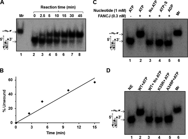
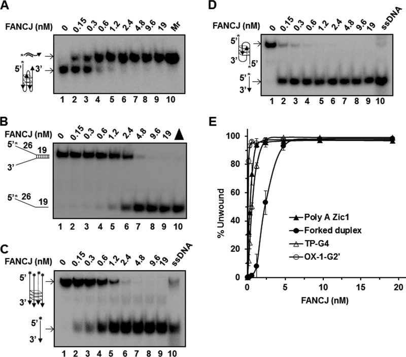
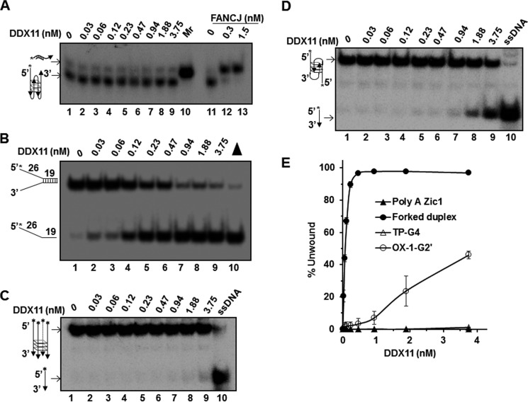
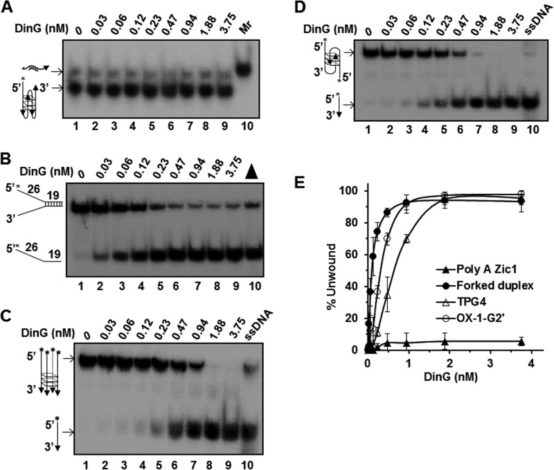
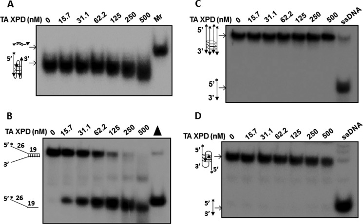
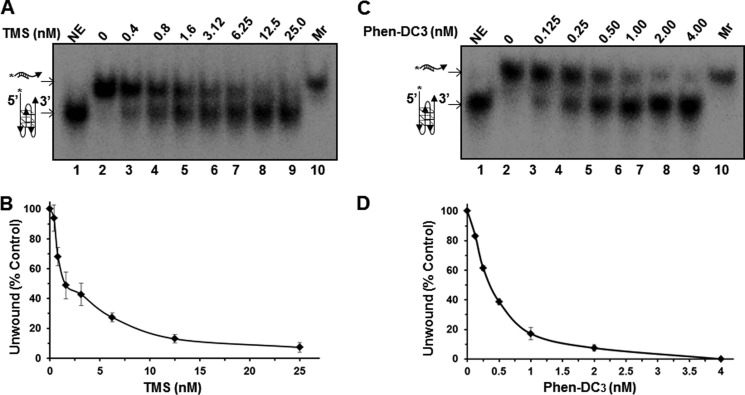
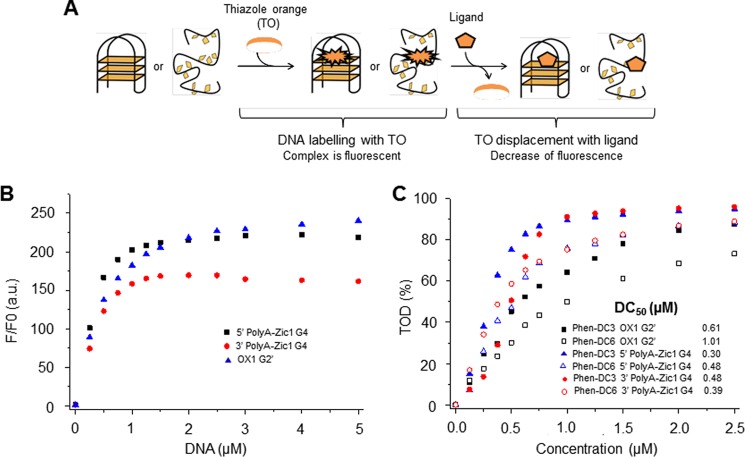
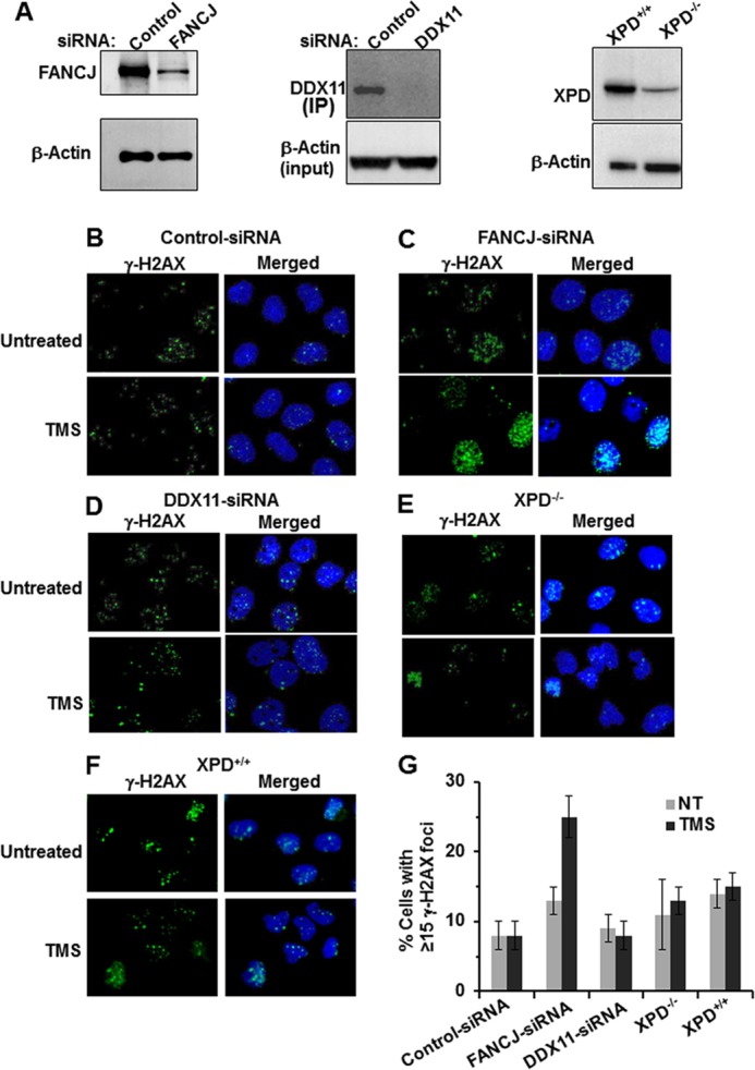
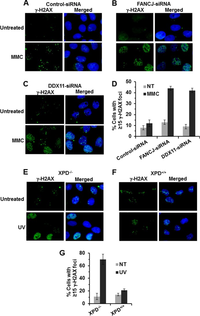
G-quadruplex (G4) DNA, an alternate structure formed by Hoogsteen hydrogen bonds between guanines in G-rich sequences, threatens genomic stability by perturbing normal DNA transactions including replication, repair, and transcription. A variety of G4 topologies (intra- and intermolecular) can form in vitro, but the molecular architecture and cellular factors influencing G4 landscape in vivo are not clear. Helicases that unwind structured DNA molecules are emerging as an important class of G4-resolving enzymes. The BRCA1-associated FANCJ helicase is among those helicases able to unwind G4 DNA in vitro, and FANCJ mutations are associated with breast cancer and linked to Fanconi anemia. FANCJ belongs to a conserved iron-sulfur (Fe S) cluster family of helicases important for genomic stability including XPD (nucleotide excision repair), DDX11 (sister chromatid cohesion), and RTEL (telomere metabolism), genetically linked to xeroderma pigmentosum/Cockayne syndrome, Warsaw breakage syndrome, and dyskeratosis congenita, respectively. To elucidate the role of FANCJ in genomic stability, its molecular functions in G4 metabolism were examined. FANCJ efficiently unwound in a kinetic and ATPase-dependent manner entropically favored unimolecular G4 DNA, whereas other Fe-S helicases tested did not. The G4-specific ligands Phen-DC3 or Phen-DC6 inhibited FANCJ helicase on unimolecular G4 ∼1000-fold better than bi- or tetramolecular G4 DNA. The G4 ligand telomestatin induced DNA damage in human cells deficient in FANCJ but not DDX11 or XPD. These findings suggest FANCJ is a specialized Fe-S cluster helicase that preserves chromosomal stability by unwinding unimolecular G4 DNA likely to form in transiently unwound single-stranded genomic regions.

Keywords: DNA Helicase; DNA Repair; DNA Replication; Fanconi Anemia; G-quadruplex; Genetic Diseases; Genomic Instability.


Figures

FIGURE 1.
FIGURE 1.
Unwinding of unimolecular G4 DNA by FANCJ is dependent on time, hydrolysable ATP, and intrinsic ATPase activity. Panel A, FANCJ (0.3 nm) was incubated with 5′ Poly(A) Zic1 unimolecular G4 DNA substrate (0.25 nm) for the indicated period of time (min). Lane 1, Mr, marker prepared by annealing a 3-fold excess of peptide-nucleic acid complementary oligonucleotide with unimolecular G4 forming single-stranded DNA after heating at 95 °C and slow cooling to room temperature; lanes 2–8, FANCJ helicase reactions incubated for the indicated times. Panel B, quantitative analysis of FANCJ helicase activity on 5′ Poly(A) Zic1 unimolecular G4 DNA substrate as a function of time in the linear range. Panel C, FANCJ (0.3 nm) was incubated with 5′ Poly(A) Zic1 unimolecular G4 DNA substrate (0.25 nm) and the indicated nucleotide for 15 min as described under “Experimental Procedures.” Lane 1, no enzyme control; lane 6, Mr, marker. ATPγS, adenosine 5′-O-(thiotriphosphate). Panel D, the indicated purified recombinant FANCJ protein (wild-type (WT); FANCJ-K52R (K52R), or FANCJ-A349P (A349P)) was incubated with 5′ Poly(A) Zic1 unimolecular G4 DNA substrate (5 fmol) and ATP (1 mm) for 15 min. Lane 1, no enzyme; lane 6, Mr, marker prepared by incubating heat-denatured 5′ Poly(A) Zic1 with 3-fold excess of peptide-nucleic acid complementary oligonucleotide and slow cooling to room temperature. NE, no enzyme control.
FIGURE 2.
FIGURE 2.
FANCJ efficiently unwinds unimolecular G4 DNA. FANCJ was tested for helicase activity on various DNA substrates. Helicase reactions (20 μl) were performed at 30 °C for 15 min under standard helicase conditions as described under “Experimental Procedures”. Panel A, the indicated concentration of FANCJ was incubated with 5′ Poly(A) Zic1 unimolecular G4 DNA substrate (0.25 nm) in the presence of a 20- fold excess of peptide-nucleic acid-complementary oligonucleotide. Lane 1, no enzyme control; lanes 2–9 indicated concentrations of FANCJ; lane 10, Mr, marker, prepared as described in the Fig. 1 legend. Panel B, the indicated concentration of FANCJ was incubated with a 19-bp forked duplex DNA substrate (0.25 nm) under standard helicase conditions as described under “Experimental Procedures.” Lane 1, no enzyme control; lanes 2–9 indicated concentrations of FANCJ; lane 10, heat-denatured DNA substrate control (filled triangle). Panels C and D, the indicated concentrations of FANCJ were incubated with 0.25 nm TP-G4 tetramolecular G4 DNA substrate (C) or bimolecular OX-I G2′ G4 DNA substrate (D) under similar conditions as described above. Lane 1, no enzyme control; lanes 2–9, indicated concentrations of FANCJ; lane 10, single-stranded DNA (ssDNA) as a marker control (Mr). Panel E, quantitative analysis of helicase activity on all DNA substrates is shown with S.D. indicated by error bars. Filled triangles, 5′ Poly(A) Zic1 G4; filled circles, 19 bp forked duplex; open triangles, TP-G4; open circles, OX-1 G2′.
FIGURE 3.
FIGURE 3.
The Fe-S helicase DDX11 fails to unwind unimolecular G4 DNA. DDX11 was tested for helicase activity on various DNA substrates. Helicase reactions (20 μl) were performed at 37 °C for 15 min under standard helicase conditions as described under “Experimental Procedures.” The indicated concentration of DDX11 was incubated with 5′ Poly(A) Zic1 unimolecular G4 DNA substrate (0.25 nm) (panel A), 19-bp forked duplex (0.25 nm) (panel B), TP-G4 tetramolecular DNA substrate (0.25 nm) (panel C), or OX-1 bimolecular G4 DNA substrate (0.25 nm) (panel D). Descriptions of the respective lanes for helicase reactions with the indicated DNA substrates are the same as those described above. Panel E, quantitative analysis of helicase activity on all DNA substrates are shown with S.D. indicated by error bars.
FIGURE 4.
FIGURE 4.
DinG efficiently unwinds bimolecular and tetramolecular G4 DNA but fails to unwind unimolecular G4 DNA. DinG was tested for helicase activity on various DNA substrates. Helicase reactions (20 μl) were performed at 37 °C for 15 min under standard helicase conditions as described under “Experimental Procedures.” The indicated concentration of DinG was incubated with 5′ Poly(A) Zic1 unimolecular G4 DNA substrate (0.25 nm) (panel A), 19-bp forked duplex (0.25 nm) (panel B), TP-G4 tetramolecular DNA substrate (0.25 nm) (panel C), or OX-1 bimolecular G4 DNA substrate (0.25 nm) (panel D). ssDNA, single-stranded DNA. Descriptions of the respective lanes for helicase reactions with the indicated DNA substrates are the same as those described above. Panel E, quantitative analysis of helicase activity on all the DNA substrates are shown with S.D. indicated by error bars.
FIGURE 5.
FIGURE 5.
XPD fails to unwind all three topological G4 DNA substrates. T. acidophilum XPD was tested for helicase activity on various DNA substrates. Helicase reactions (20 μl) were performed at 37 °C for 15 min under standard helicase conditions as described under “Experimental Procedures”. The indicated concentration of T. acidophilum XPD was incubated with 5′ Poly(A) Zic1 unimolecular G4 DNA substrate (0.25 nm) (Panel A), 19-bp forked duplex (0.25 nm) (Panel B), TP-G4 tetramolecular DNA substrate (0.25 nm) (Panel C), or OX-1 bimolecular G4 DNA substrate (0.25 nm) (Panel D). Descriptions of the respective lanes for helicase reactions with the indicated DNA substrates are the same as those described above. ssDNA, single-stranded DNA.
FIGURE 6.
FIGURE 6.
G4 ligand Phen-DC3 potently inhibits FANCJ helicase activity on unimolecular G4 DNA. Panels A and C, increasing concentrations of TMS (A) or Phen-DC3 (C) were incubated with FANCJ (0.6 nm) and the 5′ Poly(A) Zic1 unimolecular G4 DNA substrate (0.25 nm) under standard FANCJ helicase reaction conditions as described under “Experimental Procedures.” Lane 1, NE, no enzyme control; lanes 2–9, indicated concentrations of G4 ligand; lane 10, Mr, marker prepared as described above. Panels B and D, quantitative analyses of helicase data from helicase reactions with FANCJ and 5′ Poly(A) Zic1 unimolecular G4 DNA substrate containing TMS (B) or Phen-DC3 (D). Data represent the mean of at least three independent experiments with S.D. indicated by error bars.
FIGURE 7.
FIGURE 7.
G4 DNA binding by Phen-DC compounds as measured by fluorescence-induced displacement. Panel A, schematic principle of the FID assay. Panel B, data from fluorimetric titrations with TO and the specified uni- or bimolecular G4 substrates are shown. a.u., absorbance units. Panel C, data from G4-FID experiments are shown. The DC50 values corresponding to ligand concentrations required for 50% TO displacement (TOD) are indicated. See “Experimental Procedures” for details.
FIGURE 8.
FIGURE 8.
Cellular deficiency in FANCJ, but not DDX11 or XPD, sensitizes human cells to the G4 ligand TMS. Panel A, whole cell extracts from U2 OS cells transfected with control-siRNA, FANCJ-siRNA, or DDX11-siRNA were probed by Western blotting with antibodies against FANCJ, DDX11, or actin (as a loading control for input). For DDX11 Western blot detection, whole cell lysates were immunoprecipitated (IP) with rabbit anti-DDX11 antibody due to the low cellular abundance of DDX11. Also shown in Panel A is Western blot detection of XPD from whole cell extracts of XP-D mutant and corrected cells. Panels B–F, γ-H2AX foci were detected to measure the effect of TMS on DNA damage induction in human cells that are deficient in FANCJ, DDX11, or XPD helicases. FANCJ siRNA-treated, DDX11 siRNA-treated, and control siRNA-treated U2 OS cells (panels B–D) or XP-D mutant and corrected cells (panels E and F) were either exposed to 5 μm TMS for 3 h or left untreated, fixed with formaldehyde, permeabilized, and stained with anti-γ-H2AX antibody or DAPI. The merged images show cells stained with anti-γ-H2AX (green) and DAPI (blue). Panel G, quantitative analysis of γ-H2AX immunofluorescence foci data from at least three independent experiments with S.D. indicated by error bars. NT, not treated.
FIGURE 9.
FIGURE 9.
Cellular deficiency in DDX11 or XPD confers sensitivity to MMC and UV irradiation, respectively. Panels A–C, γ-H2AX foci were detected to measure the effect of MMC (panels A–D) or UV irradiation (panels E–G) on DNA damage induction in human cells that are deficient in FANCJ or DDX11 (panels A–D) or XPD (panels E–G). FANCJ siRNA-treated, DDX11 siRNA-treated, and control siRNA-treated U2 OS cells were either exposed to 100 nm MMC for 3 h or left untreated, fixed with formaldehyde, permeabilized, and stained with anti-γ-H2AX antibody or DAPI. XPD mutant and corrected cells were irradiated with UV light (2.5 J/m2), fixed, permeabilized, and stained with anti-γ-H2AX antibody or DAPI. The merged images show cells stained with anti-γ-H2AX (green) and DAPI (blue). Panels D and G, quantitative analysis of γ-H2AX immunofluorescence foci data from at least three independent experiments with S.D. indicated by error bars. NT, not treated.

Similar articles

Cited by

References

    1. Bochman M. L., Paeschke K., Zakian V. A. (2012) DNA secondary structures. Stability and function of G-quadruplex structures. Nat. Rev. Genet. 13, 770–780 - PMC - PubMed
    1. Wu Y., Brosh R. M., Jr. (2010) G-quadruplex nucleic acids and human disease. FEBS J. 277, 3470–3488 - PMC - PubMed
    1. Paeschke K., Capra J. A., Zakian V. A. (2011) DNA replication through G-quadruplex motifs is promoted by the Saccharomyces cerevisiae Pif1 DNA helicase. Cell 145, 678–691 - PMC - PubMed
    1. Ribeyre C., Lopes J., Boulé J. B., Piazza A., Guédin A., Zakian V. A., Mergny J. L., Nicolas A. (2009) The yeast Pif1 helicase prevents genomic instability caused by G-quadruplex-forming CEB1 sequences in vivo. PLoS Genet. 5, e1000475. - PMC - PubMed
    1. Piazza A., Boulé J. B., Lopes J., Mingo K., Largy E., Teulade-Fichou M. P., Nicolas A. (2010) Genetic instability triggered by G-quadruplex interacting Phen-DC compounds in Saccharomyces cerevisiae. Nucleic Acids Res. 38, 4337–4348 - PMC - PubMed

Publication types

MeSH terms

LinkOut - more resources