. 2013 Sep 15;191(6):2999-3005.
doi: 10.4049/jimmunol.1301483. Epub 2013 Aug 9.

Neuromyelitis optica IgG causes placental inflammation and fetal death

Affiliations

Neuromyelitis optica IgG causes placental inflammation and fetal death

Samira Saadoun et al. J Immunol. .

Abstract

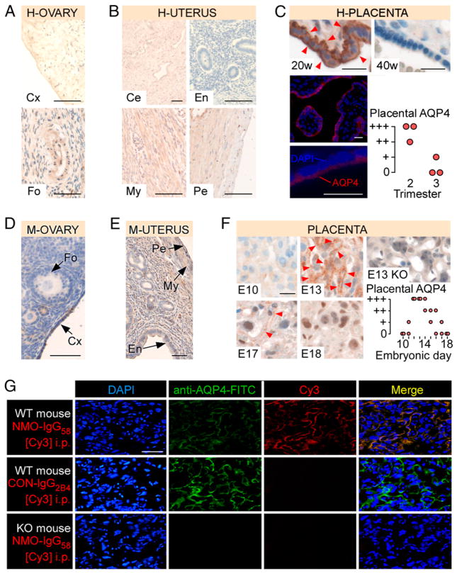
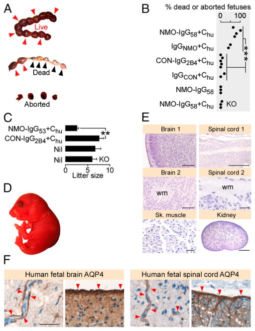
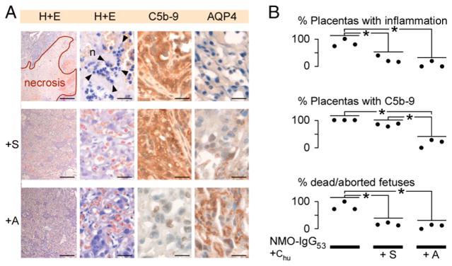
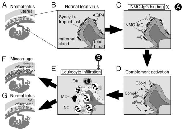
Neuromyelitis optica (NMO) is an inflammatory demyelinating disease of the CNS and affects women of childbearing age. Most patients with NMO have circulating Abs, termed NMO-IgG, against the astrocytic water channel protein aquaporin-4. In the CNS, NMO-IgG causes complement-mediated astrocyte damage, inflammatory cell infiltration, and myelin loss. In this study, we show that aquaporin-4 is expressed in the syncytiotrophoblast of human and mouse placenta. Placental aquaporin-4 expression is high during mid-gestation and progressively decreases with advancing pregnancy. Intraperitoneally injected NMO-IgG binds mouse placental aquaporin-4, activates coinjected human complement, and causes inflammatory cell infiltration into the placenta and placental necrosis. There was no damage to maternal organs that express aquaporin-4, including the brain, spinal cord, kidneys, and skeletal muscle. In control experiments, no placentitis was found in mice injected with NMO-IgG without complement, non-NMO-IgG with human complement, or in aquaporin-4 null mice injected with NMO-IgG and human complement. The infiltrating cells were primarily neutrophils with a few scattered eosinophils and macrophages. NMO-IgG and human complement-induced placentitis caused fetal death, but some fetuses were born normal when lower amounts of NMO-IgG and human complement were injected. Sivelestat, a neutrophil elastase inhibitor, and aquaporumab, a nonpathogenic IgG that competes with NMO-IgG for aquaporin-4 binding, significantly reduced NMO-IgG and human complement induced placentitis and fetal death. Our data suggest that NMO-IgG can cause miscarriage, thus challenging the concept that NMO affects only the CNS. These findings have implications for the management of NMO during pregnancy.


Conflict of interest statement

Disclosures

The authors have no financial conflicts of interest.

Figures

FIGURE 1
FIGURE 1
AQP4 is expressed in human and mouse placenta. AQP4 immunoreactivity in (A) human ovary (H-OVARY), (B) human uterus (H-UTERUS), and (C) human placenta (top; H-PLACENTA; 20 and 40 wk gestation), AQP4 immunofluorescence in 20 wk human placenta (bottom left), and human placental AQP4 versus gestational age (bottom right). AQP4 immunoreactivity in (D) mouse ovary (M-OVARY), (E) mouse uterus (M-UTERUS), (F) mouse placenta (left; M-PLACENTA; E10, E13, E17, and E18). KO is AQP4 null mouse. Red arrowheads show AQP4. Mouse placental AQP4 versus gestational age is shown (right). (G) Binding of i.p. injected NMO-IgG58[Cy3] or CON-IgG2B4[Cy3] to placenta (WT or KO mouse). Tissue was double-stained (FITC) with commercial anti-AQP4: blue (DAPI), green (FITC), red (Cy3), and yellow (Merge). Scale bars, 20 μm (C, F), 50 μm (A, D, E, G), and 100 μm (B). Ce, Cervix; Cx, cortex; En, endometrium; Fo, follicle; My, myometrium; Pe, perimetrium.
FIGURE 2
FIGURE 2
NMO-IgG causes complement-mediated placental inflammation in mice. WT pregnant mice were injected i.p. at E12 and reinjected at E13 with NMO-IgG58 plus Chu (n = 4), IgGNMO plus Chu (n = 2), CON-IgG2B4 plus Chu (n = 5), IgGCON plus Chu (n = 2) or NMO-IgG58 (n = 3) and killed at E14. KO pregnant mice were similarly injected with NMO-IgG58 plus Chu (n = 3). (A) E14 placentas stained with H&E (top), and immunostained for C5b-9 (middle) and AQP4 (bottom). Neutrophils are indicated by black arrowheads; eosinophil is indicated by a purple arrowhead; AQP4 is indicated by red arrowheads. (B) Percent of placentas with (left) inflammation, (middle) C5b-9 immunoreactivity, and (right) normal AQP4 immunoreactivity. Each dot is a pregnant mouse. (C) E14 placentas (H&E). (D) Inflammatory cells within placental lesions; neutrophils (n, black arrowheads), eosinophils (e, purple arrowheads), macrophages (m, green arrowheads), and T lymphocytes (T). (E) A WT pregnant mouse was injected with NMO-IgG58 plus Chu at E7, reinjected at E8, and killed at E9. E9 placenta is stained with H&E. (F) Brain, spinal cord, kidney, and skeletal muscle from a pregnant mother. Insets show H&E and AQP4 immunostain. Scale bars, 10 μm (D), 20 μm (A), 50 μm [(E, F), insets], 200 μm [(C, F), sk. Muscle], 500 μm [(F), brain, sp. Cord, kidney]. fRBC, Fetal RBC; mRBC, maternal RBCs.**p < 0.01, ***p < 0.001.
FIGURE 3
FIGURE 3
NMO-IgG causes complement-mediated fetal death in mice. (A) Live (red arrowheads), dead in utero plus spontaneously aborted (blue arrowheads) fetuses. (B) Dead (in utero plus spontaneously aborted) fetuses per pregnant mouse after injection of NMO-IgG58 plus Chu (n = 4), IgGNMO plus Chu (n = 2), CON-IgG2B4 plus Chu (n = 5), IgGCON plus Chu (n = 2), CON-IgG58 (n = 3), or NMO-IgG58 plus Chu in KO mice (n = 3). Each dot is a pregnant mouse. (C) Litter size delivered after injection of NMO-IgG53 plus Chu (n = 3) or CON-IgG2B4 plus Chu (n = 5), or without injection (Nil) in WT (n = 7) and KO (n = 5) mice. (D) Macroscopic appearance and (E) H&E staining of sections of brain, spinal cord, skeletal muscle, and kidney of P1 mouse from a mother that received i.p. NMO-IgG53 plus Chu. (F) AQP4 immunoreactivity (red arrowheads) in human fetal (left) frontal lobe and (right) spinal cord. Scale bars, 30 μm [(E) Sp. Cord 2, Sk. Muscle, (F)], 100 mm [(E), brain 1, brain 2], 500 μm [(E) kidney, Sp. Cord 1]. wm, White matter. **p < 0.01, ***p < 0.001.
FIGURE 4
FIGURE 4
Sivelestat and aquaporumab reduce NMO-IgG–mediated placental damage. (A) Placentas (H&E, C5b-9 immunostain, AQP4 immunostain) after i.p. injection of NMO-IgG53 plus Chu (n = 3), NMO-IgG53 plus Chu plus sivelestat (+S, n = 3) or NMO-IgG53 plus Chu plus aquaporumab (+A, n = 3). (B) Percentage of placentas with neutrophil infiltration and C5b-9 deposits and percentage of dead fetuses per pregnant mouse. Scale bars, 30 μm [(A) H&E right, C5b-9, AQP4], 300 μm [(A) H&E left]. *p < 0.01.
FIGURE 5
FIGURE 5
Proposed mechanism of NMO-IgG–induced placental inflammation. (A) Normal fetus in uterus with a (B) magnified view of a normal fetal villus showing AQP4 within the syncytiotrophoblast plasma cell membrane. (C) NMO-IgG binds extracellular epitopes on AQP4 and (D) activates complement causing the deposition of membrane attack complexes (C5b-9) in the syncytiotrophoblast plasma membrane. (E) Leukocytes infiltrate the placenta, primarily neutrophils (NΦ) with some eosinophils (EΦ) and macrophages (MΦ). (F) Severe placental inflammation causes fetal death, but (G) mild placental inflammation allows normal fetal growth. Aquaporumab (A) inhibits NMO-IgG binding, and sivelestat (S) inhibits neutrophil-mediated damage.

References

    1. Papadopoulos MC, Verkman AS. Aquaporin 4 and neuromyelitis optica. Lancet Neurol. 2012;11:535–544. - PMC - PubMed
    1. Jacob A, McKeon A, Nakashima I, Sato DK, Elsone L, Fujihara K, de Seze J. Current concept of neuromyelitis optica (NMO) and NMO spectrum disorders. J Neurol Neurosurg Psychiatry. 2013;84:922–930. - PubMed
    1. Waters PJ, McKeon A, Leite MI, Rajasekharan S, Lennon VA, Villalobos A, Palace J, Mandrekar JN, Vincent A, Bar-Or A, Pittock SJ. Serologic diagnosis of NMO: a multicenter comparison of aquaporin-4-IgG assays. Neurology. 2012;78:665–671. discussion 669. - PMC - PubMed
    1. Lennon VA, Kryzer TJ, Pittock SJ, Verkman AS, Hinson SR. IgG marker of optic-spinal multiple sclerosis binds to the aquaporin-4 water channel. J Exp Med. 2005;202:473–477. - PMC - PubMed
    1. Lennon VA, Wingerchuk DM, Kryzer TJ, Pittock SJ, Lucchinetti CF, Fujihara K, Nakashima I, Weinshenker BG. A serum autoantibody marker of neuromyelitis optica: distinction from multiple sclerosis. Lancet. 2004;364:2106–2112. - PubMed

Publication types

LinkOut - more resources