. 2013;9(7):e1003456.
doi: 10.1371/journal.ppat.1003456. Epub 2013 Jul 25.

A multi-targeted drug candidate with dual anti-HIV and anti-HSV activity

Affiliations

A multi-targeted drug candidate with dual anti-HIV and anti-HSV activity

Jan Balzarini et al. PLoS Pathog. 2013.

Abstract

Human immunodeficiency virus (HIV) infection is often accompanied by infection with other pathogens, in particular herpes simplex virus type 2 (HSV-2). The resulting coinfection is involved in a vicious circle of mutual facilitations. Therefore, an important task is to develop a compound that is highly potent against both viruses to suppress their transmission and replication. Here, we report on the discovery of such a compound, designated PMEO-DAPym. We compared its properties with those of the structurally related and clinically used acyclic nucleoside phosphonates (ANPs) tenofovir and adefovir. We demonstrated the potent anti-HIV and -HSV activity of this drug in a diverse set of clinically relevant in vitro, ex vivo, and in vivo systems including (i) CD4⁺ T-lymphocyte (CEM) cell cultures, (ii) embryonic lung (HEL) cell cultures, (iii) organotypic epithelial raft cultures of primary human keratinocytes (PHKs), (iv) primary human monocyte/macrophage (M/M) cell cultures, (v) human ex vivo lymphoid tissue, and (vi) athymic nude mice. Upon conversion to its diphosphate metabolite, PMEO-DAPym markedly inhibits both HIV-1 reverse transcriptase (RT) and HSV DNA polymerase. However, in striking contrast to tenofovir and adefovir, it also acts as an efficient immunomodulator, inducing β-chemokines in PBMC cultures, in particular the CCR5 agonists MIP-1β, MIP-1α and RANTES but not the CXCR4 agonist SDF-1, without the need to be intracellularly metabolized. Such specific β-chemokine upregulation required new mRNA synthesis. The upregulation of β-chemokines was shown to be associated with a pronounced downmodulation of the HIV-1 coreceptor CCR5 which may result in prevention of HIV entry. PMEO-DAPym belongs conceptually to a new class of efficient multitargeted antivirals for concomitant dual-viral (HSV/HIV) infection therapy through inhibition of virus-specific pathways (i.e. the viral polymerases) and HIV transmission prevention through interference with host pathways (i.e. CCR5 receptor down regulation).


Conflict of interest statement

Jan Balzarini and Antonin Holý are co-inventors of the acyclic nucleoside phosphonates. This did not alter our adherence to all PLOS Pathogens policies on sharing data and materials. The other authors declare no competing financial interests.

Figures

Figure 1
Figure 1. Structural formulae of the acyclic nucleoside phosphonates.
Adefovir and tenofovir are purine (adenine) analogues. PMEO-DAPym is a (2,4-diamino)pyrimidine analogue , . Molecular modeling revealed that the PMEO-derivatives are structural mimics of the corresponding purine (2-aminoadenine) analogues , and it was recently shown that PMEO-DAPym also functionally behaves as an adenine analogue like adefovir and tenofovir .
Figure 2
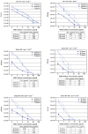
Figure 2. Antiherpetic activity of test compounds in cell culture.
Confluent HEL cell cultures were exposed to 100 TCID50 of clinical virus isolates [wild-type HSV-1 (panel A), wild-type HSV-2 (panel B), TK HSV-1 (panel C) and TK HSV-2 (panel D)] in the presence of drugs at different concentrations and incubated for 3 days at 37°C. Then, the cytopathicity was determined microscopically and the EC50 values determined. aEC50, 50% effective concentration or compound concentration required to reduce virus-induced cytopathicity (CPE) by 50%. Data shown are the means of at least two independent experiments. The HSV-1, HSV-2, HSV-1 TK, and HSV-2 TK clinical isolates have been described in reference 19, including the nature of the mutations in the TK gene of the acyclovir-resistant virus strains.
Figure 3
Figure 3. Inhibitory activities of tenofovir, adefovir, and PMEO-DAPym against laboratory HSV-1 and HSV-2 strains in organotypic epithelial raft cultures.
Panels A (HSV-1) and B (HSV-2): Drugs at different concentrations were added to the raft cell cultures on the day of infection (10 days after initiation of differentiation). The drugs remained in the presence of the cells for 5 days until the rafts were frozen for determination of virus production with a plaque assay in HEL cell cultures. Error bars represent S.D.
Figure 4
Figure 4. Inhibitory activity of PMEO-DAPym against clinical HSV isolates in HEL cell cultures.
Different PMEO-DAPym concentrations were exposed to several wild-type clinical HSV-1 (RV-174) and HSV-2 (NS and RV-194) isolates in HEL cell cultures at different multiplicities of infection (m.o.i.; 10−3 (left graph) or 10−4 (right graph)). Virus yield was determined at 24, 48, and 72 h post infection, and EC90 and EC99 values were calculated from the graphical plots. Error bars represent S.D.
Figure 5
Figure 5. Suppression of HSV-2 in infected human ex vivo tonsillar tissue by adefovir and PMEO-DAPym.
Blocks of human tonsillar tissue were inoculated ex vivo with HSV-2 (G) and treated or not with adefovir or PMEO-DAPym. We monitored HSV-2 (G) replication by measuring viral DNA in culture media at different times throughout the culture period. Presented are means ± SEM of cumulative HSV-2 (G) replication in tissues from two to six donors. For each donor, data represent pooled viral release from 27 tissue blocks.
Figure 6
Figure 6. Suppression of HSV-2 in human cervico-vaginal tissues by adefovir and PMEO-DAPym.
Blocks of human cervico-vaginal tissues were inoculated ex vivo with HSV-2 (G) or co-infected with HIV-1 and HSV-2 and treated or not with adefovir (1 µg/ml) or PMEO-DAPym (1 µg/ml). We monitored HSV-2 (G) replication by measuring viral DNA accumulated in culture media at different times throughout the culture period. Presented is cumulative HSV-2 (G) replication, with data representing pooled viral release from 16 tissue blocks.
Figure 7
Figure 7. Suppression of HSV-2 in single-infected and HIV-1 co-infected human ex vivo tonsillar tissue by adefovir and PMEO-DAPym.
Panels A and B: Blocks of human tonsillar tissue from five different donors were co-inoculated ex vivo with HSV-2 (G) and HIV-1 (LAI) and treated or not with adefovir or PMEO-DAPym (1 µg/ml). For tissue from each donor, we monitored replication of both viruses by evaluating their accumulation in the culture media bathing 27 tissue blocks at different times throughout the culture. We evaluated HIV-1 replication by measuring p24gag and HSV-2 (G) replication by measuring HSV-2 viral DNA. Panel A: Presented are kinetics of HSV-2 (G) and HIV-1 (LAI) replication (insert). Each point represents the mean ± SEM of viral replication in tissues from five donors. Panel B: Presented are means ± SEM of cumulative HSV-2 (G) and HIV-1 (LAI) replication (insert).
Figure 8
Figure 8. Effects of adefovir and PMEO-DAPym on mortality in mice inoculated with HSV-1 or HSV-2.
Groups of five nu/nu mice were inoculated with HSV-1 (panel A) or HSV-2 (panel B) on the lumbosacral area. Each cohort was then subjected to topical treatment twice daily for 5 consecutive days, starting on the day of viral infection. The placebo groups received a similar treatment with the test formulation without drug. Mortality was recorded over a period of 30 days. Animals were euthanized when more than 30% loss in body weight or development of paralysis occurred. We estimated survival rates according to the Kaplan-Meier method and compared them using the log-rank test (Mantel-Cox using GraphPad Prism). Several curves proved statistically significant (p<0.01) in comparison of each treatment with placebo.
Figure 9
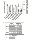
Figure 9. Expression of CC-chemokines, CCR5 and chemokine mRNA expression in PBMC cultures after drug treatment.
Panel A: Production of CC-chemokines by PBMC and the effects of adefovir, tenofovir, and PMEO-DAPym on the expression of the CCR5 receptor. Freshly isolated PBMCs from four healthy donors were incubated for 24 h with medium only (NC) or with adefovir, tenofovir, or PMEO-DAPym. We collected the supernatants and measured the concentrations of MIP-1α (▪), MIP-1β (▴), and RANTES (•) using a Bioplex system (Bio-Rad, Hercules, CA). Shown are means ± SEM. The cells were also collected, and the expression of the CCR5 receptor (□) was measured with flow cytometry using the PE-labeled CCR5 (clone 2D7) mAb; it is shown as percentage of control (± SEM) from four independent representative experiments. Panel B: Chemokine mRNA expression upon PMEO-DAPym exposure to PBMC. RT-PCR analysis of MIP-1α, MIP-1β, RANTES, and GAPDH (control) expression in PBMC treated with 100-µg/ml PMEO-DAPym for 4 h. Similar results were obtained with PBMC from three different donors. Results from one representative donor are shown.
Figure 10
Figure 10. Expression of activation markers on PBMCs treated with adefovir, tenofovir, or PMEO-DAPym.
PBMCs were incubated with adefovir, tenofovir, or PMEO-DAPym for 3 days. The mitogenic lectin PHA (at 2 µg/ml) was included as a positive control. We measured cell surface expression of CD4 and the activation markers CD69 (top panel), CD25 (middle panel), and HLA-DR (bottom panel) using flow cytometry with the fluorescein isothiocyanate-labeled CD4-specific mAb (clone SK3) and the phycoerythrin-labeled CD69, CD25, HLA-DR-specific mAbs. Shown are means ± SEM from two independent experiments.
Figure 11
Figure 11. Expression of the HIV-1 coreceptor CCR5 on PBMCs after treatment with adefovir, tenofovir, or PMEO-DAPym.
We treated PBMC with the drugs for 24 h and collected and analyzed the cells using flow cytometry. Cell surface expression of CD4 and the chemokine receptor CCR5 were measured with the fluorescein isothiocyanate-labeled CD4 specific mAb (clone SK3) and the phycoerythrin-labeled CCR5 (clone 2D7). The percentages of positive cells in each quadrant of the dot plots are given. The data shown are from one representative experiment that was independently repeated at least four times.
Figure 12
Figure 12. Effects of the supernatants collected after incubation of PBMCs with adefovir, tenofovir, and PMEO-DAPym on the expression of CCR5.
PBMCs were treated with medium alone or with various concentrations of adefovir, tenofovir, or PMEO-DAPym, and the supernatants were collected after 24 h. Then, freshly isolated PBMCs were incubated at 37°C for 1 h with these various supernatants, with medium alone, or with LD78β (control) at 100 ng/ml, 10 ng/ml, and 1 ng/ml. We measured the expression of the CCR5 receptor using flow cytometry with the phycoerythrin-labeled CCR5 (clone 2D7) mAb: shown are the percentages (means ± SEM for two independent experiments) of CCR5+ cells.

References

    1. Blower S, Ma L (2004) Calculating the contribution of herpes simplex virus type 2 epidemics to increasing HIV incidence: treatment implications. Clin Infect Dis 39, Suppl 5: S240–S247. - PubMed
    1. Corey L (2007) Synergistic copathogens–HIV-1 and HSV-2. N Engl J Med 356: 854–856. - PubMed
    1. Buvé A (2010) Can we reduce the spread of HIV infection by suppressing herpes simplex virus type 2 infection? F1000 Med Rep 2: 41. - PMC - PubMed
    1. Freeman EE, Weiss HA, Glynn JR, Cross PL, Whitworth JA, et al. (2006) Herpes simplex virus 2 infection increases HIV acquisition in men and women: systematic review and meta-analysis of longitudinal studies. AIDS 20: 73–83. - PubMed
    1. Martinelli E, Tharinger H, Frank I, Arthos J, Piatak M Jr, et al. (2011) HSV-2 infection of dendritic cells amplifies a highly susceptible HIV-1 cell target. PLoS Pathog 7: e1002109. - PMC - PubMed

Publication types

MeSH terms