. 2013;9(8):e1003513.
doi: 10.1371/journal.ppat.1003513. Epub 2013 Aug 1.

Rab18 binds to hepatitis C virus NS5A and promotes interaction between sites of viral replication and lipid droplets

Affiliations

Rab18 binds to hepatitis C virus NS5A and promotes interaction between sites of viral replication and lipid droplets

Shadi Salloum et al. PLoS Pathog. 2013.

Abstract

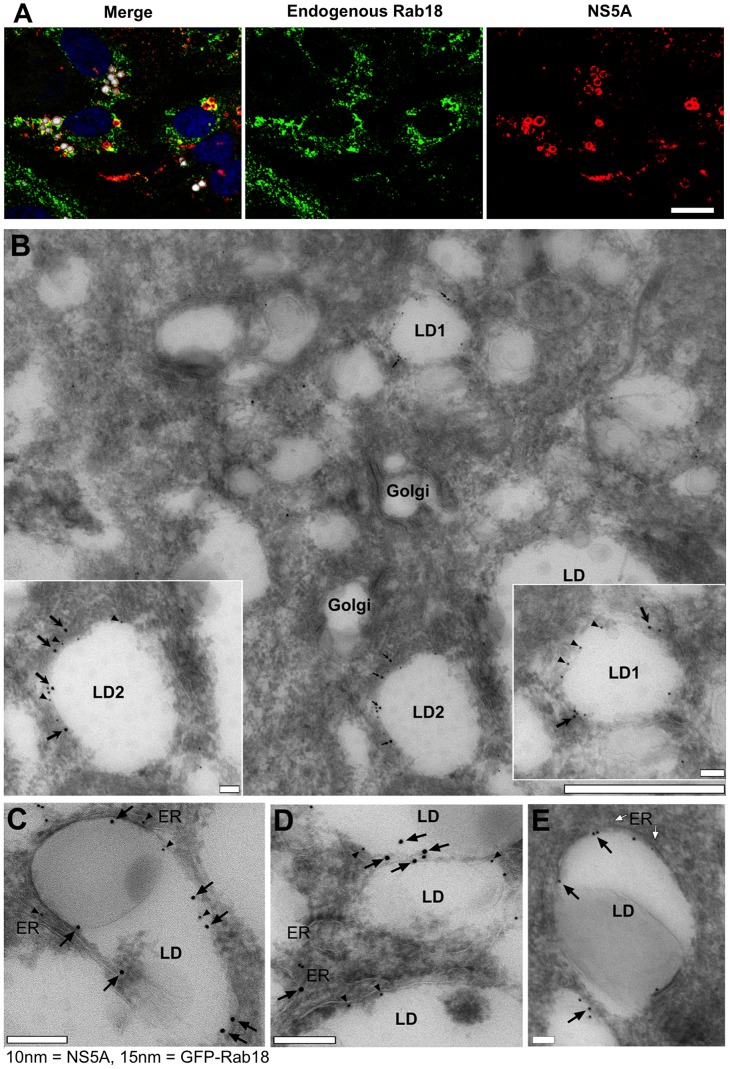
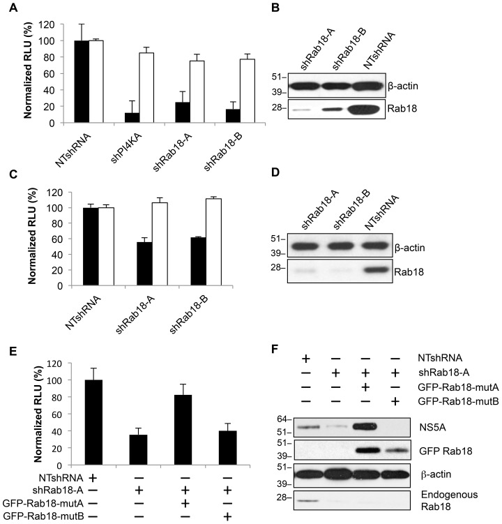
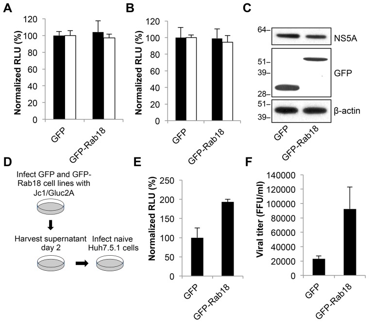
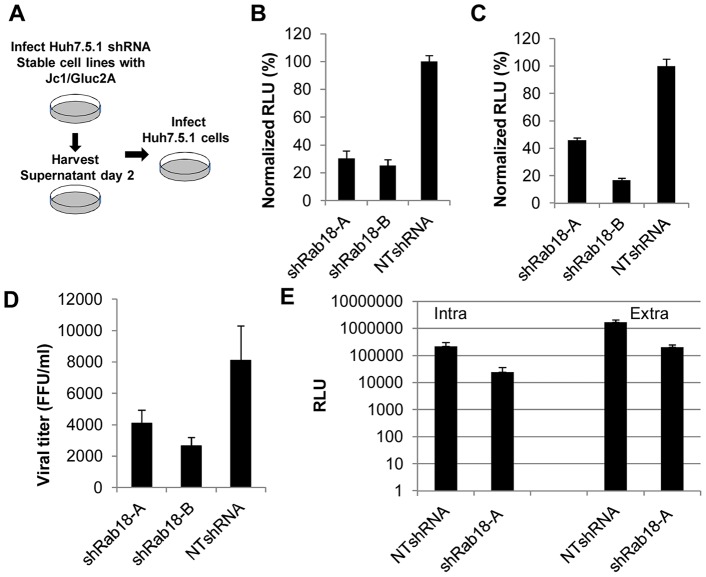
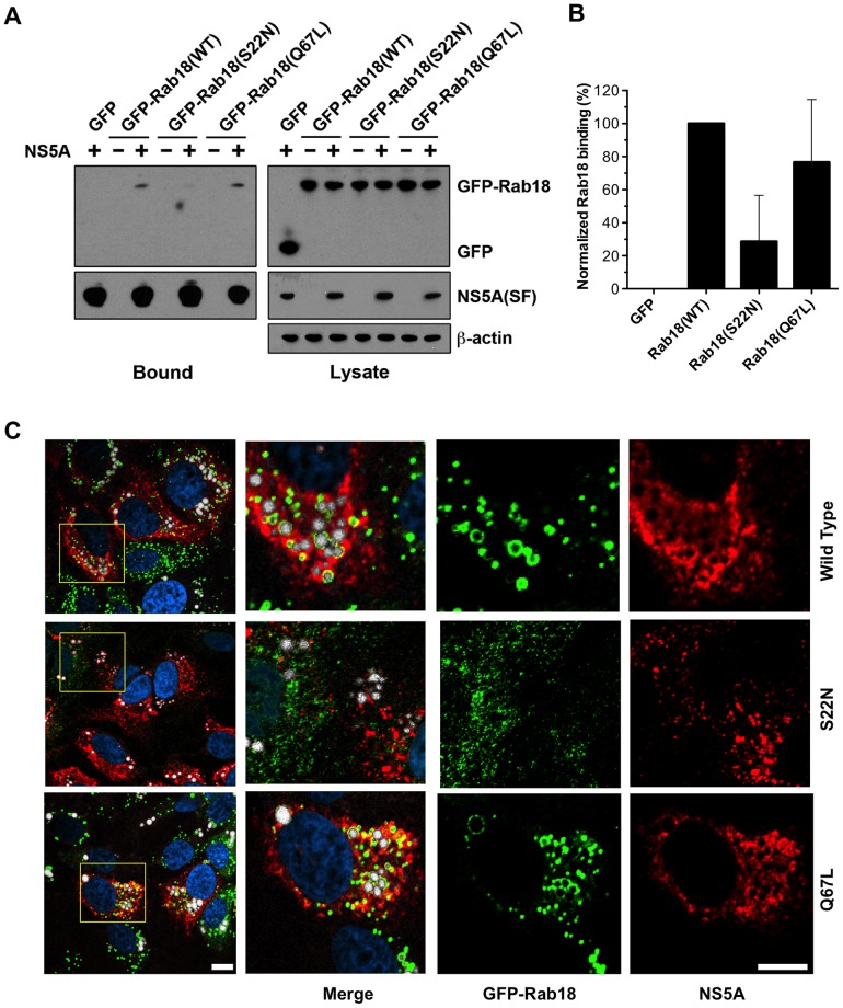
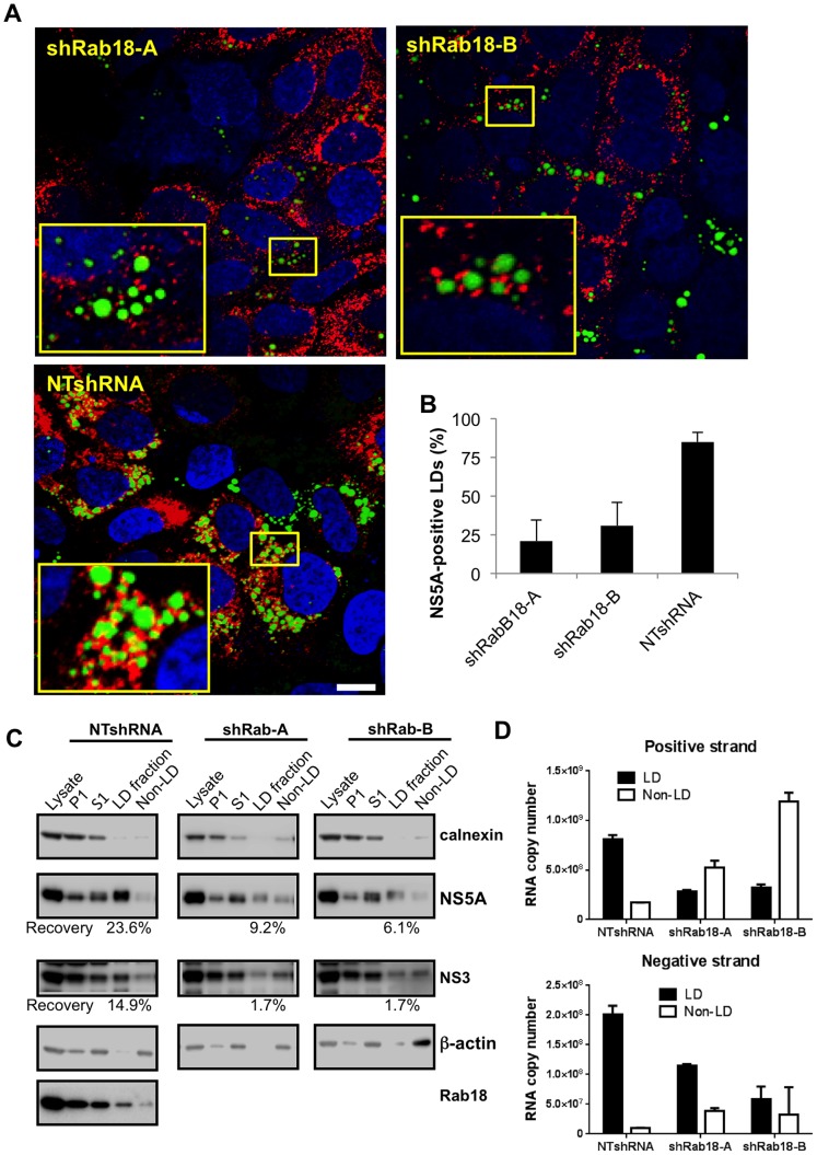
Hepatitis C virus (HCV) is a single-stranded RNA virus that replicates on endoplasmic reticulum-derived membranes. HCV particle assembly is dependent on the association of core protein with cellular lipid droplets (LDs). However, it remains uncertain whether HCV assembly occurs at the LD membrane itself or at closely associated ER membranes. Furthermore, it is not known how the HCV replication complex and progeny genomes physically associate with the presumed sites of virion assembly at or near LDs. Using an unbiased proteomic strategy, we have found that Rab18 interacts with the HCV nonstructural protein NS5A. Rab18 associates with LDs and is believed to promote physical interaction between LDs and ER membranes. Active (GTP-bound) forms of Rab18 bind more strongly to NS5A than a constitutively GDP-bound mutant. NS5A colocalizes with Rab18-positive LDs in HCV-infected cells, and Rab18 appears to promote the physical association of NS5A and other replicase components with LDs. Modulation of Rab18 affects genome replication and possibly also the production of infectious virions. Our results support a model in which specific interactions between viral and cellular proteins may promote the physical interaction between membranous HCV replication foci and lipid droplets.


Conflict of interest statement

The authors have declared that no competing interests exist.

Figures

Figure 1
Figure 1. Identification of NS5A-binding host proteins using a proteomic strategy.
A. Diagram of Jc1(SF) showing location of FLAG and tandem Strep-Tags in domain III of NS5A. B. Detection of proteins specifically associated with NS5A(SF). Huh7.5.1 cells were infected with Jc1(SF) (left lane) and untagged wild-type Jc1 (right lane). Cell lysates were incubated with Streptactin-Sepharose and the affinity matrix was washed extensively followed by elution with biotin. Eluted proteins were separated by SDS-PAGE and visualized by colloidal Coomassie Blue staining. The position of NS5A(SF) is indicated by the gray arrowhead. Proteins that specifically associate with NS5A(SF) are indicated by black arrows. C. Schematic showing strategy of affinity purification combined with Stable Isotope Labeling with Amino Acids in Cell Culture (SILAC). Huh 7.5.1 cells were metabolically labeled with medium containing normal “light” arginine and lysine amino acids (LAA) or “heavy” L-arg-13C6, 15N4 and L-lys-13C6, 15N2 (HAA) for 6 days before infecting LAA-labeled cells with Jc1 and HAA-labeled cells with Jc1(SF) virus. Cells were harvested at day 6 post infection. Equal amounts of protein from HAA and LAA-labeled lysates were mixed and then subjected to Streptactin affinity purification as described above. D. Lysates from LAA-labeled, Jc1-infected cells and HAA-labeled, Jc1(SF)-infected cells were mixed and subjected to Streptactin affinity purification. The entire eluate was concentrated by precipitation, separated by SDS-PAGE and stained with Coomassie Blue prior to gel slice excision for mass spectrometry. The expected position of Rab18 is indicated with an arrow, although no discrete band is visible on this gel by Coomassie Blue staining.
Figure 2
Figure 2. Colocalization of NS5A with Rab18-positive LDs in HCV-infected cells.
A. Huh7.5.1 hepatoma cells were infected with JFH-1 and then immunolabeled for endogenous Rab18 (green) and NS5A (red). LDs were visualized by HCS LipidTOX Deep Red neutral lipid staining (false-colored white in these panels) and nuclei were counterstained with DAPI (blue). Bar, 10 µm. (B–E). Immunogold transmission electron microscopy of HCV-infected (B–D) or control uninfected (E) Huh7.5.1 cells stably expressing GFP-Rab18. Insets in panel B represent zoomed-in views of the lipid droplets LD1 and LD2. GFP-Rab18 is labeled with 15 nm gold particles (arrows), while NS5A is labeled with 10 nm gold particles (arrowheads). The white arrows in panel E point to putative ER membranes in close apposition to the LD surface. Bar in panel B, 1 µm; C–E and insets, 100 nm.
Figure 3
Figure 3. Rab18 silencing inhibits HCV replication.
A. OR6 cells harboring a full-length genotype 1b replicon with a Renilla luciferase reporter were transduced with lentiviral shRNA vectors encoding a negative control nontargeting shRNA (NTshRNA), a positive control shRNA targeting PI4KA (shPI4KA), or two independent shRNAs targeting Rab18 (shRab18-A and -B). HCV replication was assessed by quantitation of Renilla luciferase activity (black bars) and cell viability was assessed by cellular ATP content (white bars). All values are normalized to OR6 cells transduced with NTshRNA and represent means ± SD of three independent experiments. B. Immunoblotting of the OR6 cells shown in panel A was performed for Rab18 and β-actin to confirm silencing of Rab18. C. Jc1/Gluc2A encoding a Gaussia luciferase reporter was used to infect Huh7.5.1 cells stably expressing NTshRNA, shRab18-A, or shRab18-B at an MOI of 1. 96 hr post-infection, Gaussia luciferase activity (black bars) and cellular ATP levels (white bars) were measured. All values are normalized to cells stably expressing NTshRNA and represent means ± SD of three independent experiments. D. Immunoblotting of the Huh7.5.1 cells shown in panel C was performed for Rab18 and β-actin to confirm silencing of Rab18. E. OR6 cells stably expressing GFP (first two bars), GFP-Rab18-mutA (resistant to silencing by shRab18-A; third bar), or GFP-Rab18-mutB (resistant to shRab18-B; fourth bar) were transduced with lentiviral shRNA vectors encoding a nontargeting shRNA (first bar) or shRab18-A (bars 2–4). HCV replication was assessed by quantitation of Renilla luciferase activity. All values are normalized to OR6 cells transduced with NTshRNA and represent means ± SD of three independent experiments. F. Immunoblotting of the OR6 cells shown in panel E was performed to confirm endogenous Rab18 knockdown and expression of GFP-Rab18 constructs.
Figure 4
Figure 4. Rab18 overexpression enhances HCV infectious particle production.
A. OR6 replicon cells were transduced with lentiviral vectors expressing either GFP or GFP-Rab18. 96 hr post-transduction, the cells were harvested for viral Renilla luciferase activity (black bars) and cell viability (white bars) measurement. All values are normalized to OR6 cells transduced with GFP alone and represent means ± SD of three independent experiments. B. Huh7.5.1 cells stably expressing GFP or GFP-Rab18 were infected with Jc1/Gluc2A at an MOI of 1. 96 hr post-infection, Gaussia luciferase activity (black bars) and cellular viability (white bars) were measured. All values are normalized to cells transduced with GFP and represent means ± SD of three independent experiments. C. Cell lysates from the previous experiment were subjected to immunoblotting for NS5A, GFP, and β-actin. D and E. GFP or GFP-Rab18 stable Huh7.5.1 cell lines were infected with Jc1/Gluc2A at an MOI of 1. Cell culture medium was harvested at day 4 post-infection, then used to infect naïve Huh7.5.1 cells. Gaussia luciferase signal was measured at 72 hr post-infection. All values are normalized to cells stably expressing GFP and represent means ± SD of three independent experiments. F. Stable cell lines expressing GFP or GFP-Rab18 were infected with JFH-1 at an MOI of 3. Five days post-infection, the secreted virus titer was determined using a focus-forming assay in naive Huh7.5.1 cells. All values represent means ± SD of three independent experiments.
Figure 5
Figure 5. Rab18 silencing reduces the production of infectious HCV particles.
A. Schematic of viral assembly/secretion assay. B. Huh 7.5.1 cell lines stably expressing shRab18-A, -B, or NTshRNA were infected with Jc1/Gluc2A at an MOI of 1. Cell culture medium was collected at day 2 postinfection, then used to infect naïve Huh7.5.1 cells. Gaussia luciferase activity was measured at 72 hr post-infection. These values were normalized to Huh7.5.1 cells infected with supernatant from NTshRNA-expressing cells. Values represent means ± SD of three independent experiments. C. Huh7.5.1 cells lines stably expressing shRab18-A, -B, or NTshRNA were transfected with in vitro transcribed Jc1/Gluc2A RNA. Relative quantitation of infectious particle release was performed as described above. D. Effect of Rab18 silencing on wild type JFH-1 secretion. Stable shRNA-expressing cell lines were infected with the JFH-1 strain of HCV at an MOI of 3. Five days post-infection, the secreted virus titer was determined using a focus-forming assay in naive Huh7.5.1 cells. Values represent means ± SD of three independent experiments. E. Huh 7.5.1 cell lines stably expressing shRab18-A, -B, or NTshRNA were infected with Jc1/Gluc2A at an MOI of 1. At 72 hr post-infection, the cell culture supernatant was collected for quantitation of released extracellular virus as described above. The infected cell monolayer was washed, trypsinized, and then the pellet was subjected to three rounds of freeze-thawing to release intracellular virus particles, which were then quantitated by infection of naïve Huh7.5.1 cells as described for extracellular infectious virus.
Figure 6
Figure 6. HCV NS5A binds to active forms of Rab18.
(A) 293T cells were co-transfected with expression plasmids encoding NS5A(SF) with tandem Strep and FLAG tags (see Figure 1A) and either GFP, GFP-Rab18, GFP-Rab18(S22N), or GFP-Rab18 (Q67L). NS5A(SF) was pulled down from cell lysates with Streptactin-Sepharose at 48 hr post-transfection. Cell lysates (right panels) and eluted proteins (left panels) were subjected to Western blotting for NS5A, GFP, and β-actin. B. Quantitation of bound GFP-Rab18 was performed by quantitation of chemiluminescent signals on immunoblots. Results are normalized to wild-type GFP-Rab18 and represent means ± SD of four independent experiments. C. Distribution of NS5A and GFP-Rab18 (wild-type, S22N, or Q67L) in stable cell lines infected with JFH-1. Cells were immunostained for GFP (green) and NS5A (red) with counterstaining for lipid droplets (HCS LipidTox Deep Red, false-colored white) and DNA (DAPI, blue). Bar, 10 µm.
Figure 7
Figure 7. Rab18 modulates the association of multiple HCV components with lipid droplets.
A. Huh7.5.1 cells stably expressing a nontargeting shRNA or two independent Rab18 shRNAs were infected with JFH-1 and then processed for immunofluorescence staining of NS5A (red). LDs were visualized with BODIPY 493/503 (green) and nuclei were counterstained with DAPI (blue). Bar, 10 µm. B. Quantitation of the percentage of LDs (visualized by BODIPY 493/503 staining) positive for NS5A in cells expressing Rab18 shRNAs or a nontargeting shRNA. All values represent means ± SD of two independent experiments in which a minimum of 1000 LDs were counted from each cell line. C. Association of NS5A and NS3 with LDs in Rab18-silenced cells. Stable cell lines expressing a nontargeting shRNA (left panels) or shRNAs targeting Rab18 (middle and right panels) were infected with JFH-1 at an MOI of 3. Five days later, cells were homogenized and a postnuclear supernatant was centrifuged at 16,000× g for 15 min, resulting in a P1 pellet and an S1 supernatant. The S1 supernatant was separated by sucrose density gradient centrifugation. LDs and associated proteins float to the top of the gradient, while cytosol and other denser cellular components remain at the bottom of the gradient. Samples from the P1, S1, top and bottom fractions were subjected to immunoblotting for NS5A, NS3, calnexin as an ER marker, β-actin as a cytoskeletal/cytosolic marker, and Rab18. Chemiluminescent immunoblot detection was used to quantitate NS5A and NS3 band intensities; the recovery of NS5A and NS3 in the LD-associated top fractions is expressed as a percentage of the total input NS5A and NS3 in the S1 supernatant. This is a representative immunoblot from four independent experiments. D. Total positive- and negative-strand HCV RNA in the LD and non-LD fractions was quantitated by strand-specific quantitative RT-PCR. Samples were diluted to approximately 106 copies of input RNA to maximize specificity. Values are means ± SD of triplicate measurements.

Similar articles

Cited by

References

    1. Appel N, Zayas M, Miller S, Krijnse-Locker J, Schaller T, et al. (2008) Essential role of domain III of nonstructural protein 5A for hepatitis C virus infectious particle assembly. PLoS Pathog 4: e1000035. - PMC - PubMed
    1. Brass V, Bieck E, Montserret R, Wölk B, Hellings JA, et al. (2002) An amino-terminal amphipathic alpha-helix mediates membrane association of the hepatitis C virus nonstructural protein 5A. J Biol Chem 277: 8130–8139. - PubMed
    1. Elazar M, Cheong KH, Liu P, Greenberg HB, Rice CM, et al. (2003) Amphipathic helix-dependent localization of NS5A mediates hepatitis C virus RNA replication. J Virol 77: 6055–6061. - PMC - PubMed
    1. Penin F, Brass V, Appel N, Ramboarina S, Montserret R, et al. (2004) Structure and function of the membrane anchor domain of hepatitis C virus nonstructural protein 5A. J Biol Chem 279: 40835–40843. - PubMed
    1. Tellinghuisen TL, Marcotrigiano J, Gorbalenya AE, Rice CM (2004) The NS5A protein of hepatitis C virus is a zinc metalloprotein. J Biol Chem 279: 48576–48587. - PubMed

Publication types

MeSH terms