. 2013;9(7):e1003640.
doi: 10.1371/journal.pgen.1003640. Epub 2013 Jul 25.

Oxidative stress and replication-independent DNA breakage induced by arsenic in Saccharomyces cerevisiae

Affiliations

Oxidative stress and replication-independent DNA breakage induced by arsenic in Saccharomyces cerevisiae

Ireneusz Litwin et al. PLoS Genet. 2013.

Abstract

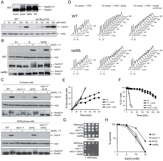
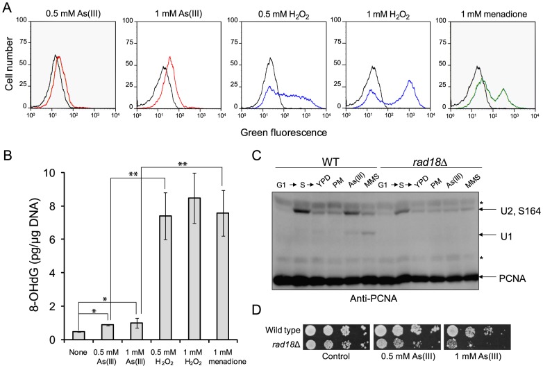
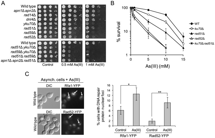
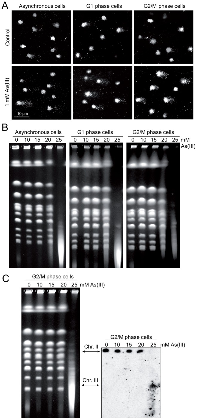
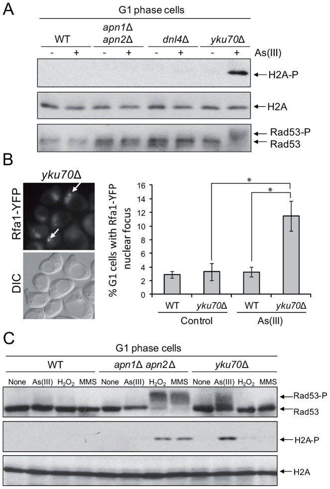
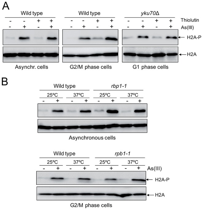
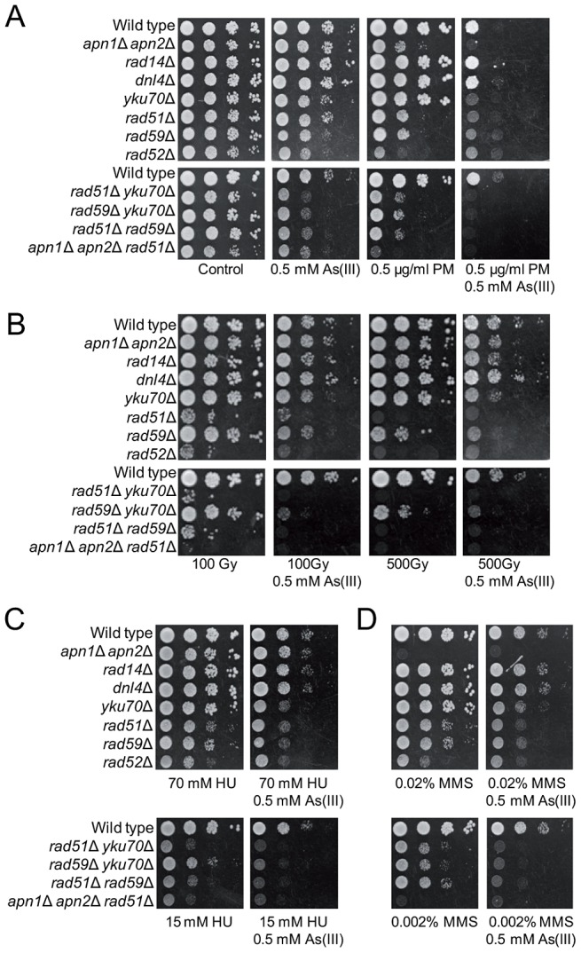
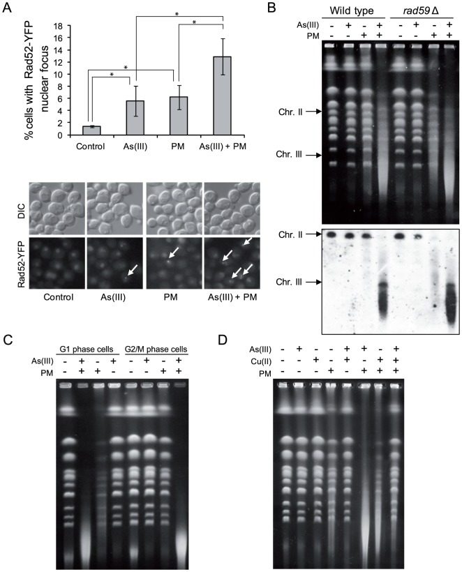
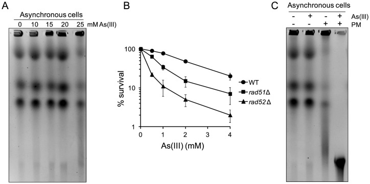
Arsenic is a well-established human carcinogen of poorly understood mechanism of genotoxicity. It is generally accepted that arsenic acts indirectly by generating oxidative DNA damage that can be converted to replication-dependent DNA double-strand breaks (DSBs), as well as by interfering with DNA repair pathways and DNA methylation. Here we show that in budding yeast arsenic also causes replication and transcription-independent DSBs in all phases of the cell cycle, suggesting a direct genotoxic mode of arsenic action. This is accompanied by DNA damage checkpoint activation resulting in cell cycle delays in S and G2/M phases in wild type cells. In G1 phase, arsenic activates DNA damage response only in the absence of the Yku70-Yku80 complex which normally binds to DNA ends and inhibits resection of DSBs. This strongly indicates that DSBs are produced by arsenic in G1 but DNA ends are protected by Yku70-Yku80 and thus invisible for the checkpoint response. Arsenic-induced DSBs are processed by homologous recombination (HR), as shown by Rfa1 and Rad52 nuclear foci formation and requirement of HR proteins for cell survival during arsenic exposure. We show further that arsenic greatly sensitizes yeast to phleomycin as simultaneous treatment results in profound accumulation of DSBs. Importantly, we observed a similar response in fission yeast Schizosaccharomyces pombe, suggesting that the mechanisms of As(III) genotoxicity may be conserved in other organisms.


Conflict of interest statement

The authors have declared that no competing interests exist.

Figures

Figure 1
Figure 1. Cell cycle phase-dependent activation of DNA damage checkpoints by As(III) in budding yeast.
(A) As(III) triggers activation of DNA damage response in yeast. Exponentially growing wild type (WT) cells were treated with 0.5 mM sodium arsenite [As(III)], 0.01% methyl methanesulfonate (MMS) or 5 µg/ml phleomycin (PM) for 1 h before protein extraction (upper panel). WT and the acr3Δ ycf1Δ mutant lacking arsenic detoxification transporters were exposed to indicated concentrations As(III) for 1 h (lower panel). (B) As(III) promotes histone H2A phosphorylation and Rad9-dependent hyperphosphorylation of Rad53 in S and G2/M but not in G1 phase. Cells were treated with 0.5 mM As(III) for 1 h. (A–B) Total protein extracts were analyzed by Western blot with anti-Rad53 antibodies to detect unmodified (Rad53) and hyperphosphorylated (Rad53-P) forms of the checkpoint effector kinase Rad53 as well as with anti-phospho-S129 H2A antibodies and anti-H2A antibodies as a loading control. (C) As(III) induces Mec1 and Tel1-dependent activation of DNA damage checkpoints. Experiments were performed as in (B). (D) Flow cytometry analysis of cell cycle progression during 0.5 mM As(III) treatment reveals a partial lack of DNA synthesis slowing in rad9Δ compared to WT. (E) Duration of G2/M checkpoint arrest is partially dependent on DNA damage signalling pathway during exposure to As(III). Cells were synchronized in G2/M with nocodazole and released in fresh media in the presence or absence of 0.5 mM As(III). (F) G1/S transition delay in the presence of 0.5 mM As(III) is not maintained by DNA damage checkpoint as shown by the α-factor-nocodazole trap assay. (G, H) DNA damage checkpoint mutants showed increased sensitivity to As(III). Serial dilutions of indicated strains were plated on rich media in the presence or absence of As(III) at 30°C and photographed after 3 days (G) or cells were exposed to indicated concentrations of As(III) for 6 h in liquid minimal media before plating on YPD plates to score viability (H). (E,F,H) Results are shown as means with standard deviations from three independent experiments.
Figure 2
Figure 2. As(III) treatment induces low level of oxidative stress and replication-associated DNA damage.
(A) Wild type cells were exposed to indicated concentrations of As(III) (red line), H2O2 (blue line) and menadione (green line) for 2 h or left untreated (black line). Levels of ROS were determined by measuring green fluorescence of rhodamine 123 (RH3) formed by oxidation of dihydrorhodamine 123 (DHR123) using flow cytometry. (B) Oxidative DNA damage in the form of 8-hydroxy-2′-deoxyguanosine (8-OHdG) induced by indicated concentrations of As(III), H2O2 and menadione after 2 h exposure. Standard deviations are derived from three independent experiments (*p<0.01, **p<0.001; Student's t-test). (C) PCNA ubiquitylation is slightly increased in response to As(III). Wild type (WT) and rad18Δ cells were synchronized in G1 with α-factor (G1) and released in fresh medium for 30 min to reach middle S phase (S). Then cells were left untreated (YPD) or treated with either 5 µg/ml phleomycin (PM), 0.5 mM As(III) or 0.01% methanesulfonate (MMS) for 1 h followed by protein extraction. Analysis of PCNA modifications was performed with total protein extracts and anti-PCNA antibodies. Bands corresponding to monoubiquitylated (U1), polyubiquitylated (U2) and sumoylated (S164) forms of PCNA are indicated. Non-specific bands are depicted by asterisks. (D) Cells lacking the Rad18 ubiquitin ligase involved in PCNA monoubiquitylation exhibited increased sensitivity to As(III). Serial dilutions of indicated strains were plated on rich media in the presence or absence of As(III) at 30°C and photographed after 2 days.
Figure 3
Figure 3. The role of DNA repair pathways in tolerance to As(III) in budding yeast.
(A) Homologous recombination and single-strand annealing DNA repair pathways as well as the presence of Yku70 are required for maintaining viability of yeast cells in the presence of As(III). 10-fold serial dilutions of the indicated strains were spotted on rich media that contained either no drug (control) or sodium arsenite [As(III)] and incubated at 30°C for 2 days. (B) As(III)-induced killing of wild type and indicated DNA repair mutants treated with various concentrations of As(III) in minimal media for 6 h. After treatment cells were plated on solid YPD media. The percentage is the ratio of colonies arising after As(III) exposure vs. mock treatment. Results are shown as means with standard deviations from three independent experiments. (C) Homologous recombination DNA repair centers are formed after 1 h treatment with 0.5 mM As(III) as visualized by detection of Rfa1-YFP and Rad52-YFP foci in nuclei with fluorescence microscopy. Standard deviations are derived from three independent experiments (*p<0.05, **p<0.01; Student's t-test). DIC, differential interference contrast.
Figure 4
Figure 4. DNA breakage induction by As(III).
(A) As(III) induces DNA breakage in all phases of the yeast cell cycle as revealed by the comet assay. Asynchronous, G1 or G2/M-arrested wild type cells (W303-1A) were exposed to 1 mM As(III) for 1 h or left untreated followed by a single-cell gel electrophoresis. Representative images of comets are shown. (B) DSBs induction after As(III) treatment was analyzed by PFGE. Logarithmically growing, G1 and G2/M wild type cells were treated with indicated concentrations of As(III) in minimal media for 6 h and processed for PFGE analysis. In the case of G1 and G2/M-synchronized cells, α-factor or nocodazole were also added during As(III) treatment to maintain cell cycle arrest. (C) As(III)-induced DSBs in G2/M arrested MWJ49 cells containing a circular chromosome III were measured using PFGE followed by Southern hybridization of the shown gel with a LEU2-probe to detect chromosome II and III. PFGE experiments were repeated at least two times with similar results and representative images are shown.
Figure 5
Figure 5. As(III) induces DNA damage checkpoint response in G1 phase in Yku70-deficient cells.
(A) Histone H2A and Rad53 phosphorylation induction by As(III) in G1 cells in the absence of Yku70. The indicated strains were synchronized in G1 with α-factor and treated with 0.5 mM As(III) for 1 h followed by protein extraction and Western blot analysis. (B) Accumulation of Rfa1-YFP foci in the G1-synchronized yku70Δ strain reveals existence of As(III)-induced DSBs in G1 phase which undergo resection in the absence of Yku70. The representative image of yku70Δ cells in G1 phase containing As(III)-induced Rfa1 foci (arrows) is shown. Cell treatment was as in (A). Standard deviations are derived from three independent experiments (*p<0.01; Student's t-test). DIC, differential interference contrast. (C) Analysis of DNA damage response activation in G1-synchronized cells devoid of BER (apn1Δ apn2Δ) or Yku70 reveals that As(III)-induced DNA lesions are different from those generated by H2O2 or MMS. The indicated strains were exposed to 0.5 mM As(III), 0.5 mM H2O2 or 0.03% methyl methanesulfonate (MMS) for 1 h and analyzed by western blot.
Figure 6
Figure 6. Transcription-independent DNA damage induction by As(III).
(A) To shut off transcription 3 µg/ml thiolutin, an inhibitor of RNA polymerases, was added to asynchronous and G2/M phase wild type cells and G1-arrested yku70Δ mutant for 1 h and then 0.5 mM As(III) was added to the cells for 1 h followed by protein extraction and western blotting analysis of histone H2A phosphorylation. (B) The rpb1-1 cells bearing a temperature-sensitive mutation in the catalytic subunit of RNA polymerase II grown at permissive temperature (25°C) were shifted or not to non-permissive temperature (37°C) by adding YPD pre-warmed to 45°C to block transcription and exposed to 0.5 mM As(III) for 1 h. For a control wild type cells were treated in a similar way. Protein extracts were analyzed by western blotting to detect levels of phosphorylated H2A. Total histone H2A was used as a loading control.
Figure 7
Figure 7. As(III) enhances cytotoxicity of phleomycin.
(A) Serial dilutions of the indicated strains were spotted onto control YPD and As(III)-containing YPD plates or media containing phleomycin (PM) with or without As(III). (B) Alternatively, yeast cells were irradiated with various doses of ionizing radiation before plating in the presence or absence As(III). (C, D) Cells were also cultivated on media containing hydroxyurea (HU) (C) or methyl methanesulfonate (MMS) (D) with or without As(III). Cells were incubated at 30°C for 2 days.
Figure 8
Figure 8. As(III) and phleomycin co-treatment increases formation of DSBs.
(A) Increased accumulation of Rad52-YFP nuclear foci in wild type cells after 1 h of 0.5 mM As(III) and 0.5 µg/ml phleomycin (PM) co-treatment. Standard deviations are derived from three independent experiments (*p<0.05; Student's t-test). (B) Yeast chromosome breaks in asynchronously growing cells of indicated strains containing a circular chromosome III were measured using PFGE followed by Southern hybridization of the shown gel with a LEU2-probe to detect chromosome II and III. (C) DSB induction during As(III) and PM co-treatment in wild type cells synchronized and maintained in G1 or G2/M phase. (B, C) Cells were treated with 5 mM As(III) and 10 µg/ml PM in YPD medium for 4 h. (D) PFGE analysis of S. cerevisiae chromosomes isolated from wild type cells exposed to 5 mM As(III), 10 µg/ml PM or 4 mM copper sulfate [Cu(II)] in YPD medium for 4 h. PFGE experiments were repeated at least two times with similar results and representative images are shown.
Figure 9
Figure 9. DSB induction by As(III) in Schizosaccharomyces pombe and the role of HR in surviving As(III) treatment.
(A) DSBs induction after As(III) treatment was analyzed by PFGE. Logarithmically growing (mostly G2/M phase in the case of fission yeast) wild type cells were treated with indicated concentrations of As(III) in minimal medium for 6 h and processed for PFGE analysis. (B) Survival of wild type (WT) and indicated DNA repair mutants treated with various concentrations of As(III) in minimal media for 6 h. After treatment cells were plated on solid YES plates. The percentage is the ratio of colonies arising after As(III) exposure vs. mock treatment. Results are shown as means with standard deviations from three independent experiments. (C) Induction of DSBs by combined treatment with As(III) and PM as revealed by PFGE analysis. Logarithmically growing cells were treated with 5 mM As(III) and 0.25 µg/ml PM in YES medium for 4 h. PFGE experiments were repeated at least two times with similar results and representative images are shown.

Similar articles

Cited by

References

    1. Tapio S, Grosche B (2006) Arsenic in the aetiology of cancer. Mutat Res 612: 215–246. - PubMed
    1. Dilda PJ, Hogg PJ (2007) Arsenical-based cancer drugs. Cancer Treat Rev 33: 542–564. - PubMed
    1. Murray HW, Berman JD, Davies CR, Saravia NG (2005) Advances in leishmaniasis. Lancet 366: 1561–1577. - PubMed
    1. Martinez VD, Vucic EA, Becker-Santos DD, Gil L, Lam WL (2011) Arsenic exposure and the induction of human cancers. J Toxicol 2011: 431287. - PMC - PubMed
    1. Liu JX, Zhou GB, Chen SJ, Chen Z (2012) Arsenic compounds: revived ancient remedies in the fight against human malignancies. Curr Opin Chem Biol 16: 92–98. - PubMed

Publication types

MeSH terms

Substances