. 2013;9(7):e1003644.
doi: 10.1371/journal.pgen.1003644. Epub 2013 Jul 25.

A dual role for SOX10 in the maintenance of the postnatal melanocyte lineage and the differentiation of melanocyte stem cell progenitors

Affiliations

A dual role for SOX10 in the maintenance of the postnatal melanocyte lineage and the differentiation of melanocyte stem cell progenitors

Melissa L Harris et al. PLoS Genet. 2013.

Abstract

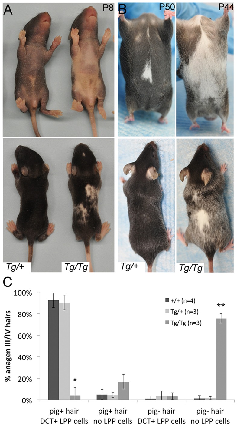
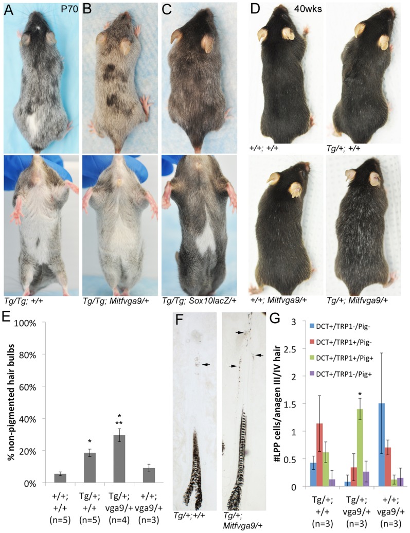
During embryogenesis, the transcription factor, Sox10, drives the survival and differentiation of the melanocyte lineage. However, the role that Sox10 plays in postnatal melanocytes is not established. We show in vivo that melanocyte stem cells (McSCs) and more differentiated melanocytes express SOX10 but that McSCs remain undifferentiated. Sox10 knockout (Sox10(fl); Tg(Tyr::CreER)) results in loss of both McSCs and differentiated melanocytes, while overexpression of Sox10 (Tg(DctSox10)) causes premature differentiation and loss of McSCs, leading to hair graying. This suggests that levels of SOX10 are key to normal McSC function and Sox10 must be downregulated for McSC establishment and maintenance. We examined whether the mechanism of Tg(DctSox10) hair graying is through increased expression of Mitf, a target of SOX10, by asking if haploinsufficiency for Mitf (Mitf(vga9) ) can rescue hair graying in Tg(DctSox10) animals. Surprisingly, Mitf(vga9) does not mitigate but exacerbates Tg(DctSox10) hair graying suggesting that MITF participates in the negative regulation of Sox10 in McSCs. These observations demonstrate that while SOX10 is necessary to maintain the postnatal melanocyte lineage it is simultaneously prevented from driving differentiation in the McSCs. This data illustrates how tissue-specific stem cells can arise from lineage-specified precursors through the regulation of the very transcription factors important in defining that lineage.


Conflict of interest statement

The authors have declared that no competing interests exist.

Figures

Figure 1
Figure 1. Differentiation status of LPP and UTP melanocytes varies with hair cycling.
(A) Schematic of hair follicles during hair morphogenesis (P2 stage 4; P2 stage 6, P6, and P14) and adult hair cycling (7dpp, and 21dpp). Grayed area represents anatomical regions quantified in B. (B) Percent of melanocytes that double-label with DCT and the indicated marker. Counts include melanocytes located within the LPP plus UTP at P2, P6, P14, and 7dpp or bulge and secondary hair germ at 21dpp (complete data available in Table S1 and Fig. S2, S3, S4, S5, S6). KIT expression is reported as KIThi being melanocytes displaying high intensity fluorescence and KIT+ being melanocytes positive for KIT independent of fluorescence intensity. LPP, lower permanent portion of the hair; UTP, upper transitory portion of the hair; dpp, days post plucking; SHG, secondary hair germ of the hair; DP, dermal papilla; SG, sebaceous gland; Sc, subcutis.
Figure 2
Figure 2. Sox10 is required by bulb melanocytes postnatally.
(A–B) Sox10fl/fl (fl/fl; +/+) and Sox10fl/fl; Tyr::CreERT2 (fl/fl; Cre/+) pups treated with TAM by IP injection to the lactating mother on P0–3 display variegated hypopigmentation on the belly and back and exhibit a white head spot upon the emergence of the morphogenetic coat (P10 shown here, n>5). (C) Number of PAX3+ melanocytes per hair bulb in skins harvested from these mice at P10 are significantly decreased in Sox10fl/fl; Tyr::CreERT2 animals compared to similarly-treated Sox10fl/fl animals (*p = 0.002). (D–E) Adult Sox10fl/fl; Tyr::CreERT2 mice treated with TAM by IP injection on 0–3dpp exhibit white hairs within the plucked region upon hair regrowth that is not visible in similarly treated Sox10fl/fl mice (brackets indicate plucked region, lower image is a magnification of plucked region). (F) Number of PAX3+ melanocytes per hair bulb in skins harvested from similarly-treated mice at 7dpp are significantly decreased in Sox10fl/fl; Tyr::CreERT2 animals compared to Sox10fl/fl animals (*p = 0.001). (G–H) Fluorescent and corresponding brightfield images of hair bulbs from mice described in D–E. Arrows and arrowheads indicate PAX3+/SOX10+ and PAX3+/SOX10 melanocytes, respectively. (I) Distribution of melanocytes double-labeled for PAX3 and SOX10 within pigmented (gray) and non-pigmented (white) hair bulbs in skins from Sox10fl/fl (n = 3) and Sox10fl/fl; Tyr::CreERT2 (n = 4) harvested on 7dpp from mice treated with TAM on 0–3dpp (*p<0.006).
Figure 3
Figure 3. Sox10 is required by LPP melanocytes postnatally.
(A) Number of KIT+ LPP melanocytes within hairs from Sox10fl/fl (fl/fl; +/+) and Sox10fl/fl; Tyr::CreERT2 (fl/fl; Cre/+) mice. P0–3/P10 indicates skins harvested from pups on P10 that were maintained by lactating mothers that were IP injected with TAM on P0–3. 0–3dpp/7dpp indicates skins harvested from adult mice on 7dpp after IP injections of TAM on 0–3dpp. (B) White hairs remain visible in adult Sox10fl/fl; Tyr::CreERT2 mice that were treated with TAM by IP injection on 0–3dpp, allowed for complete hair regeneration, replucked and allowed for a second round of hair regrowth (brackets indicate plucked/replucked region, lower image is a magnification of plucked region; mouse in 2E and 3B are the same, imaged prior to and after replucking). (C) Number of PAX3+ bulb melanocytes within hairs from Sox10fl/fl and Sox10fl/fl; Tyr::CreERT2 mice treated as described in B but harvested on 7dpp after replucking (0–3dpp/7dpp repluck). (D) Distribution of melanocytes double-labeled for PAX3 and SOX10 within pigmented (gray) and non-pigmented (white) hair bulbs in skins from Sox10fl/fl (n = 3) and Sox10fl/fl; Tyr::CreERT2 (n = 3) mice treated as described in B but harvested on 7dpp after replucking (*p<0.002). (E) Persistent hair graying is visible in Sox10fl/fl; Tyr::CreERT2 mice treated with IP TAM for pulse of five days beginning at five weeks old and imaged at one and two years old.
Figure 4
Figure 4. Tg(DctSox10) results in congenital white spotting and premature hair graying.
(A, B) Ventral and dorsal views demonstrating variable hypopigmentation in Tg(DctSox10)/+ and Tg(DctSox10)/Tg(DctSox10) mice during hair morphogenesis and adult hair cycling. (C) Frequency of pigmented (pig+) and non-pigmented (pig−) anagen III/IV (7dpp) hairs that contain (DCT+ LPP cells) or do not contain (no LPP cells) LPP melanocytes within Tg(DctSox10) or +/+ mice. The ages of mice analyzed ranged between 9–22 weeks at harvest. Significance determined by chi-square analysis (p<<0.0001) and evaluation of standardized residuals (*, z = −8.84; **, z = 12.24).
Figure 5
Figure 5. LPP melanocytes are reduced in Tg(DctSox10) homozygotes during hair morphogenesis.
(A) Brightfield images of hairs in Tg(DctSox10) and +/+ littermates at P2. (B) Number of DCT+ melanocytes within the LPP of hairs at P2 (stage 6 hairs) and P7/8. At both time points, LPP melanocytes per hair are reduced in Tg(DctSox10)/Tg(DctSox10) compared to Tg(DctSox10)/+ and +/+ mice (*p<0.017). (C, D) Quantitative immunohistochemical analysis of stage 6 hairs from P2 skins for DCT and TRP1, or DCT and KIT. The population of DCT+/TRP1+ cells is significantly reduced in Tg(DctSox10)/Tg(DctSox10) in comparison to Tg(DctSox10)/+ and +/+ mice (*p<0.008). Tg(DctSox10) also causes a switch in KIT intensity from KIThi in wild type to KITlow in Tg(DctSox10) animals (*KITlo and **KIThi comparisons made between +/+ and Tg(DctSox10)/+ or +/+ and Tg(DctSox10)/Tg(DctSox10); p<0.005).
Figure 6
Figure 6. Overexpression of Sox10 results in premature differentiation of LPP melanocytes in anagen hairs.
(A) Number of DCT+ LPP melanocytes per anagen III/IV hair follicle (independent of the presence or absence of hair pigmentation) is significantly reduced in Tg(DctSox10)/Tg(DctSox10) mice when compared to wild type and Tg(DctSox10)/+ mice (*p<0.0003). The ages of mice analyzed ranged between 9–22 weeks at harvest. (B) Eosin-stained skin sections of these hairs demonstrate the presence of ectopic pigmentation in the LPP of Tg(DctSox10)/+ and Tg(DctSox10)/Tg(DctSox10) hairs (arrows) that is not see in wild type hairs. In Tg(DctSox10)/+ LPP regions, this pigmentation often appeared in cells that were highly dendritic. (C, D) Brightfield and corresponding fluorescent images of anagen III/IV hair follicles double labeled for DCT and TRP1 (C) or KIT (D) in wild type and Tg(DctSox10)/+ animals. The intensity of KIT fluorescence expression was variable, and categorized as KITlo (arrows) or KIThi (arrowheads), and did not appear to correlate with the presence or absence of pigmentation. (E,F) Comparison of the number LPP melanocytes per anagen III/IV hair follicle in +/+ and Tg(DctSox10)/+ animals that express DCT, and TRP1 or KIT, and produce ectopic pigmentation (*p<0.008).
Figure 7
Figure 7. Alteration of the Tg(DctSox10) phenotype through the reduction of Mitf.
(A–B) Comparison of Tg(DctSox10)/Tg(DctSox10) and Tg(DctSox10)/Tg(DctSox10); Mitfvga9/+ animals at P70. Addition of the Mitfvga9/+ allele reduces the congenital hypopigmentation seen in Tg(DctSox10)/Tg(DctSox10) animals, and is evident in dorsal views (loss of back spotting) and in ventral views (reduction in belly spot size). Premature hair graying of Tg(DctSox10) homozygotes seen at p70 is retained with Mitfvga9 (n = 6). (C) Introduction of Sox10lacZ into Tg(DctSox10)/Tg(DctSox10) homozygotes partially rescues both congenital white spotting and premature hair graying (n = 2). (D) At 40 weeks of age, Tg(DctSox10)/+; Mitfvga9/+ double heterozygotes exhibit visibly increased hair graying in comparison to Tg(DctSox10)/+. (E) Hair graying severity was determined in animals 6–10 weeks of age by quantitating the number of non-pigmented anagen III/IV hair bulbs in +/+, Tg(DctSox10)/+, Tg(DctSox10)/+; Mitfvga9/+, and Mitfvga9/+ skins after plucking and harvesting at 7dpp. Tg(DctSox10)/+; Mitfvga9/+ mice exhibit a significant increase in non-pigmented hair bulbs in comparison to the single heterozygotes or +/+ animals (**p<0.0015). Tg(DctSox10)/+ animals also produce more non-pigmented hair bulbs in comparison to +/+ and Mitfvga9/+ animals (*p<0.002). (F) Tg(DctSox10)/+; Mitfvga9/+ animals (from E) display extensive ectopic pigmentation within the LPP of their hair follicles beyond what is normally observed in Tg(DctSox10)/+ animals (arrows, n = 4). (G) Number of LPP melanocytes per anagen III/IV hair follicle in Tg(DctSox10)/+, Tg(DctSox10)/+; Mitfvga9/+, Mitfvga9/+ animals (from E) that double label for DCT, TRP1, and produce ectopic pigmentation. Hairs from Tg(DctSox10)/+; Mitfvga9/+ animals exhibit significantly more TRP1+/PIG+ LPP melanocytes than in either single heterozygote (*p<0.0125).

Similar articles

Cited by

References

    1. Tobin DJ, Paus R (2001) Graying: gerontobiology of the hair follicle pigmentary unit. Exp Gerontol 36: 29–54. - PubMed
    1. Nishimura EK, Jordan SA, Oshima H, Yoshida H, Osawa M, et al. (2002) Dominant role of the niche in melanocyte stem-cell fate determination. Nature 416: 854–860 doi:10.1038/416854a - DOI - PubMed
    1. Mak S-S, Moriyama M, Nishioka E, Osawa M, Nishikawa S-I (2006) Indispensable role of Bcl2 in the development of the melanocyte stem cell. Dev Biol 291: 144–153 doi:10.1016/j.ydbio.2005.12.025 - DOI - PubMed
    1. Osawa M, Egawa G, Mak S-S, Moriyama M, Freter R, et al. (2005) Molecular characterization of melanocyte stem cells in their niche. Development 132: 5589–5599 doi:10.1242/dev.02161 - DOI - PubMed
    1. Botchkareva NV, Botchkarev VA, Gilchrest BA (2003) Fate of melanocytes during development of the hair follicle pigmentary unit. J Investig Dermatol Symp Proc 8: 76–79 doi:10.1046/j.1523-1747.2003.12176.x - DOI - PubMed

Publication types

Substances