

The significance of the subplate for evolution and developmental plasticity of the human brain
- PMID: 23935575
- PMCID: PMC3731572
- DOI: 10.3389/fnhum.2013.00423
The significance of the subplate for evolution and developmental plasticity of the human brain
Abstract
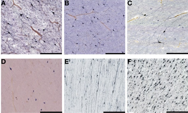
The human life-history is characterized by long development and introduction of new developmental stages, such as childhood and adolescence. The developing brain had important role in these life-history changes because it is expensive tissue which uses up to 80% of resting metabolic rate (RMR) in the newborn and continues to use almost 50% of it during the first 5 postnatal years. Our hominid ancestors managed to lift-up metabolic constraints to increase in brain size by several interrelated ecological, behavioral and social adaptations, such as dietary change, invention of cooking, creation of family-bonded reproductive units, and life-history changes. This opened new vistas for the developing brain, because it became possible to metabolically support transient patterns of brain organization as well as developmental brain plasticity for much longer period and with much greater number of neurons and connectivity combinations in comparison to apes. This included the shaping of cortical connections through the interaction with infant's social environment, which probably enhanced typically human evolution of language, cognition and self-awareness. In this review, we propose that the transient subplate zone and its postnatal remnant (interstitial neurons of the gyral white matter) probably served as the main playground for evolution of these developmental shifts, and describe various features that makes human subplate uniquely positioned to have such a role in comparison with other primates.
Keywords: cerebral cortex; life-history; metabolic cost; neuron number; subplate zone.
Figures
Similar articles
-
Developmental history of the subplate zone, subplate neurons and interstitial white matter neurons: relevance for schizophrenia.Int J Dev Neurosci. 2011 May;29(3):193-205. doi: 10.1016/j.ijdevneu.2010.09.005. Epub 2010 Sep 29. Int J Dev Neurosci. 2011. PMID: 20883772 Review.
-
Developmental history of the transient subplate zone in the visual and somatosensory cortex of the macaque monkey and human brain.J Comp Neurol. 1990 Jul 15;297(3):441-70. doi: 10.1002/cne.902970309. J Comp Neurol. 1990. PMID: 2398142
-
Populations of subplate and interstitial neurons in fetal and adult human telencephalon.J Anat. 2010 Oct;217(4):381-99. doi: 10.1111/j.1469-7580.2010.01284.x. J Anat. 2010. PMID: 20979586 Free PMC article. Review.
-
Interstitial cells of the adult neocortical white matter are the remnant of the early generated subplate neuron population.J Comp Neurol. 1989 Apr 22;282(4):555-69. doi: 10.1002/cne.902820407. J Comp Neurol. 1989. PMID: 2566630
-
Early history of subplate and interstitial neurons: from Theodor Meynert (1867) to the discovery of the subplate zone (1974).J Anat. 2010 Oct;217(4):344-67. doi: 10.1111/j.1469-7580.2010.01283.x. J Anat. 2010. PMID: 20979585 Free PMC article. Review.
Cited by
-
The fetal pain paradox.Front Pain Res (Lausanne). 2023 Mar 21;4:1128530. doi: 10.3389/fpain.2023.1128530. eCollection 2023. Front Pain Res (Lausanne). 2023. PMID: 37025166 Free PMC article. Review.
-
Plasticity of neonatal neuronal networks in very premature infants: Source localization of temporal theta activity, the first endogenous neural biomarker, in temporoparietal areas.Hum Brain Mapp. 2017 May;38(5):2345-2358. doi: 10.1002/hbm.23521. Epub 2017 Jan 23. Hum Brain Mapp. 2017. PMID: 28112458 Free PMC article.
-
Role of neuroserpin and N-Cadherin in mesenchymal stromal cell modulation of preterm brain injury.Pediatr Res. 2024 Nov 11. doi: 10.1038/s41390-024-03708-0. Online ahead of print. Pediatr Res. 2024. PMID: 39528742 No abstract available.
-
Early Diagnostics and Early Intervention in Neurodevelopmental Disorders-Age-Dependent Challenges and Opportunities.J Clin Med. 2021 Feb 19;10(4):861. doi: 10.3390/jcm10040861. J Clin Med. 2021. PMID: 33669727 Free PMC article. Review.
-
Spatio-temporal extension in site of origin for cortical calretinin neurons in primates.Front Neuroanat. 2014 Jun 26;8:50. doi: 10.3389/fnana.2014.00050. eCollection 2014. Front Neuroanat. 2014. PMID: 25018702 Free PMC article. Review.
References
-
- Aboitiz F., Montiel J., Garcia R. R. (2005). Ancestry of the mammalian preplate and its derivatives: evolitonary relicts or embryonic adaptations. Rev. Neurosci. 16, 359–376 - PubMed
-
- Aiello L. C., Wheeler P. (1995). The expensive tissue hypothesis: the brain and the digestive system in human and primate evolution. Curr. Anthropol. 36, 199–221 10.1086/204350 - DOI
LinkOut - more resources
Full Text Sources
Other Literature Sources