. 2013 Jul 18;8(7):e68171.
doi: 10.1371/journal.pone.0068171. Print 2013.

T cells induce pre-metastatic osteolytic disease and help bone metastases establishment in a mouse model of metastatic breast cancer

Affiliations

T cells induce pre-metastatic osteolytic disease and help bone metastases establishment in a mouse model of metastatic breast cancer

Ana Carolina Monteiro et al. PLoS One. .

Abstract

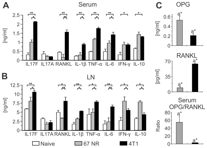
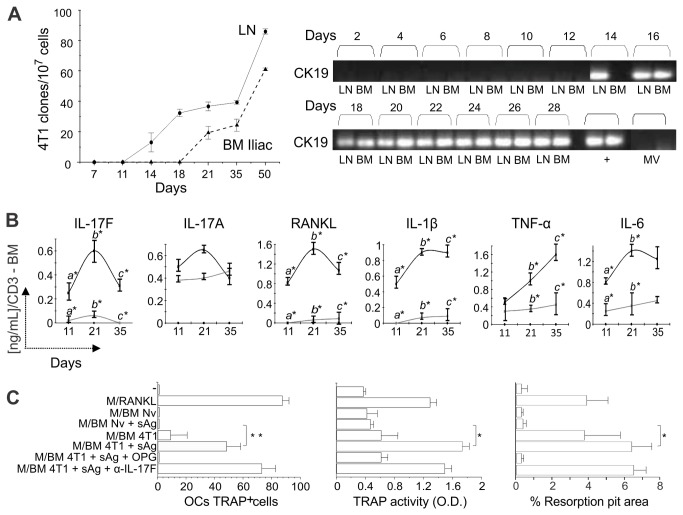
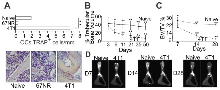
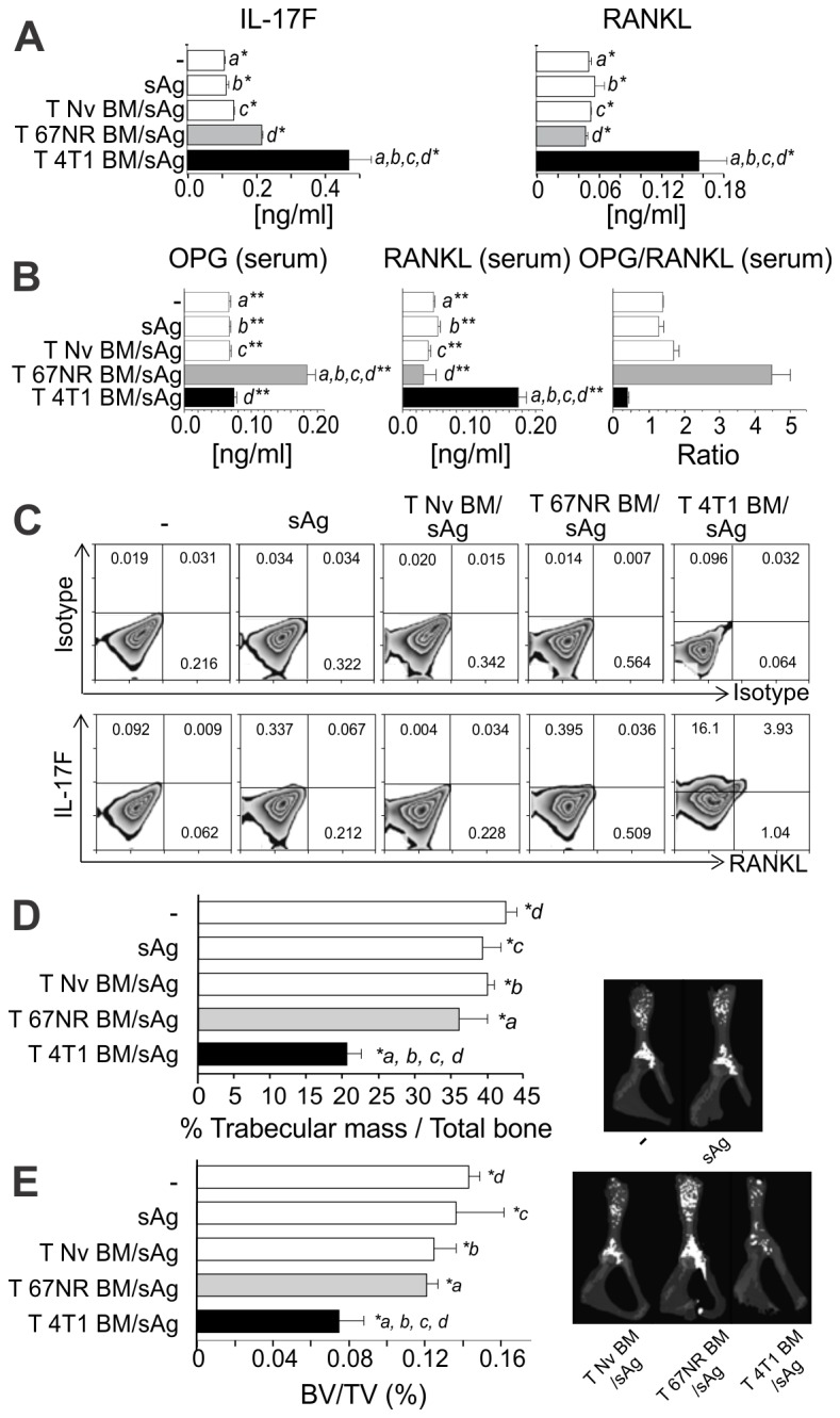
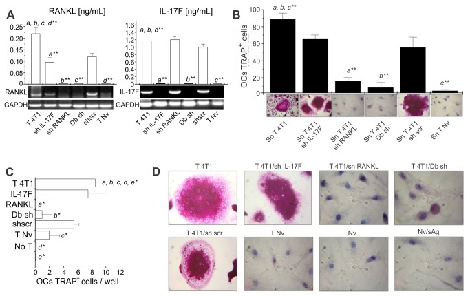
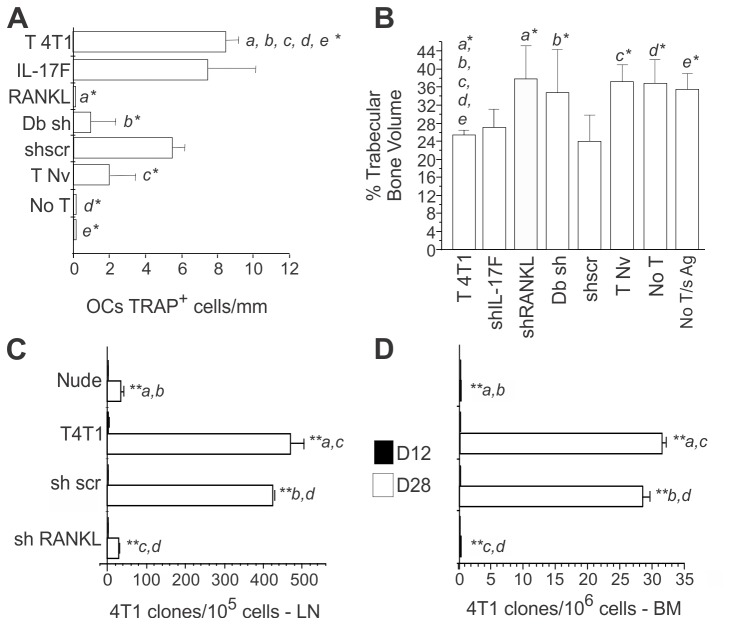
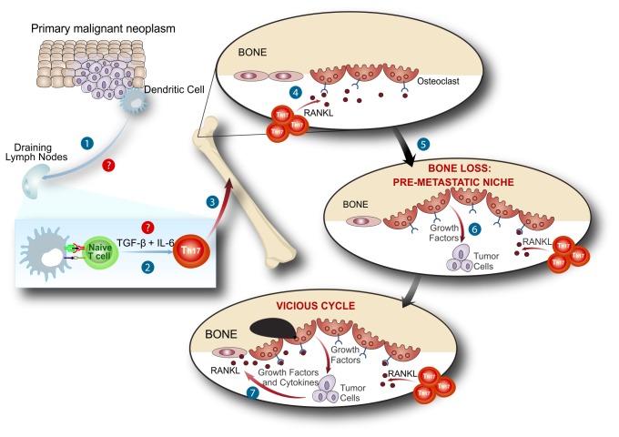
Bone metastases, present in 70% of patients with metastatic breast cancer, lead to skeletal disease, fractures and intense pain, which are all believed to be mediated by tumor cells. Engraftment of tumor cells is supposed to be preceded by changes in the target tissue to create a permissive microenvironment, the pre-metastatic niche, for the establishment of the metastatic foci. In bone metastatic niche, metastatic cells stimulate bone consumption resulting in the release of growth factors that feed the tumor, establishing a vicious cycle between the bone remodeling system and the tumor itself. Yet, how the pre-metastatic niches arise in the bone tissue remains unclear. Here we show that tumor-specific T cells induce osteolytic bone disease before bone colonization. T cells pro-metastatic activity correlate with a pro-osteoclastogenic cytokine profile, including RANKL, a master regulator of osteoclastogenesis. In vivo inhibition of RANKL from tumor-specific T cells completely blocks bone loss and metastasis. Our results unveil an unexpected role for RANKL-derived from T cells in setting the pre-metastatic niche and promoting tumor spread. We believe this information can bring new possibilities for the development of prognostic and therapeutic tools based on modulation of T cell activity for prevention and treatment of bone metastasis.


Conflict of interest statement

Competing Interests: The authors have declared that no competing interests exist.

Figures

Figure 1
Figure 1. Metastatic 4T1 tumor stimulates the production of pro-osteoclastogenic cytokines.
(AB) BALB/c female mice were orthotopically injected in the mammary fat pad with 104 metastatic 4T1 or non-metastatic 67NR tumor cells. Cytokine production was evaluated by ELISA in the sera (A) and supernatants of αCD3-stimulated cells (B) collected from inguinal draining LNs 35 days post tumor injection. Naïve animals were used as negative controls. Data are expressed as the mean ± SD of five mice/group and are representative of at least three independent experiments. *p<0.05; **p<0.001. (C) Serum concentration of OPG and RANKL, and the OPG/RANKL ratio in tumor-bearing BALB/c mice, measured by ELISA, 35 days after injection of 4T1 or 67NR tumor cells. a*p≤0.05. Data are expressed as the mean ± SD of five mice/group.
Figure 2
Figure 2. Pro-osteoclastogenic cytokine production by BM cells in response to tumor antigenic stimulation precedes metastatic colonization of the bones.
(A) Clonogenic metastatic assays were performed with cells obtained from the draining LNs and iliac BM of BALB/c mice orthotopically injected with 104 4T1 tumor cells. The number of metastatic clones was determined at different time points, using a 6-thioguanine resistant assay (left panel). Expression of cytokeratin 19 (CK19) was also determined in such samples by RT-PCR (right panel). 67NR or 4T1 tumor derived microvesicles (MV) were used as a control to ascertain that a positive RT-PCR indicates exclusively the presence of whole tumor cells in the target organ. 4T1 cells mRNA was obtained from in vitro cultures or in vivo tumors (+) and was used as a positive control in the RT-PCR assay. (B) Kinetics of pro-osteoclastogenic cytokine production in response to tumor soluble antigen (sAg) stimulation of BM cells obtained from iliac bones of mice bearing 4T1 or 67NR tumors. Naïve animals were used as controls. Supernatants of sAg stimulated iliac BM cells were harvested after 72hs and cytokine production was measured by ELISA. Data are expressed as the mean ± SD of five mice/group and are representative of at least three independent experiments. *p<0.05. (C) Osteoclastogenesis assays using naïve BM cells cultured in the presence of either recombinant M-CSF and RANKL, or M-CSF and supernatant of iliac BM cells derived from 4T1 tumor-bearing mice (11 d after tumor injection). Supernatant from iliac BM cells of naïve animals (BM Nv) stimulated or not with sAg was used as specific control. Cultures were also treated with r-OPG or α-IL17F as indicated. The number of TRAP+ multinucleated OC cells obtained in vitro was determined (left panel) and TRAP activity in such supernatants was measured by a colorimetric assay (middle panel). In the right panel, generation of functional OC cells in vitro was also determined using BD BioCoatTM OsteologicTM Bone Cell Culture System (BD Biosciences). The graphic represents the resorbed area on osteologic discs. All data are from at least two independent experiments (n=5 mice/group) and presented as mean ± SD. *p<0.05; **p<0.001.
Figure 3
Figure 3. Increased number of osteoclasts in 4T1 tumor-bearing mice relates to early bone loss.
(A) Number of TRAP+ multinucleated OC cells was determined in iliac bones, 11 days after 4T1 or 67NR tumor cells injection in mammary fat pad. Representative TRAP-stained sections are shown (original magnifications 40x). (B) Histomorphometric analysis of iliac bones from naïve and 4T1 tumor-bearing mice, at different time points after tumor injection in the mammary fat pad. Sagital sections from demineralized iliac bones were made following conventional methods and stained with H and E. All microscopic slides were scanned with a ScanScope GL equipped with a 40x objective. Trabecular bone volume was expressed as a percentage of total tissue volume. (CD) High resolution µCT analysis. BV/TV%, trabecular bone volume/tissue volume were calculated from µCT images. Results are expressed as mean ± SD and are representative of at least three independent experiments with 5 mice/group. * p<0.05; ** p<0.001.
Figure 4
Figure 4. Early bone loss in 4T1 tumor-bearing mice is T cell mediated and independent of metastatic colonization.
CD3+ T cells derived from iliac BM of BALB/c mice, 11 days after 4T1 (T 4T1) or 67NR(T 67NR) tumor cells injection into the mammary fat pad, or control T cells from naïve mice (T Nv) were transferred intravenously to athymic nude mice and challenged with the soluble fraction of tumor antigen lysate (sAg). (A) 14 days after transference, spleen cells were restimulated with sAg and IL-17F and RANKL production was evaluated by ELISA. Data are expressed as the mean ± SD of five mice/group and are representative of two independent experiments * p≤0.05. (B) Frequency of IL-17F+ RANKL+ T cells was assessed by flow cytometry, 14 days after T cells transference. Plots show data from CD3+ CD4+ gated T cells. (C) Serum concentrations of OPG and RANKL and the OPG/RANKL ratio, measured by ELISA, 14 days after T cells transference. Data are expressed as the mean ± SD of five mice/group. a, b, c ,d** p<0.001. (D) Histomorphometric analysis of the iliac bones from mice of the different groups and (E) high resolution µCT analysis of the iliac bones. Both analyses were performed as described in Figure 3. Results shown are representative of at least two independent experiments with 5 mice/group. a, b, c ,d* p<0.05.
Figure 5
Figure 5. Specific blockage of RANKL expression, but not IL17F, in T cells abolishes the in vitro pro-osteoclastogenic activity of tumor-specific T cells.
LN cells obtained from 4T1 tumor-bearing BALB/c mice (T 4T1), 11 days after tumor injection, were transfected with shRNA for IL17F (shIL17-F), RANKL (shRANKL), scramble (scr), or both RANKL and IL17F (Db sh). These cells were transferred intravenously to athymic nude mice and the recipients were challenged with soluble tumor antigen (sAg). (A) Knocked down cells were re-stimulated in vitro with sAg, and assayed for RANKL and IL17 expression by ELISA or RT-PCR, prior to injection into recipient mice. ** p<0.001. (B) The osteoclatogenic activity of supernatants obtained from knocked down cells, stimulated in vitro with sAg for 3 days, was also evaluated in osteoclastogenic assays (as described in Figure 2C). The number of TRAP+ multinucleated OCs was determined per well and the representative TRAP staining of OCs is shown under each graphic bar. (CD) Spleen cells recovered 6 days after i. v. transfer were restimulated in vitro with sAg and the osteoclastogenic activity of the supernatants obtained was also tested over naive BM cells. The number of TRAP+ multinucleated OCs was determined per well and the representative TRAP staining of OCs is shown in panel D * p≤0.05.
Figure 6
Figure 6. T cell-induced osteoclastogenesis and bone loss requires expression of RANKL in T cells which “helps” metastatic colonization
(A) LN cells obtained from 4T1 tumor-bearing BALB/c mice (T 4T1), 11 days after tumor injection, were transfected with shRNA for IL17F (shIL17-F), RANKL (shRANKL), scramble (scr), or both, RANKL and IL17F (Db sh), transferred i. v. into athymic nude mice and challenged with soluble tumor antigen (sAg). Bone sections from recipient mice were prepared and the number of TRAP+ OCs/mm of bone surface was determined. (B) High resolution µCT of iliac bones from the different groups of nude mice transferred with the indicated T cells. Results shown are representative of two experiments with 5 mice/group). * p≤0.05; ** p≤0.001. (C–D) Number of metastatic clones in the LNs and iliac BMs was assessed by clonogenic metastatic assay in the recipient mice on day 12 and 28. Nude, non-reconstituted control; T 4T1; reconstitution with 4T1 T cells; sh scr, sh Scramble T 4T1; sh RANKL, sh RANKLT 4T1. Results shown are representative of two experiments with 6 mice/group). ** p≤0.001.
Figure 7
Figure 7. RANKL+ tumor-specific T cells prepare the bone pre-metastatic niche.
After being stimulated (1) and modulated (2) by tumor cells T cells migrate to the bone marrow (3). When inside the bone marrow niche, T cell derived RANKL stimulate osteoclastogenesis (4) with bone consumption before tumor bone colonization (5). This initial bone loss induced by T cells in response to tumor antigen, prepares the bone marrow niche to receive tumor cells (6). Once inside the marrow, tumor cells will be able to establish themselves comfortable (7), at the expense of the pre-metastatic niche already set by T cell derived RANKL in response to tumor stimulation.

References

    1. Schreiber RD, Old LJ, Smyth MJ (2011) Cancer immunoediting: integrating immunity’s roles in cancer suppression and promotion. Science 331: 1565-1570. doi:10.1126/science.1203486. PubMed: 21436444. - DOI - PubMed
    1. Old LJ, Boyse EA (1964) Immunology of experimental tumors. Annu Rev Med 15: 167-186. doi:10.1146/annurev.me.15.020164.001123. PubMed: 14139934. - DOI - PubMed
    1. Matsushita H, Vesely MD, Koboldt DC, Rickert CG, Uppaluri R et al. (2012) Cancer exome analysis reveals a T-cell-dependent mechanism of cancer immunoediting.Nature 482:400-404 - PMC - PubMed
    1. DuPage M, Mazumdar C, Schmidt LM, Cheung AF, Jacks T (2012) Expression of tumour-specific antigens underlies cancer immunoediting. Nature 482: 405-409. doi:10.1038/nature10803. PubMed: 22318517. - DOI - PMC - PubMed
    1. Hanahan D, Weinberg RA (2011) Hallmarks of cancer: the next generation. Cell 144: 646-674. doi:10.1016/j.cell.2011.02.013. PubMed: 21376230. - DOI - PubMed

Publication types

MeSH terms