. 2013 Jul 31;8(7):e68399.
doi: 10.1371/journal.pone.0068399. Print 2013.

Detection of VEGF-A(xxx)b isoforms in human tissues

Affiliations

Detection of VEGF-A(xxx)b isoforms in human tissues

David O Bates et al. PLoS One. .

Abstract

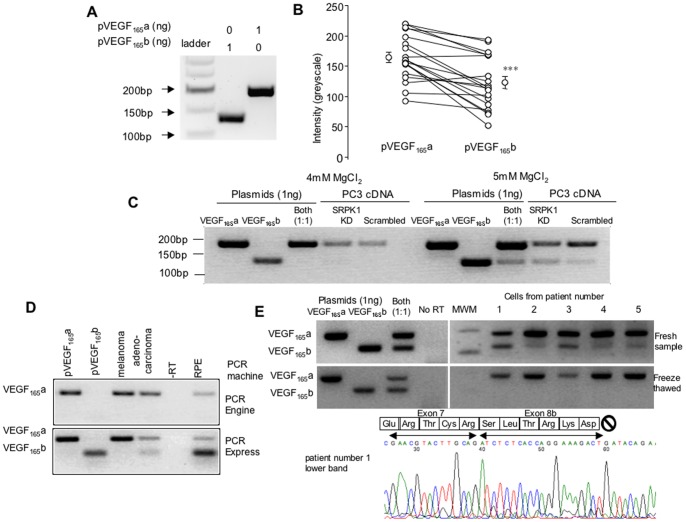
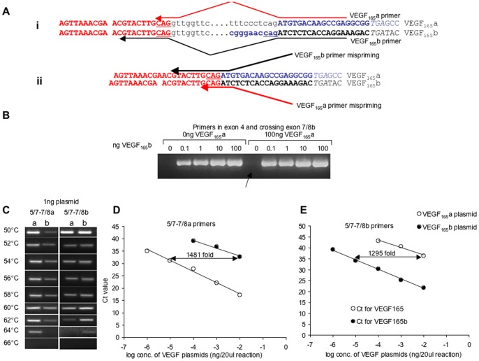
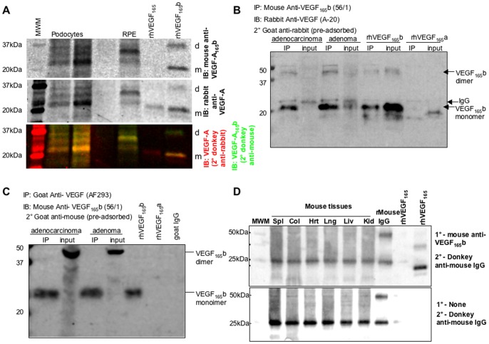
Vascular Endothelial Growth Factor-A (VEGF-A) can be generated as multiple isoforms by alternative splicing. Two families of isoforms have been described in humans, pro-angiogenic isoforms typified by VEGF-A165a, and anti-angiogenic isoforms typified by VEGF-A165b. The practical determination of expression levels of alternative isoforms of the same gene may be complicated by experimental protocols that favour one isoform over another, and the use of specific positive and negative controls is essential for the interpretation of findings on expression of the isoforms. Here we address some of the difficulties in experimental design when investigating alternative splicing of VEGF isoforms, and discuss the use of appropriate control paradigms. We demonstrate why use of specific control experiments can prevent assumptions that VEGF-A165b is not present, when in fact it is. We reiterate, and confirm previously published experimental design protocols that demonstrate the importance of using positive controls. These include using known target sequences to show that the experimental conditions are suitable for PCR amplification of VEGF-A165b mRNA for both q-PCR and RT-PCR and to ensure that mispriming does not occur. We also provide evidence that demonstrates that detection of VEGF-A165b protein in mice needs to be tightly controlled to prevent detection of mouse IgG by a secondary antibody. We also show that human VEGF165b protein can be immunoprecipitated from cultured human cells and that immunoprecipitating VEGF-A results in protein that is detected by VEGF-A165b antibody. These findings support the conclusion that more information on the biology of VEGF-A165b isoforms is required, and confirm the importance of the experimental design in such investigations, including the use of specific positive and negative controls.


Conflict of interest statement

Competing Interests: The authors hold patents on VEGF165b, and grant income from government and non government not-for-profit organisations investigating VEGF165b. This does not alter the authors’ adherence to all the PLOS ONE policies on sharing data and materials.

Figures

Figure 1
Figure 1. Positive controls are required to interpret lack of amplification of VEGF-A165b by competitive RT-PCR.
A. Plasmids containing VEGF-A165b or VEGF-A165a sequence were amplified using primers in exon 8b and exon 7. Two different sized products were generated. B. Densitometric analysis of published RT-PCR gels using plasmids containing VEGF-A165b and VEGF-A165a and primers in exon 7 and exon 8b. 13/15 show higher intensity for VEGF-A165a. p<0.001 paired t test. C. Example of failure of amplification of the VEGF-A165b isoform. Two parallel PCR reactions were run on cDNA and plasmid DNA. On the first (at 4mM MgCl2) no VEGF-A165b was generated in the cDNA, or when both VEGF-A165a and VEGF-A165b plasmids were used as positive controls. In the second (at 5mM MgCl2) VEGF-A165b was generated from cDNA from PC3 cells where SRPK1 was knocked down, and when both templates were included. D. Example of failure of amplification of the VEGF-A165b isoform in one PCR machine (and failure of template), but not in a second machine. E. Example of failure of amplification of VEGF-A165b product after freeze-thawing of cDNA derived from fresh normal human lung fibroblasts. Chromatogram confirms VEGF-A165b sequence from lower band from top gel from patient sample in lane 1 (chromatograms from the other samples also confirmed VEGF-A165b sequence).
Figure 2
Figure 2. Isoform specific PCR requires positive controls to ensure specificity.
A. Sequence of the VEGF 3′ exon sequence. (i) Exon 7 (red) contains the same last three nucleotides (underlined) as the last three nucleotides of exon 8a (blue, underlined), requiring specific PCR primers that extend into exon 7 (arrow). (ii) mispriming (VEGF-A165a -specific primers priming on VEGF-A165b, and VEGF-A165b -specific primers priming on VEGF-A165a) can occur both ways round if the conditions are not tested. B. Published control PCR gels demonstrating specificity of primer conditions. The original description of VEGF-A165b describing conditions at which VEGF-A165b is not misprimed in the presence of 100ng VEGF-A165a (lane highlighted by arrow), but still able to amplify 0.1ng VEGF-A165b. C. Annealing temperature dependence of the specificity of the isoform specific primers. Only at >62°C is specificity resolved. D. qPCR using VEGF-A165a specific primers on VEGF-A165a and VEGF-A165b plasmid E. qPCR using VEGF-A165b specific primers on VEGF-A165a and VEGF-A165b plasmid.
Figure 3
Figure 3. qRT-PCR using protocols shown in figure 2D and E can detect changes in splicing induced by splicing factor knockdown.
A. Ctmax-Ct for cDNA extracted from prostate cancer (PC3) cells with lentiviral knockdown of SRPK1 or scrambled. B. Amount of VEGF calculated from standard curves in Figure 2. C. Amount of VEGF-A165b identified by Exon 8b primers (VEGF-A165b) or that calculated from mispriming of VEGF-A165a. D. Proportion of VEGF that is VEGF-A165a or VEGF-A165b in control and knockdown cells. Values are Mean±SEM (n = 2). 3E. qPCR for VEGF-A165a on commercially available cDNAs from 2 different companies (open bars) or cDNA reverse transcribed from freshly extracted human kidney RNA (solid bar). 3F qPCR for VEGF-A165b on commercially available cDNAs from 2 different companies (open bars) or cDNA reverse transcribed from freshly extracted human kidney RNA (solid bar).
Figure 4
Figure 4. VEGF expression determined by Western blot and immunoprecipitation.
A. Western blot using LiCor Odyssey to simultaneously image pan-VEGF and VEGF-A165b probed western blot. Two different podocyte samples, and a primary RPE sample were run on a gel and probed with antibodies to VEGF-A165b (mouse monoclonal anti-CTRSLTRKD, and 680nm-donkey anti-mouse, top image) and pan-VEGF (rabbit polyclonal anti-VEGF, and 800nm-donkey anti-rabbit, middle image). The bottom image is the pseudocoloured combined image (600nm green, 800nm red). Note the red VEGF165, but yellow VEGF-A165b. MWM = molecular weight marker. d = dimer, m = monomer. B. Protein extracted from human cell lines (adenoma and adenocarcinoma(AC)) subjected to immunoprecipitation (IP) for VEGF-A165b and immunoblotting (IB) for total VEGF-A. A clear strong band was seen in the IP for both cell types at ∼23kDa and ∼46kDa, consistent with the IP for recombinant human VEGF-A165b. A weaker band was seen in the input protein (not subjected to IP), and a second band slightly higher in the AC. A weak band at approximately 56kDa and 28kDa was seen in all lanes subjected to IP, including the VEGF-A165a band, but not seen in the recombinant human VEGF-A165b not subjected to IP, indicating that this is cross reactivity with the IgG. This band was clearly above the VEGF-A165b bands. C. Protein extracted from human cell lines (adenoma and adenocarcinoma(AC)) subjected to immunoprecipitation (IP) for VEGF-A and immunoblotting (IB) for VEGF-A165b. A clear strong band was seen in the IP for both cell types at ∼23kDa, the same size as recombinant human VEGF-A165b. In the input a band at ∼46Da was seen predominantly, for both cell types, labelled as VEGF-A165b dimers. D. Mouse tissues probed with VEGF-A165b antibody detect mouse IgG due to the secondary antibody. Top image, western blot of mouse tissues, recombinant mouse IgG or human VEGF-A165b or VEGF-A165b probed with mouse anti-CTRSLTRKD, and 680nm-donkey anti-mouse IgG. Bottom image blot of same tissues, probed without primary antibody. The same bands are seen in the mouse tissues. Spl = spleen, Col = colon, Hrt = heart, Lng = lung, Liv = liver, Kid = kidney.

References

    1. Houck KA, Ferrara N, Winer J, Cachianes G, Li B, et al. (1991) The vascular endothelial growth factor family: identification of a fourth molecular species and characterization of alternative splicing of RNA. Mol Endocrinol 5: 1806–1814. - PubMed
    1. Harper SJ, Bates DO (2008) VEGF-A splicing: the key to anti-angiogenic therapeutics? Nat Rev Cancer 8: 880–887. - PMC - PubMed
    1. Bates DO, Cui TG, Doughty JM, Winkler M, Sugiono M, et al. (2002) VEGF-A165b, an inhibitory splice variant of vascular endothelial growth factor, is down-regulated in renal cell carcinoma. Cancer Res 62: 4123–4131. - PubMed
    1. Rennel E, Varey AHR, Churchill AJ, Wheatley ER, Stewart L, et al. (2009) VEGF121b, a new member of the VEGFxxxb family of VEGF-A splice isoforms, inhibits neovascularisation and tumour growth in vivo. Br J Cancer 101: 1250–1257. - PMC - PubMed
    1. Miller-Kasprzak E, Jagodzinski PP (2008) 5-Aza-2′-deoxycytidine increases the expression of anti-angiogenic vascular endothelial growth factor 189b variant in human lung microvascular endothelial cells. Biomed Pharmacother 62: 158–163. - PubMed

Publication types

MeSH terms

LinkOut - more resources