. 2013 Jul 30;8(7):e68597.
doi: 10.1371/journal.pone.0068597. Print 2013.

NF-κB regulates mesenchymal transition for the induction of non-small cell lung cancer initiating cells

Affiliations

NF-κB regulates mesenchymal transition for the induction of non-small cell lung cancer initiating cells

Manish Kumar et al. PLoS One. .

Abstract

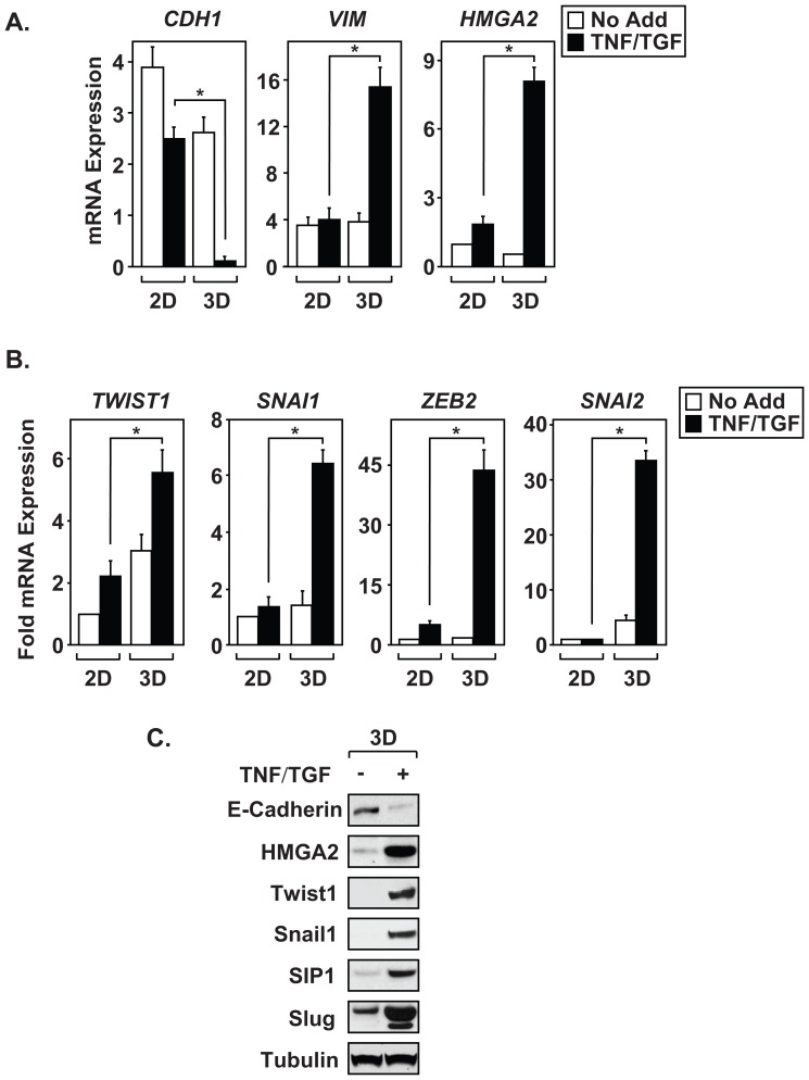
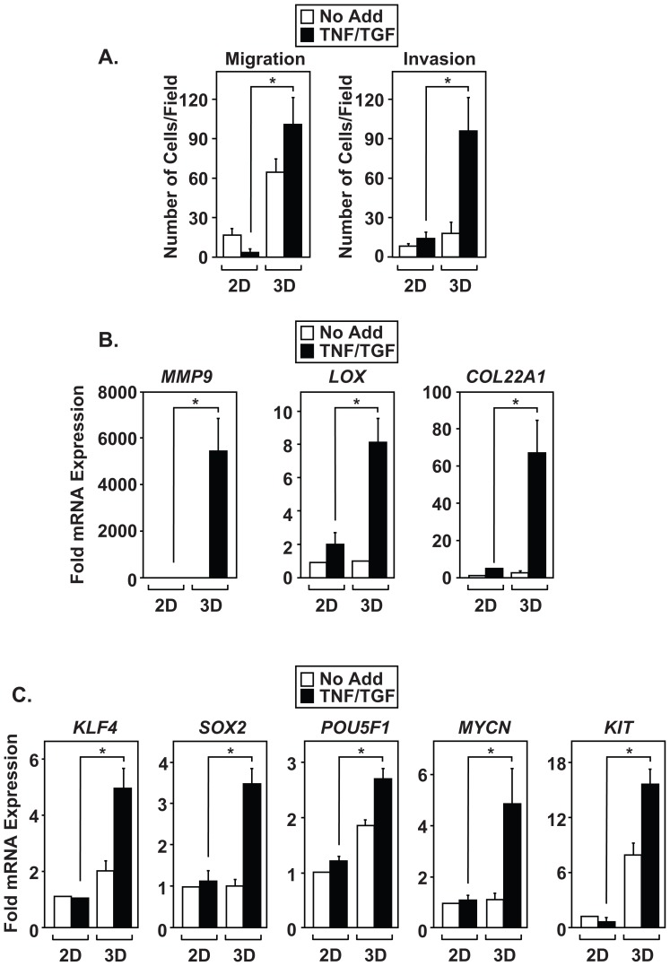
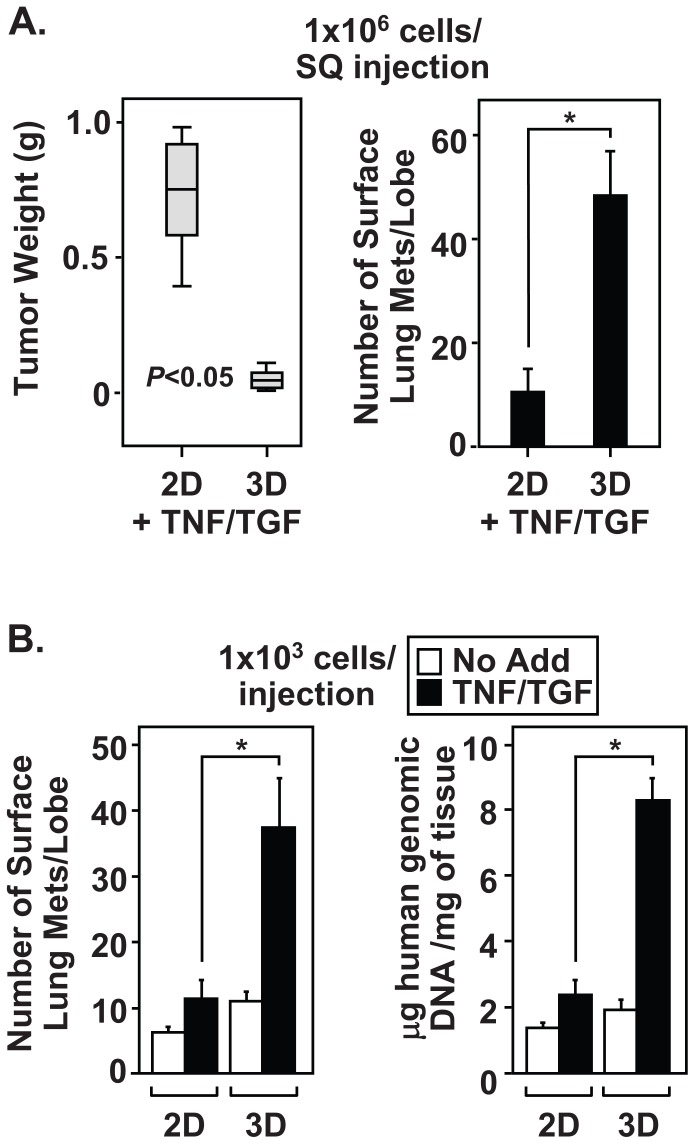
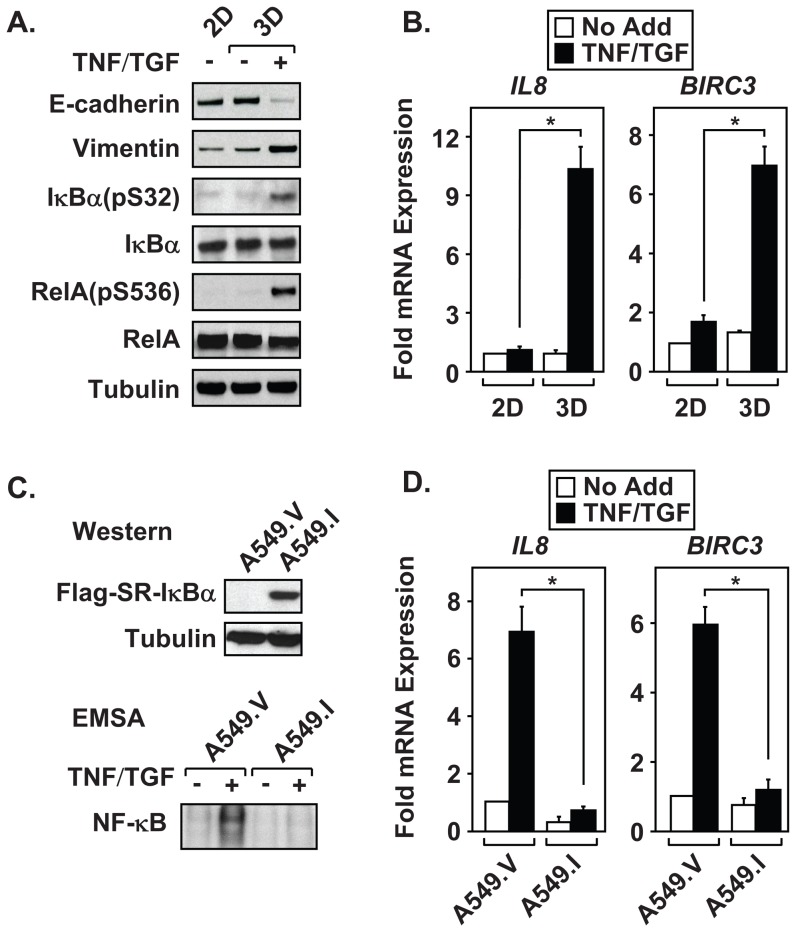
The epithelial-to-mesenchymal transition (EMT) is a de-differentiation process that has been implicated in metastasis and the generation of cancer initiating cells (CICs) in solid tumors. To examine EMT in non-small cell lung cancer (NSCLC), we utilized a three dimensional (3D) cell culture system in which cells were co-stimulated with tumor necrosis factor alpha (TNF) and transforming growth factor beta (TGFβ). NSCLC spheroid cultures display elevated expression of EMT master-switch transcription factors, TWIST1, SNAI1/Snail1, SNAI2/Slug and ZEB2/Sip1, and are highly invasive. Mesenchymal NSCLC cultures show CIC characteristics, displaying elevated expression of transcription factors KLF4, SOX2, POU5F1/Oct4, MYCN, and KIT. As a result, these putative CIC display a cancer "stem-like" phenotype by forming lung metastases under limiting cell dilution. The pleiotropic transcription factor, NF-κB, has been implicated in EMT and metastasis. Thus, we set out to develop a NSCLC model to further characterize the role of NF-κB activation in the development of CICs. Here, we demonstrate that induction of EMT in 3D cultures results in constitutive NF-κB activity. Furthermore, inhibition of NF-κB resulted in the loss of TWIST1, SNAI2, and ZEB2 induction, and a failure of cells to invade and metastasize. Our work indicates that NF-κB is required for NSCLC metastasis, in part, by transcriptionally upregulating master-switch transcription factors required for EMT.


Conflict of interest statement

Competing Interests: The authors have declared that no competing interests exist.

Figures

Figure 1
Figure 1. Establishment of three-dimensional multicellular culture model for EMT studies.
(A) A timeline illustrates the procedure used to create a three-dimensional mesenchymal cell population from confluent monolayers. (B) Spheroid cultures of A549 cells were treated, with TNF, TGFβ, or both cytokines every forty-eight hours for the indicated times. Immunoblot analysis measured changes in epithelial (E-cadherin) and mesenchymal (N-cadherin, Vimentin, and fibronectin) markers over an eight day timecourse. (C) 3D cultures of multiple NSCLC cell lines (A549, H358, H1299) were incubated for ninety-six hours in the absence or presence of TNF and TGFβ. Epithelial and mesenchymal markers were subsequently measured by immunoblot. Results from Figure 1B and 1C are representative examples from at least three independent experiments; α-tubulin acts as a protein loading control.
Figure 2
Figure 2. Three-dimensional cultures show enhanced sensitivity to cytokine treatment.
(A and B) Monolayer (2D) and 3D cultures of A549 cells were left alone (No Add) or treated with TNF and TGFβ (TNF/TGF) for ninety-six hours. Expression of epithelial markers (CDH1), mesenchymal markers (VIM, HMGA2), and EMT master-switch transcription factors (TWIST1, SNAI1, ZEB2, SNAI2) were measured by QRT-PCR. (C) Immunoblot analysis of 3D A549 cultures, left alone (No Add) or treated with TNF and TGFβ (TNF/TGF), was performed on E-cadherin, Vimentin, HMGA2, Twist1, Snail1, Sip1, Slug, and α-tubulin. Results in Figure 2A and 2B were normalized to GAPDH, and are calculated mean ± S.D, *p<0.05, N = 3. Immunoblots in Figure 2C are representative example from at least three independent experiments.
Figure 3
Figure 3. Efficient induction of EMT promotes invasion and the expression of genes required to maintain CICs.
Monolayer and 3D A549 cultures were left alone (No Add) or treated with TNF and TGFβ (TNF/TGF) for ninety-six hours. (A) Cells were disaggregated and subsequently subjected to migration and invasion assays. (B and C) Expression of invasion (MMP-9, LOX, COL22A1) and stem cell markers (KLF4, Sox2, POU5F1, MYCN, and KIT) was measured by QRT-PCR. Results in Figure 3 are calculated mean ± S.D, *p<0.05, N = 3. Results from 3B and 3C were normalized to GAPDH.
Figure 4
Figure 4. Cytokine-treated 3D cultures contain CICs with increased metastatic potential.
(A) Monolayer and 3D A549 cultures were treated with TNF and TGFβ for ninety-six hours. Cells were disaggregated and SC injected into nude mice (1×10⁶ cells/animal). Forty days later, the primary SC tumors were resected and weighed. Additionally, the lungs were excised and the number of surface metastases were determine. (B) Monolayer and 3D A549 cultures were either left untreated or treated with TNF and TGFβ and limiting cell numbers (1×10³/animal) were SC injected into nude mice to evaluate the presence of CICs. Metastasis was evaluated by surface lung tumor count and lung tumor burden was evaluated using genomic QRT-PCR to detect human DNA in total lung tissue. Weight and lung metastases data from Figure 4 are mean ± S.D. of five mice per condition, *p<0.05, N = 3 independent experiments. Genomic QRT-PCR data from Figure 4B are normalized to total lung tissue (mg).
Figure 5
Figure 5. Mesenchymal cells display constitutive NF-κB activity.
Monolayer and 3D cultures of A549 cells were incubated with cytokines for ninety-six hours. (A) Mesenchymal A549 cells display constitutive NF-κB activated pathways, as determined using phospho-specific antibodies to IκBα and RelA. (B) Untreated and TNF and TGFβ stimulated 2D and 3D cultures of A549 cells were harvested and analyzed for expression of NF-κB regulated genes by QRT-PCR. (C and D) Three dimensional cultures of A549.V (vector control) and A549.I (SR-IκB) were incubated for ninety-six hours in the absence or presence of TNF and TGFβ. (C) Immunoblots confirm the expression of the Flag-tagged SR-IκBα in the A549.I line, which successfully blocked nuclear translocation and DNA binding, as measured by EMSA. (D) QRT-PCR confirmed the inability of A549.I cell to upregulate NF-κB-regulated genes following TNF and TGFβ treatment. Immunoblots in Figure 5A are a representative example from three independent experiments. Results in Figure 5B and 5D are calculated mean ± S.D, *p<0.05, N = 3. RNA values were normalized to GAPDH.
Figure 6
Figure 6. NF-κB is required for the maintenance of CICs and lung metastasis.
(A) A549.I cells fail to show changes in mesenchymal markers, as determined by immunoblot analysis. (B) NF-κB is required to upregulate mRNA expression of master-switch transcription factors. (C) Spheroid cultures of A549 and H157 cell lines, expressing empty vector or the Flag-IκB super-repressor, were left alone (No Add) or treated with TNF and TGFβ (TNF/TGF) for ninety-six hours. The cells were disaggregated and subjected to invasion assays. (D) A549.V and A549.I 3D cultures were left alone (No Add) or treated with TNF and TGFβ (TNF/TGF) for ninety-six hours. The cells were disaggregated and SC injected into nude mice (1×10⁶/animal). Forty days later, animals were sacrificed and the number of surface lung metastasis were determined. In addition, SC tumors were excised and wet tumor weight determined. Weight and lung metastases data from Figure 6 are mean ± S.D. of five mice per condition, *p<0.05, N = 3 independent experiments. The graphs in Figure 6 are mean ± S.D., *p<0.05, of three independent experiments. Data with P values greater than 0.05 were considered not significant (ns). QRT-PCR experiments are normalized to GAPDH expression.

References

    1. Cirri P, Chiarugi P (2012) Cancer-associated-fibroblasts and tumour cells: A diabolic liaison driving cancer progression. Cancer Metastasis Rev 31: 195–208 10.1007/s10555-011-9340-x - DOI - PubMed
    1. Korkaya H, Liu S, Wicha MS (2011) Breast cancer stem cells, cytokine networks, and the tumor microenvironment. J Clin Invest 121: 3804–3809 10.1172/JCI57099 - DOI - PMC - PubMed
    1. Fuxe J, Karlsson MC (2012) TGF-beta-induced epithelial-mesenchymal transition: A link between cancer and inflammation. Semin Cancer Biol 22: 455–461 10.1016/j.semcancer.2012.05.004 - DOI - PubMed
    1. Thiery JP, Acloque H, Huang RY, Nieto MA (2009) Epithelial-mesenchymal transitions in development and disease. Cell 139: 871–890. - PubMed
    1. Yang J, Weinberg RA (2008) Epithelial-mesenchymal transition: At the crossroads of development and tumor metastasis. Dev Cell 14: 818–829 10.1016/j.devcel.2008.05.009 - DOI - PubMed

Publication types

MeSH terms