. 2013 Jul 25;8(7):e68841.
doi: 10.1371/journal.pone.0068841. Print 2013.

Orthosteric binding of ρ-Da1a, a natural peptide of snake venom interacting selectively with the α1A-adrenoceptor

Affiliations

Orthosteric binding of ρ-Da1a, a natural peptide of snake venom interacting selectively with the α1A-adrenoceptor

Arhamatoulaye Maïga et al. PLoS One. .

Abstract

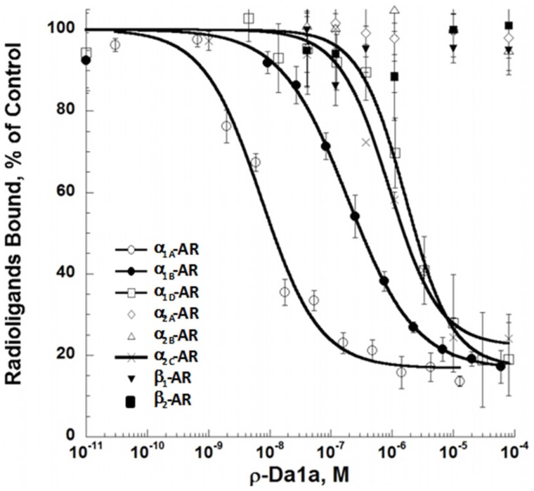
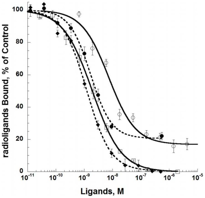
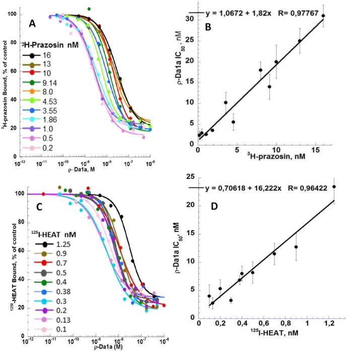
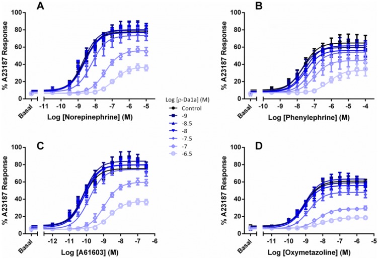
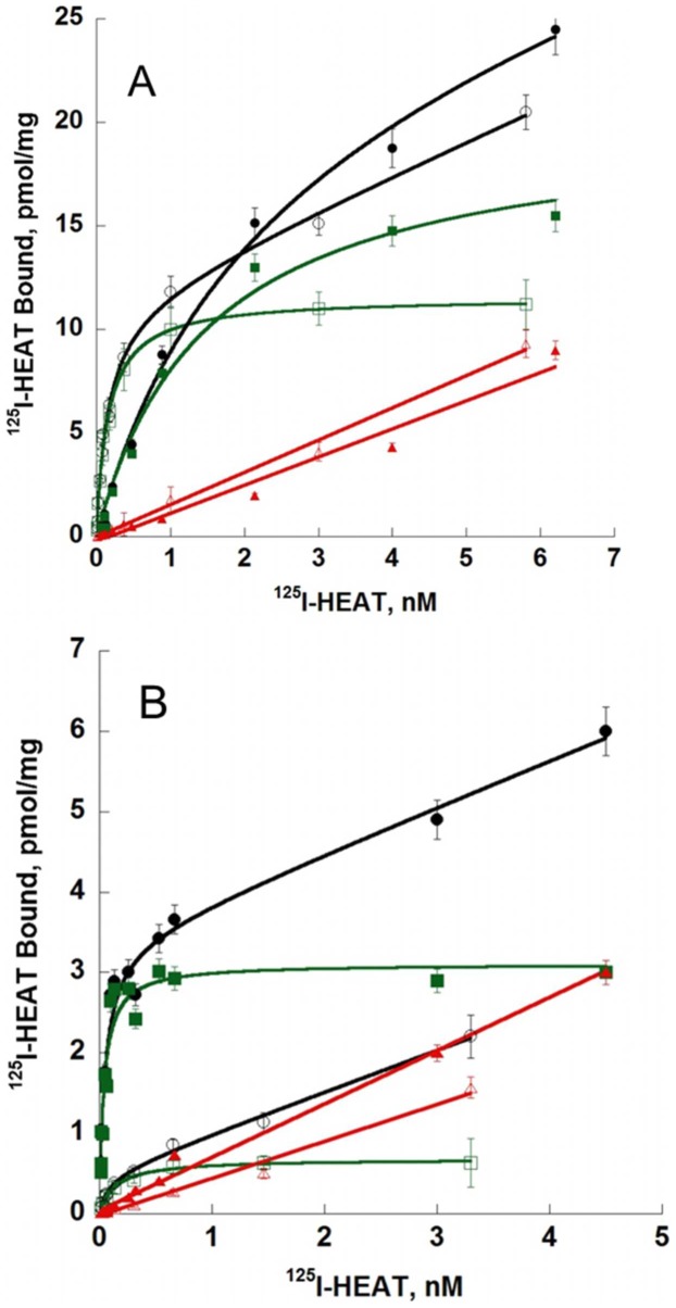
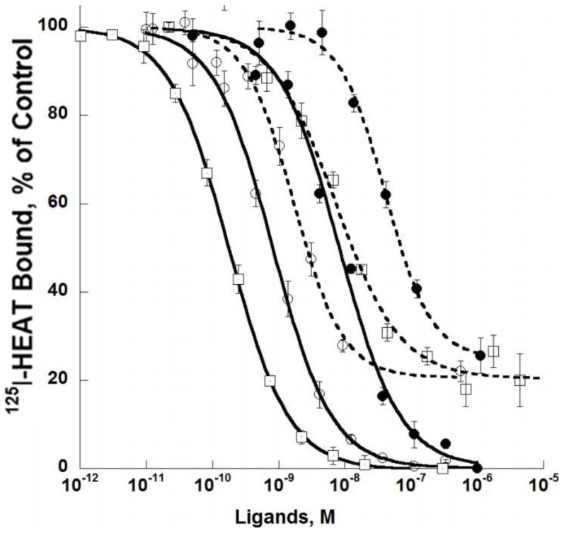
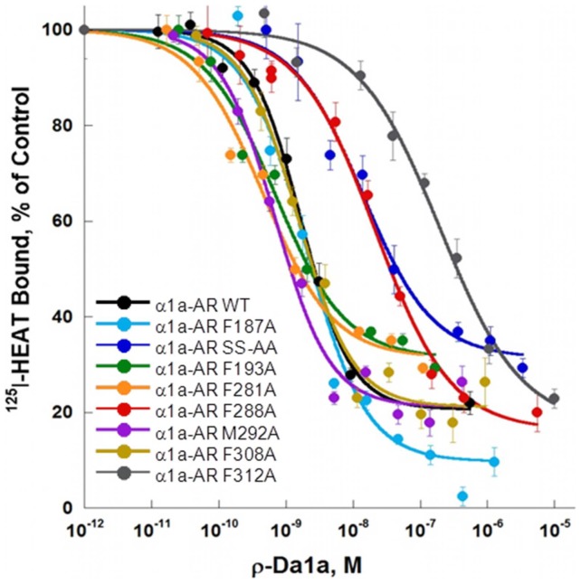
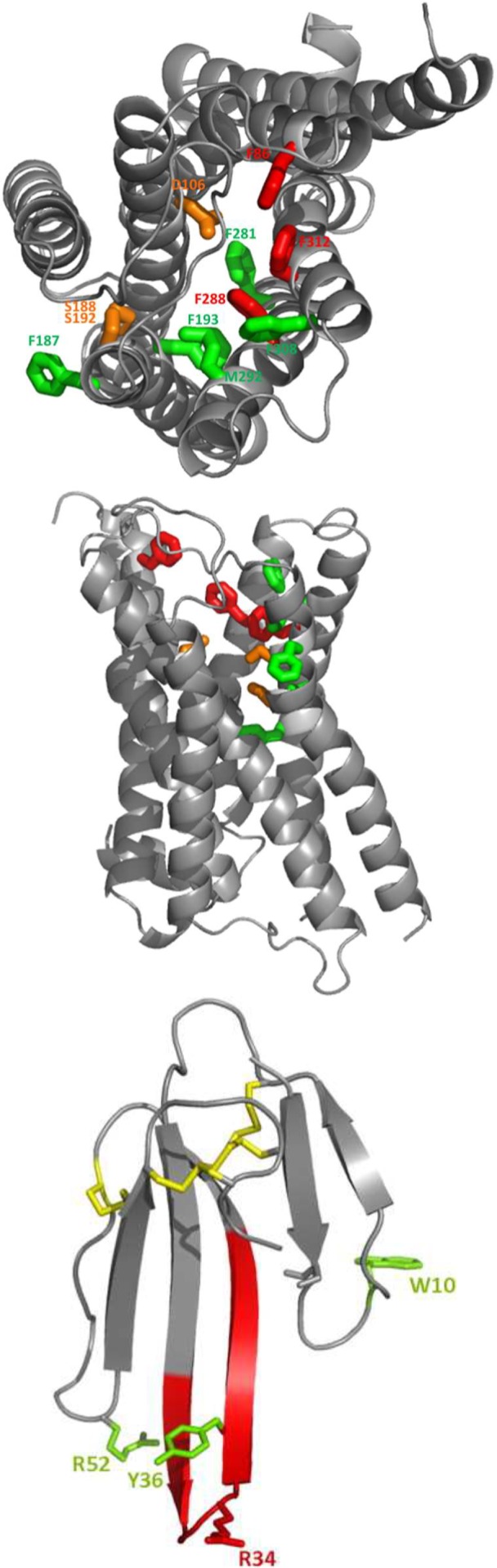
ρ-Da1a is a three-finger fold toxin from green mamba venom that is highly selective for the α1A-adrenoceptor. This toxin has atypical pharmacological properties, including incomplete inhibition of (3)H-prazosin or (125)I-HEAT binding and insurmountable antagonist action. We aimed to clarify its mode of action at the α1A-adrenoceptor. The affinity (pKi 9.26) and selectivity of ρ-Da1a for the α1A-adrenoceptor were confirmed by comparing binding to human adrenoceptors expressed in eukaryotic cells. Equilibrium and kinetic binding experiments were used to demonstrate that ρ-Da1a, prazosin and HEAT compete at the α1A-adrenoceptor. ρ-Da1a did not affect the dissociation kinetics of (3)H-prazosin or (125)I-HEAT, and the IC50 of ρ-Da1a, determined by competition experiments, increased linearly with the concentration of radioligands used, while the residual binding by ρ-Da1a remained stable. The effect of ρ-Da1a on agonist-stimulated Ca(2+) release was insurmountable in the presence of phenethylamine- or imidazoline-type agonists. Ten mutations in the orthosteric binding pocket of the α1A-adrenoceptor were evaluated for alterations in ρ-Da1a affinity. The D106(3.32)A and the S188(5.42)A/S192(5.46)A receptor mutations reduced toxin affinity moderately (6 and 7.6 times, respectively), while the F86(2.64)A, F288(6.51)A and F312(7.39)A mutations diminished it dramatically by 18- to 93-fold. In addition, residue F86(2.64) was identified as a key interaction point for (125)I-HEAT, as the variant F86(2.64)A induced a 23-fold reduction in HEAT affinity. Unlike the M1 muscarinic acetylcholine receptor toxin MT7, ρ-Da1a interacts with the human α1A-adrenoceptor orthosteric pocket and shares receptor interaction points with antagonist (F86(2.64), F288(6.51) and F312(7.39)) and agonist (F288(6.51) and F312(7.39)) ligands. Its selectivity for the α1A-adrenoceptor may result, at least partly, from its interaction with the residue F86(2.64), which appears to be important also for HEAT binding.


Conflict of interest statement

Competing Interests: The authors have declared that no competing interests exist.

Figures

Figure 1
Figure 1. Pharmacological profile of ρ-Da1a binding to various human AR subtypes expressed in eukaryotic cells.
Binding inhibition curves for 3H-prazosin (2 nM), 3H-rauwolscine (2 nM) and 3H-CGP-12177 (6 nM) on hα1A- (1 µg, ○), hα1B- (3 µg, •), hα1D- (29 µg, □), hα2A- (140 µg, ◊), hα2B- (100 µg, Δ), hα2C- (3 µg, x), β1- (3 µg,▾) and β2-AR (1.5 µg, ▪) with recombinant ρ-Da1a. n = 4.
Figure 2
Figure 2. Inhibition of 3H-prazosin (2 nM, 1 µg, open symbols) by HEAT (□), and ρ-Da1a (circle), and inhibition of 125I-HEAT (0.2 nM, 0.2 µg, full symbols) binding by prazosin (♦) and ρ-Da1a (circle) to α1A-AR.
n = 3.
Figure 3
Figure 3. Influence of various ligands on 3H-prazosin and 125I-HEAT dissociation.
Panel A: Dissociation of 3H-prazosin (2 nM) binding to α1A-AR (1 µg) in the presence of prazosin (10 µM, black), prazosin plus ρ-Da1a (2.5 µM, blue), prazosin plus adrenaline (2 mM, red) and prazosin plus EPA (150 µM, green). Panel B : dissociation of 125I-HEAT (0.4 nM) binding to α1A-AR (0.2 µg) in the presence of HEAT (5 µM, black), HEAT plus ρ-Da1a (2.5 µM, blue), HEAT plus prazosin (10 µM, red) and HEAT plus EPA (150 µM, green). n = 2.
Figure 4
Figure 4. Inhibition of the binding of a series of concentrations of 3H-prazosin and 125I-HEAT to α1A-AR by ρ-Da1a.
Panel A 3H-prazosin binding (from 0.2 to 16 nM) inhibited by ρ-Da1a. Panel B 125I-HEAT binding (from 0.1 to 1.25 nM) inhibited by ρ-Da1a. Panel C and D: Fitting, by the Cheng and Prusoff equation IC50 = Ki+Ki(L/Kd), of IC50 values as a function of the radiotracer concentrations.
Figure 5
Figure 5. Concentration-response curves for stimulation of Ca2+ release by the α1A-AR.
Agonist responses represent the difference between basal fluorescence and the peak [Ca2+]i (reached within 20 sec of agonist addition), expressed as a percentage of the response to the Ca2+ ionophore A23187 (1 µM). Concentration-dependent Ca2+ release was stimulated by noradrenaline (panel A), phenylephrine (panel B), A61603 (panel C) or oxymetazoline (panel D). Concentration response curves were performed in the presence or absence of differing concentrations of ρ-Da1a (• control, ▪ 1 nM, ▴ 3 nM, ▾ 10 nM, ♦ 30 nM, ◊ 100 nM, ○ 300 nM). Values are means ± SEM of 3–4 independent experiments.
Figure 6
Figure 6. Saturation experiments with 125I-HEAT on receptor variants.
Figure 7
Figure 7. Receptor affinities for ρ-Da1a (dash lines) and HEAT (solid lines) on mutated α1A-ARs.
Binding inhibition curves for 125I-HEAT binding to WT (200 pM, 0.2 µg, ○), D1063.32A (200 pM, 1 µg, □) and F862.64A (1.3 nM, 0.8 µg, •) receptor variants. n = 3–4.
Figure 8
Figure 8. Receptor affinities for ρ-Da1a on mutated α1A-ARs.
Binding inhibition curves for 125I-HEAT (200 pM) binding to WT (0.2 µg, black), F1875.41A (0.15 µg, light blue), the double S1885.42,S1925.46-AA (0.3 µg, dark blue), F1935.47A (0.25 µg, green), F2816.44A (0.15 µg, orange), F2886.51A (0.2 µg, red), M2926.55A (0.2 µg, purple), F3087.35A (0.1 µg, brown), F3127.39A (0.8 µg, grey), n = 3–4.
Figure 9
Figure 9. Homology modelling of the ρ-Da1a binding site in the α1A-AR and the MT7 toxin.
Views from the side of the TM bundle (Panel A), and from the top of the extracellular space (Panel B). F1875.41, F1935.47, F2816.44, M2926.55, F3087.35 in green. D1063.32 and the double S1885.42/S1925.46 in orange. F862.64, F2886.51 and F3127.39 in red. Panel C :3D structure of the three-finger fold MT7 toxin (2vlw) with the four conserved disulfide bridges in red.

Similar articles

Cited by

References

    1. Escoubas P, King GF (2009) Venomics as a drug discovery platform. Expert Rev Proteomics 6: 221–224. - PubMed
    1. Halai R, Craik DJ (2009) Conotoxins: natural product drug leads. Nat Prod Rep 26: 526–536. - PubMed
    1. Lewis RJ, Garcia ML (2003) Therapeutic potential of venom peptides. Nat Rev Drug Discov 2: 790–802. - PubMed
    1. Shen GS, Layer RT, McCabe RT (2000) Conopeptides: From deadly venoms to novel therapeutics. Drug Discov Today 5: 98–106. - PubMed
    1. Maïga A, Mourier G, Quinton L, Rouget C, Gales C, et al. (2012) G protein-coupled receptors, an unexploited animal toxin targets: Exploration of green mamba venom for novel drug candidates active against adrenoceptors. Toxicon 59: 487–496. - PubMed

MeSH terms

LinkOut - more resources